

骨组织再生中的石墨烯家族材料:前景和挑战
摘要
近年来,我们见证了石墨烯家族材料生物应用研究的大量突破。由于其纳米级尺寸、大比表面积、光致发光特性和抗菌活性,石墨烯家族材料在骨组织工程、药物/基因传递和生物传感/成像应用方面具有巨大的潜力。在此综述中,我们回顾了石墨烯研究的最新进展和成果,并对石墨烯家族材料在骨组织再生中的各种生物医学应用的生物安全性和可行性进行了批判性分析和讨论。
介绍
患有颌骨缺损的严重颌面感染、外伤、肿瘤和先天性畸形的受害者通常需要长期康复。与许多其他组织不同,骨骼在受损时具有出色的再生能力 [1, 2]。然而,人体骨骼的自我再生能力有限,使得足够大或临界尺寸的骨缺损的重建成为临床治疗的重大挑战[3]。在某些情况下,重症患者甚至需要进行大量的骨增强手术。目前的骨再生疗法包括自体移植、同种异体移植和异种移植 [4]。自体骨被认为是“金标准”的骨移植材料,具有成骨、成骨和成骨的能力,同时没有免疫原性。但自体移植在临床上仍受限于供体感染风险和恢复时间长的原因[5]。从另一个人那里获得的同种异体移植物通常被认为是次优选择。但使用同种异体移植物存在潜在风险,例如感染和免疫排斥风险显着增加 [4, 6]。异种移植材料,如酸消化脱矿骨基质和牛胶原蛋白,很容易获得和制造。现在,异种移植是临床实践中的主要方法。但它的骨诱导能力较低 [7]。目前,与骨相比,尚无具有优越甚至相同的生物学或力学性能的异源或合成骨替代品[5]。尽管这些疗法已被证明是有用的,但它们也面临着固有的挑战。因此,仍然需要研究和开发适当的骨再生疗法。诚然,骨组织工程和再生医学研究为改善骨缺损患者的预后和加速康复开辟了道路 [8]。组织工程骨结构有可能缓解因缺乏合适的自体移植物和同种异体移植物材料而引起的用于促进骨愈合的需求 [9]。为了增加骨缺损区域的骨量,已经开发了多种骨再生方法,包括支架 [1, 6]、涂层 [10] 和用于引导骨再生 (GBR) 的屏障膜 [11, 12]。
目前,石墨烯家族材料作为 2D 平面涂层或 3D 多孔支架用于将各种类型的干细胞分化为神经源性 [13,14,15]、软骨源性 [16, 17]、肌源性 [18] 的潜力已引起极大的关注。 、脂肪生成 [19] 和成骨谱系 [20、21]。因此,石墨烯家族材料更有可能成为下一代骨再生材料的候选材料。石墨烯,定义为单层或几层 sp 2 -杂化碳原子,2004 年由 Novoselov 和 Geim 首次从石墨中分离出来 [22]。随着研究兴趣的增加,石墨烯家族的材料,包括氧化石墨烯(GO)、羧基石墨烯(CXYG)、还原氧化石墨烯(rGO)和石墨烯量子点(GQDs),得到了广泛的研究。石墨烯具有优异的机械、导电、热学和光学特性 [23,24,25],已广泛应用于电子、生物技术和聚合物科学 [26]。众所周知,具有良好导电性的导电材料可增强细胞活性并刺激骨组织修复 [27, 28],同时也表现出良好的抗菌活性 [29]。氧化石墨烯(GO)和羧基石墨烯(CXYG)都是石墨烯的衍生物。由于存在含氧官能团(环氧化物、羧基和羟基),GO 和 CXYG 在亲水性溶剂中具有更好的分散性,这对于生物医学应用至关重要 [30, 31]。还原氧化石墨烯 (rGO) 可以通过在特定条件下用特定还原剂还原 GO 来合成。由于减少了一些特殊的 π-π 化学相互作用,rGO 具有某些比石墨烯和 GO 更好的物理和化学性质 [32, 33]。石墨烯量子点(GQDs)的原材料是GO。 GQD 具有很强的量子限制和光致发光特性 [34]。 GQD 的强荧光使其可用于细胞成像。由于石墨烯家族材料的优异特性,它们在药物/基因传递、生物传感/成像应用和组织工程方面具有巨大的潜力 [35,36,37,38,39]。然而,石墨烯家族材料的长期生物安全性和诱导细胞成骨分化的能力仍然存在挑战。在这里,我们全面回顾了石墨烯及其衍生物的最新进展和成就。同时,我们批判性地分析了体外和体内生物安全性,并讨论了石墨烯家族材料用于骨组织再生的各种生物医学应用的可行性。
确定石墨烯家族材料生物安全性的挑战
确定体外生物安全性的挑战
在考虑将石墨烯家族材料用于临床试验之前,应对其细胞毒性和生物相容性进行严格评估 [38]。 “石墨烯是一种生物相容性材料吗?”答案仍然存在争议。未经任何功能化的原始石墨烯是疏水的,在水性介质中很容易团聚 [34, 40]。在疏水表面上,一层致密的非特异性蛋白质可以从表面置换水并立即积聚在材料上,从而对纳米颗粒进行免疫识别 [41]。因此,化学功能化,包括氧化、还原和引入官能团,是石墨烯用于生物医学应用的先决条件,这增加了石墨烯的亲水性。具有不同功能的石墨烯家族材料具有不同的化学性质,具有不同的毒性[13]。苏门等人。发现 rGO 的毒性低于 GO。有趣的是,氧化应激随着 rGO 表面氧官能团密度的增加而增强。他们得出结论,GO 片上的官能团密度是介导细胞毒性的关键因素之一 [31]。除了表面功能化外,石墨烯家族材料的细胞毒性还受到多种因素的影响,包括它们的浓度、大小和形状 [42]。
首先,一些研究表明,石墨烯家族材料具有剂量依赖性细胞毒性,有或没有时间依赖性细胞毒性。例如,Chang 等人。据报道,在高浓度 GO(≥ 50 μg/mL)下观察到细胞活力的轻微丧失,并且 GO 可诱导细胞内积聚并在肺癌上皮细胞系(A549)中引起剂量依赖性氧化应激[43]。魏等人。证明原始 GO 在 10 μg/mL 的高浓度下抑制骨间充质干细胞 (BMSCs) 的增殖,而在 0.1 μg/mL 的低浓度下增强 BMSCs 的增殖 [44]。类似地,在 200 μg/mL GO 时可以清楚地观察到细胞数量减少,并且在 300 μg/mL GO 时报告了更大的细胞毒性作用 [45]。更重要的是,Kim 等人。发现前成骨细胞 (MC3T3-E1) 的生存能力在浓度 <62.5 μg/mL 时受 rGO 的轻微影响,但显着(p <0.05) 在较高浓度 (≥ 100 μg/mL) 时减少 [23]。此外,CXYG、GQDs 在低浓度应用时都显示出很小的细胞毒性潜力 [34, 46]。简而言之,石墨烯家族材料在低浓度下具有细胞相容性,对细胞形态、活力和增殖的负面影响很小,但浓度不是唯一的相关因素。
其次,表明层状、纳米片和薄片、带状和点状等不同形状也有助于石墨烯家族细胞毒性的复杂性[40]。塔鲁克达尔等人。评估了石墨烯纳米洋葱 (GNO)、GO 纳米带 (GONR) 和 GO 纳米血小板 (GONP) 的细胞毒性。 CD50 值遵循 GNOs> GONRs> GONPs 的趋势,表明与 GONPs 相比,GONRs 更具细胞毒性 [47]。因此,石墨烯家族纳米材料的形状也是介导细胞毒性的关键组成部分。例如,石墨烯和多壁碳纳米管 (MWNT) 具有不同的形状(石墨烯为扁平原子片,纳米管为管状),但它们的化学成分和晶体结构相似。 GO 直到 50 μg/mL 才显示出 SK-N-SH 细胞的细胞生长抑制活性。相比之下,MWCNTs 在低浓度 (6.7 μg/mL) 下抑制细胞增殖,表明其具有急性细胞毒性。对于 HeLa 细胞,即使在高达 50 μg/mL 的浓度下,GO 也表现出轻微的生长抑制活性,而 MWCNTs 对 HeLa 细胞具有中等的细胞毒性 [48]。他们依赖于这种现象的不同形状和不同的物理/化学方式。由于扁平形状,预计石墨烯家族材料与细胞膜的相互作用很小。 MWCNT 的管状形状促进了膜的渗透,导致细胞毒性 [48,49,50]。另一个重要信息是,除了功能化、浓度、大小和形状的依赖性之外,纳米结构石墨烯衍生物的细胞毒性还取决于细胞类型。作为神经细胞系,SK-N-SH细胞对纳米结构石墨烯衍生物的不利影响表现出比HeLa细胞更高的敏感性[48]。
第三,尺寸对石墨烯家族材料的生物安全性也起着重要作用。尹等人。通过基于细胞的电化学阻抗生物传感器评估了石墨烯纳米薄片的尺寸依赖性细胞毒作用。他们发现较小的石墨烯纳米薄片 (30.9 ± 5.4 nm) 由于细胞摄取较高而诱导细胞凋亡,而较大的石墨烯纳米薄片 (80.9 ± 5.5 nm) 主要聚集在细胞膜上,引起的毒性较小 [51]。众所周知,纳米材料的细胞摄取特性可能会影响细胞增殖、分化和纳米颗粒的排泄[52]。穆等人。详细阐述了蛋白质包覆的 GO 纳米片可能的尺寸依赖性摄取机制,并观察到较大的纳米片(860 ± 370 nm)首先附着在细胞表面,然后膜内陷,伪足延伸,最终主要通过吞噬作用进入细胞,而较小的纳米片( 420 ± 260 nm) 主要通过网格蛋白介导的内吞作用进入细胞 [33]。达斯等人。将人脐静脉内皮细胞 (HUVEC) 接种在 10 μg/mL 的 GO 和 rGO 中,具有不同尺寸的片层(800 nm 和 400 nm)。结果表明,在 MTT 测定中,较小尺寸的片材比较大尺寸的片材毒性更大。然后,较大尺寸的 GO 和 rGO(800 纳米)被超声处理以破碎成更小的尺寸(70 纳米)。超声处理后观察到细胞毒性增加,表明较小尺寸的 GO 和 rGO 表现出更大的毒性 [31]。类似地,将 MCF7 细胞暴露于四种大小的 GO 样品(744 ± 178 nm、323 ± 50 nm、201 ± 28 nm 和 100 ± 10 nm)。与未处理的细胞相比,即使在暴露于较大尺寸的 GO 分散体(744 ± 178 nm)72 小时后,在体外也未观察到细胞毒性,而用 100 ± 10 nm 尺寸的 GO 分散体处理导致细胞增殖减少约 50% 的未处理细胞 [53]。根据上述结果,研究了从 30 到 860 nm 的各种尺寸的石墨烯家族材料。我们似乎得出结论,较小尺寸的石墨烯家族材料比较大尺寸的材料毒性更大。但是不同的团队有不同的标准来定义石墨烯及其衍生物的尺寸尺度。因此,这个结论或许值得商榷。同时,据报道,纳米级石墨烯家族材料对于生物医学应用更加安全[54]。石墨烯家族材料的尺寸控制合成需要在后续研究中慎重考虑。
得出的结论是,石墨烯的细胞毒性与石墨烯家族的种类、化学功能化、浓度、形状和大小密切相关。未来,我们的目标是通过更好地控制浓度和尺寸,通过用各种类型的官能团修饰石墨烯家族,制造出与细胞、组织或生物体相互作用更好的生物相容性装置。
确定体内生物安全性和生物分布的挑战
为了进一步检测石墨烯家族材料是否为生物相容性材料并增强其在广泛应用中的拟议用途,体内实验是必不可少的方法。许多关于石墨烯家族材料在体内的生物相容性和生物分布的研究与其细胞研究几乎一致。乔杜里等人。将斑马鱼胚胎应用于较大尺寸的 GO 分散体,发现与对照组相比胚胎死亡率没有增加,而在较小尺寸的 GO 分散体中观察到胚胎活力降低 [53]。 GO 不会导致胚胎细胞凋亡显着增加,而 MWCNTs 即使在 25 mg/L 的相对低浓度下也会导致发育中的胚胎出现严重的形态缺陷 [48]。这些研究进一步表明,体内毒性很大程度上取决于石墨烯及其衍生物的大小、浓度和形状。此外,石墨烯家族材料通常通过静脉注射、吸入或皮下植入暴露于动物模型。因此,毒性、一般组织学和生物分布的变化各不相同。李等人。通过静脉注射评估纳米级GO在小鼠体内的毒理学,发现GO主要保留在肝、肺和脾脏中,并引起损伤、慢性肝炎和肺纤维化。 GO (GO-PEG) 的聚乙二醇 (PEG) 涂层可以减少 GO 在肝、肺和脾中的滞留,减轻急性组织损伤 [55]。杜赫等人。探索了降低石墨烯纳米材料在肺中毒性作用的策略,因为他们发现当直接施用于小鼠肺部时,GO 比聚集的石墨烯和 Pluronic 分散的石墨烯具有更高的毒性,从而导致严重和持续的肺损伤。通过液相剥离制备原始石墨烯可显着减轻毒性,并在与嵌段共聚物 Pluronic 一起分散时进一步降低毒性 [56]。查等人。确定了 3D 石墨烯泡沫 (GF) 或氧化石墨烯泡沫 (GOF) 在皮下植入大鼠模型中的短期(植入后前 2 周)和长期(7 个月)体内毒性和性能。血液分析表明,GFs 和 GOFs 在植入后不会引起明显的血液、肝或肾毒性,并且在植入至少 7 个月后没有观察到明显的降解。仅观察到植入部位长期存在肉芽肿。 HE 染色图像显示出更好的体内生物相容性(图 1)[40]。 Zha等人的原因。如前所述,获得比其他研究更积极的结果可能是给药途径不同。皮下实验是评估植入材料体内生物相容性的非常直接有效的方法[57],它可能对石墨烯家族纳米材料在体内的接触模式、沉积位置甚至降解途径产生影响[58]。控制复合材料的降解在组织工程中至关重要,
<图片>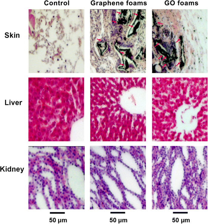
在植入后第 14 天植入石墨烯泡沫、GO 泡沫或不植入任何主要器官(从大鼠收集的植入区域、肝脏和肾脏)的代表性 HE 染色图像。未观察到明显的器官损伤或病变。转载自参考。 [40] 经纳米粒子研究杂志许可
一般来说,细胞研究在初步细胞毒性分析、了解与细胞相互作用的可能机制方面表现突出。但它是一个复杂得多的体内微环境。了解石墨烯家族材料在潮湿腐蚀性微环境中的表现也很关键。石墨烯家族材料的生物相容性与浓度、官能团种类、石墨烯家族的类型、尺寸和形状密切相关。但其机制仍需进一步深入细致地研究。然而,体内生物安全性的评估相对较少,尤其是长期的生物相容性和生物分布,需要我们多加关注。尽管一些论文引起了对生物安全性的担忧,但石墨烯家族独特的潜在多功能性使其成为生物医学应用的竞争选择。
石墨烯家族材料的抗菌活性
如果没有无菌的骨缺损微环境,骨重塑和新骨形成就不可能完全成功。事实上,感染性骨缺损的治疗仍然是一个重大挑战[59]。由于骨缺损大,感染问题多,治疗难度大,患者需要长期康复治疗。因此,石墨烯家族材料的细菌抑制能力有很大帮助。石墨烯家族材料被认为具有抗菌能力(表 1)。刘等人。提出了一种三步抗菌机制,包括(1)石墨烯基材料上的初始细胞沉积,(2)由与尖锐纳米片直接接触引起的膜应力,以及(3)随后的超氧阴离子独立氧化[60]。然而,Mangadlao 等人。认为石墨烯的表面主要负责抗菌活性,而不是边缘。当与细菌接触时,石墨烯充当电子受体,将电子泵离细菌膜,产生独立的氧化应激 [61]。与此同时,李等人。为更好地理解石墨烯薄膜的抗菌作用提供了新的见解。他们认为石墨烯家族材料的抗菌活性不是源于活性氧 (ROS) 介导的损伤,而是通过从微生物膜到石墨烯的电子转移相互作用 [62],而 Panda 等人。证明非氧化性电子转移机制和随之而来的 ROS 介导的氧化应激对细菌的协同影响诱导了天然衍生的 GO 金属薄膜的抗菌活性增强 [63]。
尽管石墨烯基片材的理化性质如何影响其抗菌活性尚不确定,但石墨烯家族材料的抗菌能力值得我们进一步研究和利用。
石墨烯家族材料介导细胞成骨分化并促进体内骨再生
许多学者指出,石墨烯不仅可以使细胞(如牙髓干细胞[64、65]、骨髓干细胞[8、20、66、67]、牙周膜干细胞[68])附着和增殖。 ]、人成骨细胞 [69]、成纤维细胞 [70]、肿瘤细胞 [43]) 没有明显的细胞毒性迹象,但也可以诱导早期细胞成骨细胞分化并产生高度矿化 [20, 64,65,66,67, 68]。目前,众多团队煞费苦心地进行了大量研究,以设计出应用石墨烯家族纳米材料作为支架或支架添加剂、作为基底材料表面的涂层、作为引导骨再生膜以及作为药物递送载体的新策略。 (图 2)。他们尝试使用石墨烯族材料进一步改善基材的某些性能,并赋予基材基复合材料生物活性。
<图片>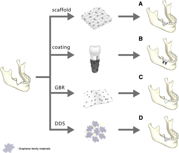
A. 石墨烯家族材料作为支架或支架中用于骨再生的增强材料。 B. 石墨烯家族材料作为涂层转移到基材上用于骨再生。 C. 石墨烯家族作为引导骨膜的添加剂。 D. 石墨烯家族材料作为药物递送系统促进骨再生
石墨烯族材料作为支架或支架中的增强材料
骨组织工程最常见的策略是模拟骨重塑和再生的自然过程。三维 (3D) 生物相容性、可生物降解和骨传导性或骨诱导性支架可以满足该策略 [3]。这种支架可以提供理想的微环境来模拟细胞外基质 (ECM),用于成骨细胞的附着、迁移、增殖和分化以及生长因子的载体 [6]。石墨烯作为一种有前途的生物相容性支架可以使大表面积可用于细胞扩散甚至基质中的成骨分化[20]。例如,用作人类间充质干细胞 (hMSC) 培养基质的 3D 石墨烯泡沫提供了证据,证明它们能够维持干细胞活力并促进成骨分化[66]。此外,3D 石墨烯 (3DGp) 支架和 2D 石墨烯 (2DGp) 涂层被证明能够通过更高水平的矿化和上调骨相关基因来诱导牙周膜干细胞 (PDLSC) 分化为成熟的成骨细胞。石墨烯上的蛋白质,使用或不使用化学诱导剂[68]。
如今,作为支架的各种生物材料如雨后春笋般涌现。用于骨再生的潜在合适的合成支架包括磷酸钙,例如羟基磷灰石 (HA) [71]; β-磷酸三钙(β-TCP)[72];合成或生物聚合物,如聚乳酸 (PLA) [73]、聚乙醇酸 (PLGA) [74]、聚己内酯 (PCL) [75]、壳聚糖 (CS) [1] 和胶原蛋白 [76] ];和上述材料的复合材料 [77, 78]。但现在,最重要的问题之一是支架的机械性能。由于天然骨表现出超弹性生物力学特性,杨氏模量值在 7-27 GPa [79] 范围内,理想的支架应该模仿天然骨的强度、刚度和机械性能。石墨烯家族材料可以作为支架中的增强材料添加,旨在增强机械性能并改善物理化学表征。例如,纯 PCL 支架的拉伸强度为 1.61 MPa,伸长率为 122%,杨氏模量为 7.01 MPa。添加 GO (2%) 后,拉伸强度显着增加到 3.50 MPa,伸长率增加到 131%,杨氏模量增加到 15.15 MPa [80]。
在成功使用石墨烯家族材料作为增强材料的刺激下,许多团队将合成或生物聚合物提供的生物相容性与石墨烯家族材料的显着物理特性相结合。他们期望获得一种理想的复合支架,具有改善的机械性能、合适的孔隙率、结构设计和优异的生物相容性,以支持和诱导新骨形成。
具有磷酸钙基材料的石墨烯家族
人体骨骼由 30% 的有机物质(主要是胶原蛋白)和 70% 的无机物质(主要是羟基磷灰石)(HA;Ca10(PO4)6(OH)2)组成 [81, 82]。合成磷酸钙基材料如HA、β-磷酸三钙(β-TCP)和磷酸钙骨水泥(CPC)是流行的支架材料,因为它们的组成和结构与骨的天然矿物相相似,而且具有良好的成骨性。能力 [83,84,85]。特别是由于HA具有良好的骨传导和骨诱导能力[86],长期以来一直被广泛用作骨科或颌面手术中的人工骨移植物,以修复骨缺损区域[11、71]。然而,HA材料固有的缺点,如成型困难,特殊的脆性,断裂韧性低[87, 88]。据报道,石墨烯家族材料增强的 HA 复合材料被开发出来,显着提高了断裂韧性和生物性能。例如,HA/石墨烯复合材料是通过放电等离子烧结(SPS)制备的,这赋予了 HA 可接受的强度 [89]。劳奇等人。以两种不同的方法将 HA 与 GO 相结合:原位溶胶-凝胶方法和仿生方法。通过原位溶胶-凝胶方法获得的 HA-GO 在不使用成骨因子的情况下增强了 hMSCs 的细胞活力并诱导了成骨细胞分化。通过仿生方法形成的 HA-GO 可维持细胞活力和增殖 [90]。此外,还原氧化石墨烯 (rGO) 还可用作 HA 的增强材料。 HA-rGO 复合材料的断裂韧性达到 3.94 MPa m 1/2 ,与纯 HA 相比增加了 203%。 HA-rGO 增强了细胞增殖和成骨细胞分化,这是通过人成骨细胞的碱性磷酸酶 (ALP) 活性评估的 [91]。此外,Nie 等人。通过自组装成功合成了 rGO 和纳米羟基磷灰石 (nHA) 3D 多孔复合材料支架 (nHA@rGO)。 GO 溶液与 nHA 水悬浮液混合,加热以诱导自组装过程。最后,将反应产物冷冻干燥以获得3D多孔支架。 nHA@rGO 支架可以显着促进大鼠骨间充质干细胞 (rBMSCs) 的细胞增殖、ALP 活性和成骨基因表达。体内实验表明,20% 掺入 nHA 的 rGO (nHA@rGO) 多孔支架可以加速兔子圆形颅骨缺损的愈合 [92]。此外,双组分和三组分均具有优异的性能,具有良好的细胞相容性和改善的亲水性和机械性能[93,94,95]。
磷酸三钙,磷酸钙的类似物,是一种三级磷酸钙,也称为骨灰[Ca3(PO4)2]。它是钙和磷的丰富来源,很容易被吸收。 β-磷酸三钙 (β-TCP) 具有高度生物相容性,可在缺损部位形成可吸收的互锁网络以促进愈合 [96]。吴等人。成功合成了 2D β-TCP-GO 圆盘和 3D β-TCP-GO 支架。与 β-TCP 和空白对照相比,2D β-TCP-GO 圆片通过激活 Wnt 相关信号通路显着增强 hBMSCs 的增殖、ALP 活性和成骨基因表达,表明 GO-改良的 β-TCP [85]。众所周知,Wnt 经典信号通路在调节细胞活动(如细胞增殖、分化和形态发生)方面发挥着重要作用 [97, 98]。体内研究表明,与纯 TCP 支架相比,3D β-TCP-GO 支架在颅骨缺损处具有更多的新骨形成(图 3)[85]。一种新型支架,磷酸钙水泥掺入 GO-Cu 纳米复合支架(CPC/GO-Cu)促进了 rBMSCs 的粘附和成骨分化,证实它们可以通过激活 Erk1/2 上调 rBMSCs 中 Hif-1α 的表达信号通路并诱导血管内皮生长因子 (VEGF) 和 BMP-2 蛋白的分泌。此外,将CPC/GO-Cu支架移植到具有临界尺寸颅骨缺损的大鼠体内,结果表明支架(CPC/GO-Cu)显着促进了缺损区域的血管生成和成骨[99]。
<图片>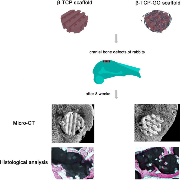
β-TCP 和 β-TCP-GO 支架的方案说明刺激了体内成骨。 β-TCP和β-TCP-GO支架植入兔颅骨缺损8周后体内骨形成能力的显微CT分析和组织学分析。转载自参考。 [85] 经《碳杂志》许可
石墨烯家族与壳聚糖
Chitosan (CS), a highly versatile biopolymer, derived from the shells of crustaceans [1, 87], has a hydrophilic surface that promotes cell adhesion and proliferation and its degradation products are nontoxic. Chitosan is biocompatible, osteoconductive, hemostatic, and can be easily converted into the desired shapes [2]. Besides, chitosan can promote bone matrix of mineralization [1] and minimize the inflammatory response after implantation [100]. All properties above make chitosan especially attractive as a bone scaffold material. But the most challenging part is the obtainment of CS-based scaffolds with good mechanical properties and processability [101]. Interestingly, CS/GO scaffolds have high water-retention ability, porosity, and hydrophilic nature [101, 102]. The CS-based 3D materials were enriched with GO in different proportions (0.5 wt% and 3 wt%). The new developed CS/GO 3 wt% scaffold was expected to be ideally designed for bone tissue engineering applications in terms of biocompatibility and properties to promote cell growth and proliferation [103]. Another CHT/GO scaffold with 0, 0.5, and 3 wt.% GO were prepared by freeze-drying method. Similarly, the CS/GO 3 wt% scaffolds significantly enhanced the ALP activity in vitro and the new bone formation in vivo, suggesting a positive contribution of 3 wt% GO to the efficiency of osteogenic differentiation process (Fig. 4) [3]. All results proved that CS/GO scaffolds could be a feasible tool for the regeneration of bone defects, and the addition of a 3 wt% of GO to material composition could have a better impact on cell osteogenic differentiation.

一 ALP activity in mice calvaria defects implanted with CHT/GO and b histomophometric analysis of Masson Goldner trichrome-stained sections. ###p < 0.001 vs CHT; **p < 0.01 vs control; ***p < 0.001 vs control.转载自参考。 [3] with permission from the Journal of Scientific Reports
Moreover, some tricomponent composites, such as CS, GO, and HA can release more Ca and P ions compared to the pure HA nanoparticles, displaying a high bioactivity of the composite scaffold [87]. Ravichandran et al. fabricated a unique composite scaffold, GO–CS–HA scaffold, and the incorporation of GO enhanced the tensile strength of CS up to 8.2 MPa and CS–HA to 10 MPa. And the results demonstrated that GO–CS–HA scaffolds facilitated cell adhesion and proliferation, meanwhile showed improved osteogenesis in in vitro tests [2]. Another tricomponent composite scaffold, containing CS, gelatin (Gn), and different concentrates of graphene oxide (0.1%, 0.25%, 0.5%, and 1% (w /v ) GO) showed better physic-chemical properties than CS/Gn scaffolds. The addition of GO at the concentration of 0.25% to CS/Gn scaffolds exhibited enhanced absorption of proteins, extensive apatite deposition. The 0.25% GO/CS/Gn scaffolds were cyto-friendly to rat osteoprogenitor cells, and they enhanced differentiation of mouse mesenchymal stem cells into osteoblasts in vitro (Fig. 5). The tibial bone defect filled with 0.25% GO/CS/Gn scaffolds showed the growth of new bone and bridging the defect area, indicating their biocompatible and osteogenic nature [104]. Thus, no matter bicomponent or tricomponent composites scaffolds, the addition of graphene family materials to chitosan can favorably improve the mechanical properties and regulate the biological response of osteoblasts, promoting osteogenic differentiation.

一 MTT assay after incubation of CS/Gn scaffolds and 0.25% GO/CS/Gn scaffolds with media for 48 h. The asterisk indicates a significant increase versus control, and the pound sign indicates a significant decrease versus control (p <0.05)。 b , c Expression of osteogenic-related genes (RUNX2, ALP, COL-1, and OC) in mMSCs cultured on CS/Gn scaffolds and 0.25% GO/CS/Gn scaffolds for 7 and 14 days measured by quantitative RT-PCR.转载自参考。 [104] with permission from the Journal of International Journal of Biological Macromolecules
Graphene Family with Other Synthetic or Bio-polymers
Sponge scaffolds of type I collagen, the major organic component of bone [81], have been clinically applied as scaffolds to regenerate bone tissue [105, 106]. Because collagen scaffolds (elastic moduli:14.6 ± 2.8 kPa) are relatively soft, the combination with GO is expected to enhance the elastic modulus of collagen scaffolds and to improve the osteogenic differentiation of MSCs for bone regeneration. The covalent conjugation of GO flakes to 3D collagen scaffolds (elastic moduli:38.7 ± 2.8 kPa) increased the scaffold stiffness by threefold and did not negatively affect the viability of BMSCs. The enhanced osteogenic differentiation observed on the stiffer scaffolds were likely mediated by BMSCs mechanosensing because the molecules involved in cell adhesion to stiff substrates were either upregulated or activated [107]. Moreover, the development of new biomaterials utilizing graphene family materials with high osteogenic capacity is urgently pursued (Table 2).
Up to now, these improved tricomponent systems for bone tissue engineering scaffolds possess good biocompatibility, which can promote cell attachment, proliferation, and have been reported mechanical properties matchable to those of natural bone. But the response to specific biological signals expressing, as well as the capabilities of enhancing cell differentiation and finally bone tissue regeneration, still needs to be explored further. Moreover, it has been reported that the pore structure (pore size, pore morphology, and pore orientation) and the elasticity of scaffolds were manipulated to regulate osteogenesis [108,109,110]. However, due to the complicated structure of porous and different elasticity accurately controlled of the scaffolds, it remains a major challenge to individually design specific pore architectures and elasticity 3D porous scaffolds that can stimulate bone regeneration. With the rapidly development of the science and technology, the emerging of the 3D-printing method may overcome this problem and open an avenue for bone tissue regeneration [85]. The in vitro bioactivity and excellent in vivo bone-forming ability of graphene family nanomaterials present a new prospect of developing a broad new type of multifunctional scaffolds for biomedical applications. Thus, we believe that the unraveled the molecular mechanisms behind will be revealed soon and graphene family materials still have attractive potential of applications in bone regeneration waiting us to explore.
Graphene Family Materials as Coating
Graphene family materials have been widely applied in diverse forms of medical applications for bone regeneration. As a coating, graphene family materials can be transferred on two dimensional (2D) flat non-metal or metal substrates to induce spontaneous osteogenic differentiation of several types of mesenchymal stem cells (MSCs) [64]. Nayak et al. transferred graphene to four 2D non-metal substrates (polydimethylsiloxane (PDMS), polyethylene terephthalate (PET), glass slide, and silicon wafer with 300 nm SiO2 (Si/SiO2).) and investigated the influence of graphene on BMSCs differentiation. They summarized that the graphene coating was cytocompatible and contributed to enhance the osteogenic differentiation of BMSCs at a rate comparable to differentiation under the influence of BMP-2 in the osteogenic medium [20]. Similarly, Elkhenany et al. found that goat BMSCs, seeded on 2D graphene-coated plates underwent osteoblastic differentiation in culture medium without the addition of any specific growth factors [8]. Simultaneously, Lee et al. tried to explain the origin of how graphene coating could accelerate stem cell renewal and differentiation. They deemed that the strong noncovalent binding abilities of graphene allowed it to serve as a preconcentration platform for osteoblastic inducers, which facilitated BMSCs osteogenic differentiation [67]. The capability of graphene in modulating osteogenic differentiation is evident. How about its derivatives? GO coatings and rGO coatings all showed favorable cytocompatibility and enhanced spontaneous osteogenic differentiation by upregulating levels of ALP activity [111, 112].
Since titanium (Ti) and medical-grade Ti alloy have been extendedly applied in the orthopedic and dental fields [113,114,115], satisfactory osseointegration for titanium and its alloys is still a major challenge and need to be explored deeply in order to help the clinicians to promote the success or survive rate of implants and diminish the likely complications encountered after their placement [114, 116, 117]. Graphene family materials coated titanium and its alloys, serving as a new method to improve their capabilities of osseointegration at the tissue-implant interface, attracted widespread attention. For example, GO-coated titanium enhanced cell proliferation, upregulated levels of ALP activity and gene expression level of osteogenesis-related markers, and promoted the protein expression of BSP, Runx2, and OCN [117].邱等人。 made different thickness GO coatings on the pure titanium surfaces respectively by cathodal electrophoretic deposition. Interestingly, with the increasing thickness of GO, the ALP-positive areas improved, ECM mineralization increased [118]. Moreover, Zeng et al. firstly fabricated GO/HA composite coatings by electrochemical deposition technique on Ti substrate. The addition of GO facilitated both the crystallinity of deposited apatite particles and the bonding strength of the as-synthesized composite coatings [119]. It is well known that hydrophilic surface is biocompatible compared to hydrophobic surface. In the case of rGO coating, the rapid adsorption of serum protein improves hydrophilia of graphene surface and enhances cell adhesion.贾等人。 used evaporation-assisted electrostatic assembly and one-pot assembly to fabricate 2D GO-coated Ti and rGO-coated Ti, with tailored sheet size and surface properties. Compared to the contact angle of titanium (60.4°), the contact angle of GO-coated Ti and rGO-coated Ti were 20° and 14.2°, respectively, indicating the successful interfacial assembly of graphene and excellent wettability properties. The rGO-coated Ti elicited better cell adhesion and growth than bulk GO, while the latter evoked higher activity of osteogenic differentiation [120].
Osseointegration is a complicated biological process determined by the surface properties of implants [114]. The graphene-based coatings above all lack 3D morphology. The 3D porous surface structure of coating can mimic the special macrostructures of the nature bone tissues [115].邱等人。 first synthesized 3D porous graphene-based coating on the pure titanium plates (GO@Ti and rGO@Ti). Water contact angles showed super hydrophilic surfaces of GO@Ti and rGO@Ti. Surface wettability exerts great effect on the biocompatibility of materials, which is strongly related to biomolecules adsorption [121]. GO@Ti and rGO@Ti both showed the excellent cytocompability and the optimal capability of osteoinduction [39]. Morin and his co-workers even transferred single or double chemical vapor deposition (CVD) grown graphene coatings onto 3D objects with differences in 3D geometries and surface roughness, such us dental implant, locking compression plate and mandible plate (Fig. 6) [64]. CVD is a very stable coating fabrication method, with substrate-independent properties and versatile surface functionalization. Besides, surface active CVD coatings are good platforms for immobilizing biomolecules, which is very important to bone regeneration [122].

一 The calvarial defects of rats were enclosed with a GO-Ti membrane. b New bone formation of the rat calvarial defects after the implantation of Ti or GO-Ti membrane at postoperative week 8. *p < 0.05 vs control; #p < 0.05 vs Ti. c Images of HE staining of the rat calvarial defects after the implantation of Ti or GO-Ti membrane at postoperative week 8. Reproduced from ref. [128] with permission from the Journal of Applied Spectroscopy Reviews
Overall, the strategy of applying graphene family materials as coating onto a surface is charming. Through currently available techniques or methods, such as CVD [123], electrochemical deposition [119], with diverse substrates (e.g. polymers, metals), graphene, and its derivatives can be obtained efficiently, with dimensions ranging from nanometer to macroscopic scales [120]. Then, graphene family nanomaterials can be transferred onto the substrate, either as 2D coatings/films/sheets or 3D porous structures of coating, to enable the binding of biomolecules, absorb the serum protein, and facilitate osteogenic differentiation of stem cells. But the different physical and chemical properties of the substrates and the type or frequent use of chemical inducers for osteogenic differentiation (e.g., dexamethasone, bone morphogenetic protein-2) that may cover up the effects exerted by graphene family materials alone [65]. Therefore, these methods still require to be well-directly improved and further studied.
Graphene Family as an Additive in Guided Bone Membrane
Barrier membranes are standardly used in oral surgical procedures, applying in guided tissue regeneration (GTR) and guided bone regeneration (GBR), for the treatment of periodontal bone defects and peri-implant defects, as well as for bone augmentation [124, 125]. GBR is considered to be one of the most promising methods for bone tissue regeneration. The concept of GBR is using a non-resorbable or absorbable membrane serving as a barrier to prevent the ingrowth of soft connective tissue into the bone defect and offer a space to “guide” the bone reconstruction [126, 127]. An ideal GBR membrane should have excellent biocompatibility and mechanical property to promote the regeneration of bone tissues and prevent soft-tissue ingrowth. Ti membrane is a non-resorbable membrane with excellent mechanical properties for the stabilization of bone grafts.帕克等人。 fabricated GO-coated Ti (GO-Ti) membranes, with increased roughness and higher hydrophilicity. GO endowed the pure Ti membranes better biocompatibility and enhanced the attachment, proliferation, and osteogenesis of MC3T3-E1 in vitro. Moreover, GO-Ti membranes were implanted into rat calvarial defects (Fig. 6) and new bone formation significantly in full-thickness calvarial defects without inflammatory responses was observed [128].
However, non-resorbable membranes need to be removed by a second operation. Thus, a resorbable membrane is recommended owing to avoid a second intervention during operation, which can diminish the risk of infection and the loss of the regenerated bone. But the resorbable membranes made of collagen or chitosan usually has poor mechanical property. The addition of graphene family materials improves the weaknesses of resorbable membrane. For instance, De et al. attempted to prepare absorbable collagen membranes enriched with different concentrations of GO. The presence of GO on the membrane altered the mechanical features of the membrane, by conferring lower deformability, improving stiffness, and increasing roughness [129].田等人。 made 3D rGO (3D-rGO) porous films, which can accelerate cell viability and proliferation, as well as significantly enhanced ALP activity and osteogenic-related gene expressions [130].
Although pristine graphene is basically incompatible with organic polymer to form homogeneous composite, and even decrease the cell viability in some cases if the amount of graphene is excessive [131]. The incorporation of graphene family materials can enhance the bioactivity and mechanical properties of composite membranes. Because of the potent effects on altering mechanical drawbacks, stimulating osteogenic differentiation, and exhibiting superior bioactivity, graphene family material-modified membranes can be applied effectively to GBR.
Graphene Family Materials as Drug Delivery System (DDS)
Due to their small size, intrinsic optical properties, large specific surface area, low cost, and useful noncovalent interactions with aromatic drug molecules, graphene family materials exhibit excellent efficacy as delivery vehicles of genes and biomolecules. Moreover, simple physisorption via π-π stacking, hydrogen bonding, and electrostatic interaction is able to assist in high drug loading of hydrophobic drugs without compromising potency or efficiency [38]. The therapeutic efficacy of drugs is always related to the drug delivery carrier, which should enable the loading of large doses, controlled release, and retention of the bioactivity of the therapeutic proteins [132]. At present, anticancer drugs, including doxorubicin [133,134,135,136,137], paclitaxel [138, 139], cisplatin [140], and methotrexate [141, 142] loaded by graphene family nanomaterials showed amazing cancerous effect for the selective killing of cancer cells.
For better bone regeneration, we sometimes need the help of osteogenic drug or macromolecular osteogenic protein. It was reported that the adsorbed drugs or loaded growth factors on graphene or its derivatives could enhance the osteogenic differentiation of cells due to the increased local concentration [143]. For example, simvastatin (SIM) chosen as a model drug was loaded on the 3D porous scaffolds, which were made of silk fibroin (SF) and GO. SIM is an inhibitor of the competitive 3-hydroxy-3-methyl coenzyme A (HMG-CoA) reductase [144]. The effects of SIM on bone formation are associated with an increase in the expression of bone morphogenetic protein-2 (BMP-2) mRNA and enhanced the vascular endothelial growth factor (VEGF) expression [145, 146]. SIM can release sustainedly (30 days), and the release rate was relevant to the GO content within the scaffolds. In vitro, compared with the blank scaffolds, the SF/GO/SIM showed better biocompatibility, and the cells cultured on them exhibited faster proliferation rate [147]. Dexamethasone (DEX) is an osteogenic drug for which can facilitate osseointegration.荣格等人。 firstly loaded DEX on rGO-coated Ti by π-π stacking. The loading efficiency of DEX on rGO-Ti was 31% after drug loading for 24 h and only 10% of total loaded DEX was released for 7 days, indicating that the drug delivery system can induce a long-term stimulation of stem cells for osteogenic differentiation. The DEX/rGO-Ti significantly facilitated MC3T3-E1 cells growth and differentiation into osteoblasts [143]. Similarly, Ren et al. also employed the GO-Ti and rGO-Ti as drug vehicles to absorb DEX. The presence of DEX-GO and DEX-rGO helped to promote the cell proliferation and largely enhanced osteogenic differentiation [115]. The graphene family materials coating on Ti alloys with controlled drug delivery can stimulate and enhance cellular response around implant surface to reduce the osseointegration time, expected to be applied for various dental and biomedical applications [143].
Not only small molecular osteogenic drug, but also macromolecular proteins can be loaded by graphene family materials for bone regeneration. Bone morphogenetic proteins (BMPs) are the most potent osteoinductive protein for bone regeneration. Thus, BMP-2 was loaded on the surface of Ti/GO through π-π stacking and the interaction between negatively charged carboxylic groups at the edges of GO and positively charged amino acid residues of BMP-2 [132]. Ti/GO/BMP-2 exhibited the high loading and the sustained release of BMP-2 with preservation of its 3D conformational stability and bioactivity. In vitro, the capability of Ti/GO/BMP-2 is to enhance osteogenic differentiation of hBMSCs. In a mouse calvarial defect model, compared to Ti/BMP-2 implants, Ti/GO/BMP-2 implants around had much more extensive bone formation [132].谢等人。 used GO-modified hydroxyapatite (HA) and GO-modified tricalcium phosphate (TCP) as an anchor for adsorbing BMP-encapsulated BSA- nanoparticles (NPs) respectively. The charge balance and BMP-2 sustained release capability of the new scaffolds synergistically improved BMSCs proliferation, differentiation, and bone regeneration in vivo [148]. Poor osteointegration and infection are the most serious complications leading to failures of Ti implantation [10].韩等人。 incorporated GO onto polydopamine (PDA)-modified Ti scaffolds. Then, BMP-2 and vancomycin (Van) were separately encapsulated into gelatin microspheres (GelMS). After that, drug-containing GelMS were loaded on GO/Ti scaffolds and anchored by the functional groups of GO (Fig. 7). The new scaffolds were endowed with dual functions of inducing bone regeneration and preventing bacterial infection [149]. Substance P (SP) is a highly conserved 11 amino acid neuropeptide [150], involved in many processes, such as the regulation of inflammation, wound healing, and angiogenesis, and it is expected to promote MSC recruitment to the implants [151]. Therefore, apart from BMP-2, La et al. added this peptide, SP, on the surface of GO-coated Ti. The dual delivery system via GO-coated Ti showed sustained release of BMP-2 and SP and the potential of SP for inducing migration of MSCs. In vivo, Ti/GO/SP/BMP-2 group showed the greater new bone formation in the mouse calvaria than Ti/GO/BMP-2 group may be due to the MSCs recruitment by SP to the implants [152].

Schematics and scanning electron micrographs of the preparation the new GO/Ti scaffold:BMP2- and Van-loaded CGelMS were immobilized on the GO/Ti scaffold through electrostatic interactions between the functional groups of GO and CGelMS.转载自参考。 [149] with permission from the Journal of Biomaterials Science
Currently, more and more teams get down to designing new drug delivery system to improve the practical applications. The loading of large doses, controlled release, and retention of the bioactivity of the therapeutic proteins are still difficulty in research on drug delivery system.
结论
Studies on the graphene family materials on biological applications is emerging rapidly, especially their potential applications for bacteria inhibition and inducing stem cell osteogenic differentiation. Before their biological applications are considered for clinical trial, the biocompatibility of graphene family materials is of vital importance. However, the challenges exist and must be overcome. These challenges include a thorough understanding of the graphene-cell (or tissue, organ) interaction and cellular uptake mechanism as well as mechanism(s) of potential toxicity. We summarize and analyze several articles and conclude that the cytotoxicity and in vivo biocompatibility of graphene family materials are influenced by numerous factors, including surface functionalization, concentration, size, and shape. At low concentration, graphene family materials are cytocompatible, with little negative influence on cell morphology, viability, and proliferation. Furthermore, it was reported that graphene family materials with flat shapes having better biocompatibility, because the flat shape materials were expected to have minor interaction with the cellular membranes [47]. Although the different criteria were used to define the size scale and shape of graphene and its derivatives, it was true that nano-sized graphene family materials were much safer for biomedical applications [54]. Size-control synthesis of graphene family materials needs to be considered prudently in subsequent researches. Moreover, the major challenge for researchers lies in understanding how graphene family materials behave in complicated microenvironment and establishing the long-term biocompatibility of graphene and its derivatives. Thus, researchers should spare no efforts to keeping studying the bio-safety of graphene family materials in vivo, as well as in vitro, to further understand the intricate interaction between cells and the materials. Although some papers raise concerns about bio-safety, after better control of the modifying of graphene family materials during synthesis, the potential versatility that graphene family uniquely offers has made it a competitive candidate of option for biomedical applications.
On the one hand, a lot of researches have pointed out that graphene family materials possess the capability of bacteria inhibition, due to their functional chemical groups, sharp edges, and synergistic effect with other drugs. Besides, bone remolding and regenerating successfully in an infective bone defect area is challenging. Peri-implant infection and poor osseointegration are also major challenges we confront. The use of graphene family materials in the design and development of antimicrobial bone regeneration application will capture tremendous attention in the future.
On the other hand, lots of teams painstakingly did researches to design and fabricate the new strategies of applying graphene family materials in bone tissue engineering. 3D graphene-based scaffold is a promising biocompatible scaffold, which can enhance pre-osteoblasts or stem cells osteoblastic differentiation. Graphene family materials also can be added as a reinforced material aiming to strengthen the composite scaffold mechanical properties and improve physicochemical characterization. In addition, the strategy of applying graphene or its derivatives as coating onto a surface is charming, which is expected to possess the antibacterial activity and better osseointegration, especially the 3D coating. It has been generally hypothesized that the surface characteristics of graphene family materials including nanostructures, surface roughness, protein absorption ability, electrostatic interactions, and surface hydrophilicity, exert an enormous effect on the molecular pathways which control the fate of stem cells [39, 115]. The 3D structure of scaffold or coating allows nutrients to be freely delivered, which influences the biocompatibility of the graphene family. But the manufacturing method of 3D scaffold or coating is relatively difficult and complicated. However, with the rapidly development of the science and technology, the emerging of the 3D-printing method may overcome this problem and open an avenue for bone tissue regeneration.
Moreover, graphene family materials show great potential in GBR and DDS as well. Graphene family materials improve poor mechanical property of the resorbable membranes made of collagen or chitosan without compromising their intrinsic property. Osteogenic drug or macromolecular osteogenic protein can be adsorbed on graphene or its derivatives via π-π stacking, hydrogen bonding, and electrostatic interaction with high loading and good efficiency. Taking the varied merits into consideration, graphene family materials hold great potential to bone tissue regeneration.
Considering that many supreme properties graphene and its derivatives have, especially in vitro osteogenesis enhancing ability and excellent in vivo bone-forming ability, although they still have drawbacks, graphene family materials still are promising candidates used for bone regeneration applications.
缩写
- ALP:
-
碱性磷酸酶
- BMP-2:
-
Bone morphogenetic protein-2
- BMSC:
-
Bone mesenchymal stem cells
- CPC:
-
Calcium phosphate cements
- CVD:
-
化学气相沉积
- CXYG:
-
Carboxyl graphene
- DEX:
-
Dexamethasone
- ECM:
-
细胞外基质
- GBR:
-
Guided bone regeneration
- GelMS:
-
Gelatin microspheres
- GNOs:
-
Graphene nano-onions
- 开始:
-
氧化石墨烯
- GONPs:
-
Graphene oxide nanoplatelets
- GONRs:
-
Graphene oxide nanoribbons
- GQDs:
-
石墨烯量子点
- HA:
-
Hydroxyapatite
- HUVEC:
-
Human umbilical vein endothelial cells
- MC3T3-E1:
-
A murine pre-osteoblastic cell line
- MWNTs:
-
Multi-walled carbon nanotubes
- OPE:
-
Oxygen plasma etching
- PCL:
-
Polycaprolactone
- PDA:
-
Polydopamine
- PDLLA:
-
Poly (d, l-lactic acid)
- PDLSC:
-
Periodontal ligament stem cells
- PDMS:
-
Polydimethylsiloxane
- PEG:
-
Polyethylene glycol
- PET:
-
聚对苯二甲酸乙二醇酯
- PLA:
-
Poly-lactic acid
- PLGA:
-
Poly-glycolic acid
- PPY:
-
聚吡咯
- rGO:
-
还原氧化石墨烯
- ROS:
-
活性氧
- SIM:
-
Simvastatin
- SP:
-
Substance P
- SPS:
-
Spark plasma sintering
- Ti:
-
Titanium
- Van:
-
Vancomycin
- VEGF:
-
Vascular endothelial growth factor
- β-TCP:
-
β-Tricalcium phosphate
纳米材料