

脂质体纳米载体在大鼠内耳鼓室内给药后的生物相容性
摘要
脂质体纳米载体(LPNs)由于其高载药量和在微创鼓室内给药后在内耳中的有效吸收而成为内耳治疗的潜在未来。然而,缺乏关于内耳中 LPN 生物相容性的信息。本研究的目的是记录鼓室内分娩后内耳中 LPN 的生物相容性。通过鼓室注射将含有或不含钆-四氮杂环-十二烷-四乙酸 (Gd-DOTA) 的 LPNs 递送给大鼠。使用 MRI 在体内跟踪中耳和内耳中含有 Gd-DOTA 的 LPN 的分布。使用钆增强 MRI 评估中耳和内耳屏障的功能。使用听觉脑干反应(ABR)测量听觉功能。通过分析内耳中的糖胺聚糖和透明质酸分泌以及 CD44 和 TLR2 表达来研究潜在的炎症反应。使用末端转移酶 (TdT) 分析潜在的细胞凋亡,以用 TMR-dUTP(TUNEL 染色)标记凋亡细胞 DNA 链中的游离 3'OH 断裂。结果,LPNs 在经鼓室注射后有效地进入内耳。经鼓室注射含或不含 Gd-DOTA 的 LPN 既不会破坏中耳和内耳屏障的功能,也不会引起大鼠的听力障碍。内耳中的关键炎症生物学标志物,包括糖胺聚糖和透明质酸分泌以及 CD44 和 TLR2 表达,不受 LPN 给药的影响。没有与 LPN 给药相关的显着细胞死亡。经鼓室注射LPNs对内耳是安全的,LPNs可作为给药基质应用于感音神经性听力损失的临床治疗。
背景
脂质体纳米载体 (LPN) 具有高载药量和内耳在微创鼓室内给药后的有效吸收,因此可能成为内耳治疗的未来 [1,2,3,4]。鼓室内方法被耳科医生广泛接受为一种合理的靶向给药方法,因为它避免了治疗药物在非靶向区域不必要的积累,这是临床治疗梅尼埃病和突发性感音神经性听力损失的优先策略使用庆大霉素和皮质类固醇。耳蜗中模型治疗的分子靶向通过鼓室内施用特定的肽功能化 LPN [5]。此外,使用由渗透泵和高性能聚酰亚胺管组成的新型装置实现了通过中耳自动持续输送 LPN 到内耳 [6]。作为临床上最古老的纳米治疗平台,LPNs 在治疗癌症、传染病、炎症、疼痛等方面是安全的 [7,8,9]。然而,LPNs在中耳和内耳的生物相容性尚不清楚,在临床应用于耳科之前需要澄清。
耳朵由外耳、中耳和内耳组成(图 1),在鼓室内分娩后可能会暴露于 LPN。中耳是暴露于最高浓度 LPN 的主要部位,内耳是治疗部位,也是对有害物质最敏感的器官,外耳道有可能被从外耳道流出的物质刺激。中耳腔。生物屏障是第一道防御系统,限制了药物的生物利用度,存在于皮肤、粘膜和神经周围结构中。内耳中的屏障系统在维持对内耳生理活动至关重要的离子稳态方面起着关键作用。使用钆增强磁共振成像 (Gd-MRI) 可以准确评估这些屏障的功能改变。听觉功能的损害可以通过听觉脑干反应 (ABR) 精确测量。因此,耳朵(包括外耳、中耳和内耳)本身就是一个很好的纳米毒理学模型[10, 11]。
<图片>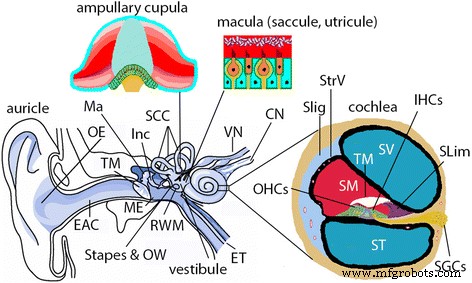
哺乳动物耳朵的插图。哺乳动物的耳朵(包括人和大鼠)由外耳、中耳和内耳组成。外耳 (OE ) 由耳廓和外耳道 (EAC )。中耳 (ME ) 由鼓膜 (TM ) 和容纳听骨链的腔,包括锤骨 (Ma ), incus (Inc ) 和镫骨。中耳腔是通过咽鼓管 (ET ) 并通过椭圆形窗口 (OW ) 和圆窗膜 (RWM )。内耳由耳蜗和前庭系统组成。耳蜗是听觉的感觉器官,有三个腔室,即鼓阶(ST ) 和前庭阶梯 (SV ),以及中阶的内淋巴室 (SM )。 SM侧壁上有血管纹(StrV ) 和螺旋韧带 (SLig )。在 SM 的底部,有包含内毛细胞 (IHCs ) 和外毛细胞 (OHCs ), 盖膜 (TM ), 和螺旋边缘 (Slim )。螺旋神经节细胞(SGCs ) 激发与毛细胞的机电转导相对应的动作电位,并提供大脑的所有听觉输入。前庭系统负责平衡,由三个半规管 (SCC ) 和前厅。 SCC 内的壶腹杯检测旋转加速度,前庭球囊和椭圆囊内的黄斑检测线性加速度。 中文 耳蜗神经,SP 螺旋突出,VN 前庭神经,VS 螺旋血管。 (改编自 Zou J. Focal Drug Delivery in Inner Ear Therapy:in Focal Controlled Drug Delivery。编辑:Domb AJ 和 Khan W. Springer,伦敦,英国。ISBN:978-1-4614-9433-1,2014;p215- 224)
透明质酸(hyaluronan)是一种天然存在的聚阴离子生物聚合物,是基底膜细胞外基质的主要成分。透明质酸由 D-葡萄糖醛酸和 N-乙酰-D-葡萄糖胺组成,它们通过交替的 β-1、4 和 β-1、3 糖苷键连接。透明质酸的积累可能导致肾缺血再灌注损伤中通透性增加和微循环炎症 [12]。在我们之前的报告中,银纳米粒子的耳毒性作用被证明与透明质酸在大鼠耳蜗中的积累有关 [11]。透明质酸与组织中的 CD44 和 Toll 样受体 2/4 (TLR2/4) 结合并触发生物反应 [13, 14]。 CD44 与透明质酸结合后介导的生物活性主要是通过与调节和衔接分子相互作用,如 SRC 激酶、Rho GTPases、VAV2、生长因子受体结合蛋白 2 相关结合蛋白 1 (GAB1)、锚蛋白、和埃兹林 [15,16,17]。除了将 T 细胞募集到炎症部位和调节 T 细胞介导的内皮损伤外,CD44 还通过细胞摄取和降解的方法介导透明质酸的代谢 [18]。据报道,抗内皮细胞抗体对内耳内皮细胞的细胞毒性可能在引起免疫介导的突发性感音神经性耳聋的血管纹损伤中起作用[19]。据报道,TLR2 依赖性核因子-κB 激活参与了内耳螺旋韧带纤维细胞中不可分型流感嗜血杆菌诱导的单核细胞趋化蛋白 1 上调,这可能是继发于慢性中耳炎的内耳功能障碍的关键步骤。 20]。如果LPNs在中耳给药后引起内耳损伤,TLR2介导的信号通路应该是重要的机制。
我们旨在评估经鼓室注射后内耳中 LPN 的生物相容性。在不同时间点使用 Gd-MRI 测量皮肤(外耳道)、粘膜(中耳腔)和内耳隔室中生物屏障的功能。使用 ABR 测量评估听觉功能。最后,通过测量耳蜗中糖胺聚糖和透明质酸的积累、CD44和TLR2的表达以及DNA片段化来分析潜在的组织病理学变化。
结果
LPNs 并未引起大鼠耳蜗的功能变化
在阳性对照组中,两侧耳蜗 (Coch) 和前庭 (Vest) 的外淋巴(L、R)(图 2a、b)中的明亮信号表明 Gd-DOTA 的摄取。经鼓膜注射银纳米粒子 (AgNPs) 后,外淋巴室中的信号强度显着增加,同时在外耳道皮肤、中耳粘膜中也检测到极强的信号,表明与 AgNP 给药相关的 Gd-DOTA 摄取增强。图 2a、b) 中的 L(表 1)。因此,评估系统得到了验证。在接受LPN + Gd-DOTA经鼓室注射的动物中,注射后3小时在听骨链、前庭阶、鼓阶和前庭上检测到亮信号,表明LPN在这些区域的明显分布(图2c) , d)。基底转角前庭阶的信号强度明显强于鼓室阶,表明 LPN 通过当前动物的椭圆窗有效进入 [21]。注射后 6 小时,基底转部的前庭阶和鼓阶之间的信号强度变得相似,整个耳蜗显示出几乎均匀的信号,但前庭中没有显着变化(图 2e,f)。在经鼓室注射空白 LPNs 后接受静脉注射 Gd-DOTA 的动物中,除了在接受经鼓室注射空白 LPNs 的中耳有强烈信号外,两侧显示出相似的信号强度,怀疑 LPNs 在听骨链表面积累(图. 2g,h)。听骨链中的黑洞表示镫骨的中空区域(图 2h)。两侧信号强度相等表明经鼓室注射 LPNs 后,双耳血液-外淋巴屏障的 Gd-DOTA 转运特性没有改变(图 2g,h)(表 1)。
<图片>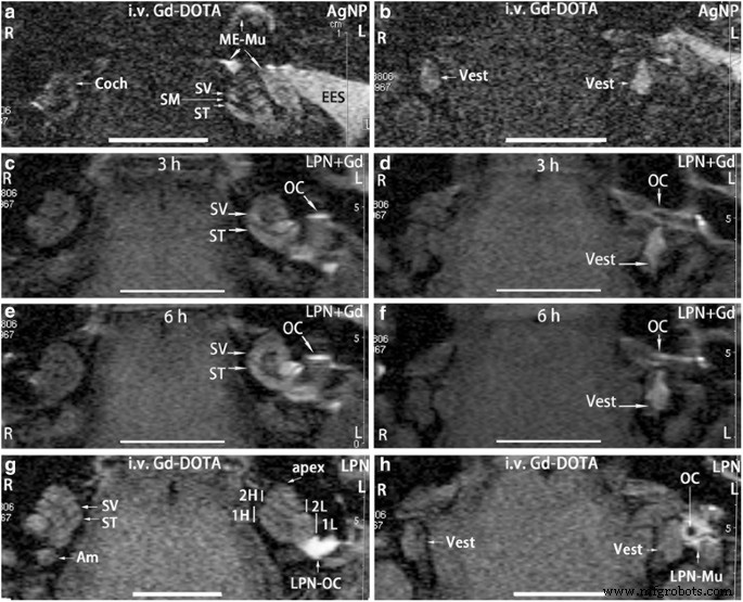
脂质体纳米载体 (LPN) 给药后大鼠内耳的钆增强 MRI。在所有动物中,纳米材料被注射到左中耳腔的内壁上。在静脉注射 Gd-DOTA 后 2 小时,继发于提前 5 小时经鼓室注射银纳米粒子 (AgNPs) 后,对阳性对照进行成像 (a , b )。 c显示了中耳和内耳LPN的动态分布 , d , e , f 通过经鼓室注射含有 Gd-DOTA 的 LPN,无需静脉注射 Gd-DOTA。空 LPN 对生物屏障的影响在静脉注射 Gd-DOTA(i.v. Gd-DOTA)后 2 小时通过 MRI 显示,大鼠预先接受经鼓室注射 LPN 5 小时(g , h )。 我 后半规管壶腹,Coch 耳蜗,EES 外耳皮肤,L 左耳,LPN-Mu 中耳粘膜中的 LPN,LPN-OC 听骨链上的 LPN (OC ), ME-Mu ,中耳粘膜,R 右耳,SM scala media , ST scala tympani, SV 前庭阶梯,背心 前庭,1H 耳蜗基底高转,1L 耳蜗基底下转,2H 第二个较高的耳蜗,2L 耳蜗的第二下转。 比例尺 =5 毫米
LPN + Gd-DOTA 和 LPN 均未引起显着的听力损失,表现为 ABR 阈值偏移,该阈值偏移是使用 2、4、8、16 和 32 kHz 频率下的咔嗒声和音调脉冲刺激测量的,频率为 2、4 和给药后 7 天,与接受去离子水 (dH2O) 经鼓室注射的耳朵相比(图 3)。
<图片>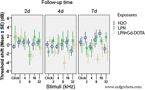
通过听觉脑干反应测量经鼓室注射脂质体纳米载体对大鼠听力功能的影响。听力损失表示为阈值变化。组间差异不显着(p> 0.05,单向方差分析)。 n 每组=6。 H2O 阴性对照组经鼓室注射去离子水,LPN 空脂质体纳米载体,LPN + Gd-DOTA 包含 Gd-DOTA 的 LPN,2d , 4d , 和 7d 注射后 2、4 和 7 天
LPN 不会诱导大鼠耳蜗中糖胺聚糖的积累
苏木精和伊红染色未显示所有分析动物耳蜗中的白细胞和纤维蛋白有任何炎症浸润,包括 LPN 通过的镫骨和椭圆窗(图 4)。高碘酸希夫氏染色表明,接受经鼓室注射 dH2O 的动物耳蜗的骨壁、螺旋缘、螺旋韧带、盖膜、赖斯纳膜、骨螺旋层和血管纹中存在糖胺聚糖。信号强度从基底转向顶点呈梯度增加,血管纹差异显着(图 5 和图 6)。经鼓室注射LPNs和LPN + Gd-DOTA的动物耳蜗中的信号梯度没有变化(图5和6)。
<图片>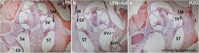
暴露于脂质体纳米载体的大鼠耳蜗的苏木精-伊红染色。接受LPN给药的耳蜗中没有炎症浸润(a ), LPN+Gd (b ) 和 H2O (c )。 圆形区域 指示选择用于强度测量的感兴趣区域 (a )。 LPN 空脂质体纳米载体,LPN + Gd 含 Gd-DOTA 的 LPN。 萨 球囊,SFP 镫骨足板,SVJ 镫骨前庭关节 SM Scala 媒体,ST scala tympani, SV 前庭阶梯,Ut 椭圆囊。 比例尺 =1 毫米
<图片>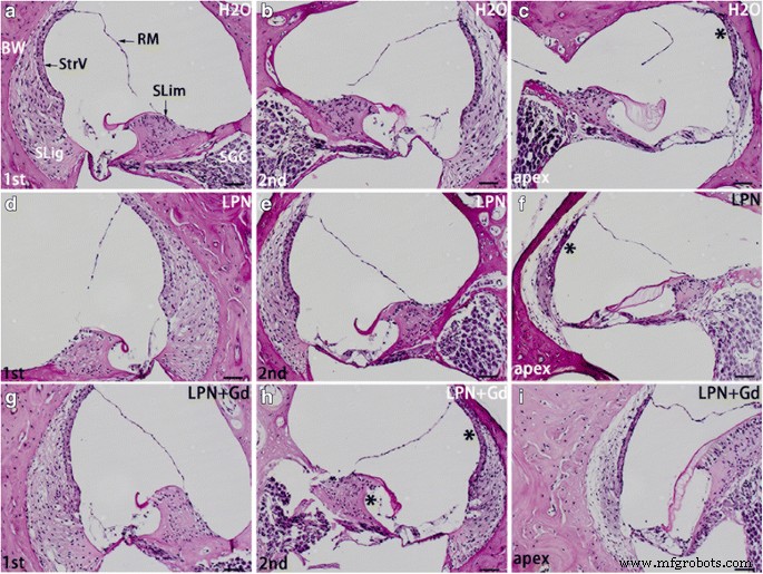
使用高碘酸希夫染色光学显微镜检测暴露于脂质体纳米载体的大鼠耳蜗中的糖胺聚糖分泌。螺旋缘 (SLim ) 和骨壁 (BW ) 的耳蜗在阴性对照 (H2O ) (a –c ), 空脂质体纳米载体 (LPN) (d –f ), 和包含 Gd-DOTA 的 LPN (LPN + Gd ) (g –我 )。 c中以*表示强度明显较高的染色区域 , f , 和 h 与左列相比。 RM 赖斯纳膜,SGC 螺旋神经节细胞,SLig 螺旋韧带,StrV 血管纹,第一 基础转弯,第二 第二轮。 比例尺 =50 μm
<图片>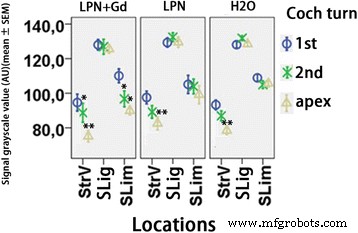
使用高碘酸希夫染色检测暴露于脂质体纳米载体的大鼠耳蜗中糖胺聚糖分泌的定量。 n 每组=3。 AU 任意单位,H2O 阴性对照,LPN 空脂质体纳米载体,LPN + Gd 包含 Gd-DOTA 的 LPN,SLig 螺旋韧带,纤细 螺旋缘,StrV 血管纹,第一 基础转弯,第二 第二轮。 *p <0.05, **p <0.01(使用 LSD 检验作为事后分析的单向方差分析)
LPNs对大鼠耳蜗透明质酸分泌的影响很小
在接受经鼓室注射 dH2O 的大鼠耳蜗中,主要在螺旋神经节细胞、纹状基底细胞、外沟细胞和毛细血管内皮细胞等细胞中检测到透明质酸阳性染色(图 7)。基底和第二圈螺旋韧带纤维细胞的信号强度明显高于顶点。随着LPNs和LPN + Gd-DOTA的应用,这些差异在大鼠耳蜗中变得不显着,表明螺旋韧带纤维细胞的透明质酸分泌受到LPN给药的影响(图8)。 LPN + Gd-DOTA 还减少了基底转弯螺旋韧带纤维细胞的染色。然而,经鼓室注射LPNs和LPN + Gd-DOTA对大多数耳蜗细胞透明质酸的分泌没有影响(图7和图8)。
<图片>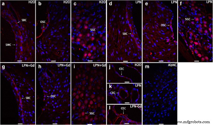
用免疫荧光共聚焦显微镜检测暴露于脂质体纳米载体的大鼠耳蜗中的透明质酸分泌。在纹状基底细胞(SBC ), 外沟细胞 (OSC ), 螺旋神经节细胞 (SGC ) 和毛细血管内皮细胞 (CEC ) 阴性对照 (H2O ) (a –c , j ), 空脂质体纳米载体 (LPN ) (d –f , k ), 和包含 Gd-DOTA 的 LPN (LPN + Gd ) (g –我 , l )。抗体中没有染色省略阴性对照(AbNC ) (米 )。 CEC 毛细血管内皮细胞,ISC 内沟细胞,SBC 纹状基底细胞,SL-I 螺旋韧带纤维细胞 I 型,SLSF 螺旋缘卫星纤维细胞,SMC 血管纹边缘细胞。 比例尺 =16 μm
<图片>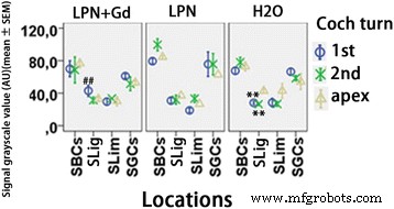
使用免疫荧光共聚焦显微镜检测暴露于脂质体纳米载体的大鼠耳蜗中透明质酸分泌的定量。 n 每组=3。 AU 任意单位,H2O 阴性对照,LPN 空脂质体纳米载体,LPN + Gd 含 Gd-DOTA 的 LPN,SBCs 纹状基底细胞,SGCs 螺旋神经节细胞,SLig , 螺旋韧带, Slim 螺旋边缘,第一 基础转弯,第二 第二轮。 **p <0.01(与顶点相比),##p <0.01(与基底转的LPN和H2O组相比)(单向方差分析,LSD检验用作事后分析)
LPN 没有改变大鼠耳蜗中的 CD44 细胞群
在暴露于 dH2O 的耳蜗中,纹状中间细胞、纹状基底细胞、螺旋韧带纤维细胞、螺旋神经节细胞、Corti 器官中的 Deiters 细胞以及耳蜗和螺旋韧带中的毛细血管内皮细胞显示出 CD44 的强染色。耳蜗转动之间的信号强度差异不显着。 CD44阳性群体和表达强度不受LPN + Gd-DOTA或LPNs的鼓室注射的影响(图9和10)。
<图片>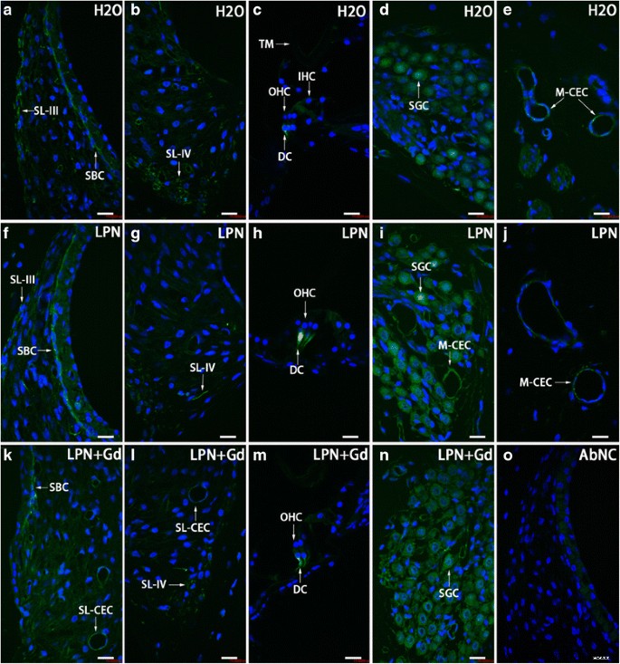
使用免疫荧光共聚焦显微镜证明暴露于脂质体纳米载体的大鼠耳蜗中 CD44 阳性细胞分布。 CD44阳性细胞主要在纹状基底细胞(SBC ), 螺旋韧带 (SL )、Dieter 细胞 (DC ), 螺旋神经节细胞 (SGC ) 和毛细血管内皮细胞 (CEC ) 在阴性对照 (H2O ) (a –e ), 空脂质体纳米载体 (LPN ) (f –j ), 和包含 Gd-DOTA 的 LPN (LPN + Gd ) (k –n )。抗体中没有染色省略阴性对照(AbNC ) (o )。 IHC 内毛细胞,M-CEC 血管内皮细胞,SL-CEC 螺旋韧带毛细血管内皮细胞:螺旋神经节细胞,SL-III 螺旋韧带纤维细胞 III 型,SL-IV 螺旋韧带纤维细胞 IV 型,TM 盖膜,OHC 外毛细胞。 比例尺 =16 μm
<图片>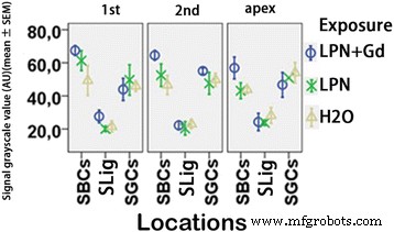
使用免疫荧光共聚焦显微镜检测暴露于脂质体纳米载体的大鼠耳蜗中 CD44 蛋白水平的量化。组间差异不显着(p> 0.05,单向方差分析)。 n 每组=3。 n 每组=3。 AU 任意单位,H2O 阴性对照,LPN 空脂质体纳米载体,LPN + Gd 含 Gd-DOTA 的 LPN,SBCs 纹状基底细胞,SGCs 螺旋神经节细胞,SLig 螺旋韧带,第一 基础转弯,第二 第二回合
LPN 没有改变大鼠耳蜗中的 TLR2 表达
在暴露于 dH2O 的耳蜗中,纹状基底细胞、螺旋韧带纤维细胞、根细胞、螺旋神经节细胞、Corti 器官的柱细胞和耳蜗中的毛细血管内皮细胞显示出 TLR2 的强烈染色。耳蜗转动之间的信号强度差异不显着。 TLR2阳性群体和表达强度不受LPN + Gd-DOTA或LPNs经鼓室注射的影响(图11和12)。
<图片>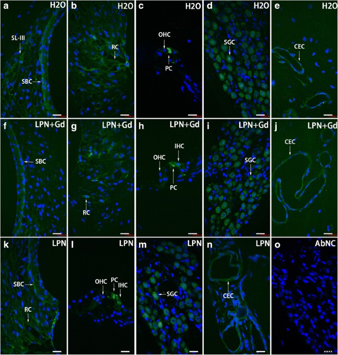
使用免疫荧光共聚焦显微镜证明暴露于脂质体纳米载体的大鼠耳蜗中 TLR2 阳性细胞分布。 TLR2阳性细胞主要在纹状基底细胞(SBC ), 螺旋韧带 (SL ), 根单元 (RC ), 柱状电池 (PC ), 螺旋神经节细胞 (SGC ) 和毛细血管内皮细胞 (CEC ) 在阴性对照 (H2O ) (a –e ), 空脂质体纳米载体 (LPN ) (k –n ), 和包含 Gd-DOTA 的 LPN (LPN + Gd ) (f –j )。在去除抗体的阴性对照 (AbNC ) (o )。 IHC 内毛细胞,SL-III 螺旋韧带纤维细胞 III 型,OHC 外毛细胞。 比例尺 =16 μm
<图片>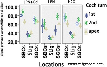
使用免疫荧光共聚焦显微镜检测暴露于脂质体纳米载体的大鼠耳蜗中 TLR2 蛋白水平的量化。组间差异不显着(p> 0.05,单向方差分析)。 n 每组=3。 AU 任意单位,H2O 阴性对照,LPN 空脂质体纳米载体,LPN + Gd 含 Gd-DOTA 的 LPN,SBCs 纹状基底细胞,SGCs 螺旋神经节细胞,SLig 螺旋韧带,第一 基础转弯,第二 第二回合
LPN 不会导致大鼠耳蜗细胞死亡
有稀疏的凋亡细胞随机分布在未治疗大鼠的耳蜗中。出人意料的是,镫骨足底和卵圆窗龛内有丰富的凋亡细胞。 LPNs和LPN + Gd-DOTA对凋亡细胞的数量和分布模式没有影响(图13)。
<图片>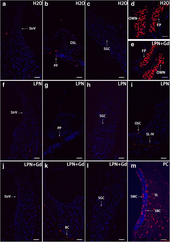
使用 TUNEL 染色共聚焦显微镜证明暴露于脂质体纳米载体的大鼠耳蜗中的细胞凋亡。阴性对照组(H2O ) (a –c ), 空脂质体纳米载体 (LPN ) (f –我 ), 和包含 Gd-DOTA 的 LPN (LPN + Gd ) (j –l )。镫骨足底有大量凋亡细胞(FP ) 和椭圆窗壁龛 (OWN ) 两组 (d , e )。在阳性对照 (PC) 中,在螺旋韧带纤维细胞 (SL ), 纹状基底细胞 (SBC ) 和边缘纹细胞 (SMC )。 PP 螺旋神经节细胞的外围过程(SGC ), OC 骨细胞,OSC 外沟细胞,SL-IV IV型螺旋韧带纤维细胞,SLim 螺旋缘,StrV 血管纹。 比例尺 一 –l =32 μm,m =16 μm
讨论
LPNs 在经鼓膜注射后有效进入内耳,MRI 证明使用 Gd-DOTA 作为药物模拟物封装在 LPNs 内(图 2c-f)。虽然之前的研究表明圆窗是 LPNs 进入内耳的主要途径 [6],但目前的观察表明,椭圆窗通道比圆窗更有效地将 LPNs 从中耳运输到内耳。耳朵。该结果表明,在靶向中耳内壁给药后,这两种途径在 LPN 的内耳负荷中都很重要。本研究采用最有效的钆增强内耳 MRI 体内方法评估生物屏障和频率特异性 ABR 评估听力功能,本研究表明经鼓室注射 LPNs 和 LPN + Gd-DOTA 均不会破坏听力功能。内耳屏障也不会导致大鼠听力受损。通过分析先前证明的关键炎症生物学标志物 [11, 22],LPNs 和 LPN + Gd-DOTA 不会诱导耳蜗中的炎症反应。尽管未评估圆窗膜,但镫骨和卵圆窗无炎症排除了圆窗膜中明显的炎症反应,因为本研究表明卵圆窗通路优于 LPN 的圆窗通路。
静脉注射钆螯合物后的内耳 MRI 能够检测氧化应激介导的线粒体毒素诱导的血液外淋巴和血液内淋巴屏障的破坏 [23]。据报道,AgNPs 通过产生活性氧 (ROS) 和 Jun 氨基末端激酶 (JNK) 的激活导致细胞损伤,导致细胞色素 C 释放到细胞质中以及 Bax 易位到线粒体 [24 ]。在经鼓膜注射中,AgNPs 进入内耳并引起大鼠内耳生物屏障的渗透性变化 [11, 25]。 LPNs 在经鼓膜注射后也以大小依赖的模式进入大鼠内耳,95 nm 直径的 LPNs 在穿过中内耳屏障方面表现出最高的功效 [3]。在本研究中,LPN 的平均尺寸为 100 到 115 nm,略大于最有效的尺寸。 LPN + Gd-DOTA表明这种大小的LPNs进入了内耳,这与之前的报道一致[3]。然而,LPNs 进入内耳并没有引起血液-外淋巴和血液-内淋巴屏障的通透性变化。该结果表明 LPN 对内耳是安全的。 ABR结果表明听力正常,支持MRI结果。
在肾缺血再灌注损伤中,透明质酸分泌与通透性变化和微循环炎症之间存在关联[12]。 The previous study also showed that AgNPs caused the accumulation of hyaluronic acid in the rat cochlea [11]. CD44 and toll-like receptor 2/4 (TLR2/4) work as receptors of hyaluronic acid and trigger biological reactions [13, 14]. CD44 also mediates the metabolism of hyaluronic acid through cellular uptake and degradation in addition to recruiting T cells to inflammatory sites and regulating T cell-mediated endothelial injury [18]. In the present study, hyaluronic acid, CD44, and TLR2 were detected in the rat cochlea. LPN + Gd-DOTA reduced the secretion of hyaluronic acid in the spiral ligament fibrocytes. The expressions of CD44 and TLR2 were not changed after the transtympanic injection of either LPNs or LPN + Gd-DOTA. The total glycosaminoglycan, which contains hyaluronic acid in the cochlea was not affected by the administrations of LPNs and LPN + Gd-DOTA. The impact of LPNs on the hyaluronic acid distribution in rat cochlea did not cause either permeability change or hearing loss, indicating that the modification is unharmful. It was reported that macrophages undergo phenotypic changes dependent on molecular weight of hyaluronan that correspond to either pro-inflammatory response for low molecular weight hyaluronic acid or anti-inflammatory response for high molecular weight hyaluronic acid [26]. The observed minor changes of hyaluronic acid distribution in the cochlea might have high molecular weight and anti-inflammatory function. Therefore, there was no hint of an inflammatory reaction in the rat cochlea.
The observed apoptosis in the stapes footplate cells might be normal biological activity. A balance between survival and apoptosis in the stapes footplate cells was reportedly as necessary to inactivate the otosclerosis [27]. Administration of LPN + Gd-DOTA did not affect apoptosis in the rat stapes.
Conclusions
The present study demonstrated that the transtympanic injection of liposome nanocarriers neither impaired the biological barriers of the inner ear nor caused hearing loss in the rats. The critical inflammatory mechanism was not activated by the administration of liposome nanocarriers, either. The results suggested that transtympanic injection of liposome nanocarrier is safe for the cochlea of rat.
Methods
Materials
Sphingosine (Sph), 1-stearoyl-2-oleoyl-sn-glycero-3-phosphocholine (SOPC), and 1, 2-distearoyl-sn-glycero-3-phosphoethanolamine-N-[methoxy(polyethyleneglycol)-2000] (ammonium salt) [DSPE-PEG-2000] were purchased from Avanti polar lipids (Alabaster, USA). DiI (Vybrant DiI cell-labeling solution, 1 mM in solvent) and N-(6-tetramethylrhodaminethiocarbamoyl)-1,2-dihexadecanoyl-sn-glycero-3-hosphoethanolamine, triethylammonium salt (TRITC-DHPE) were purchased from Thermo Fisher Scientific (Waltham, USA). Gd-DOTA (DOTAREM) was from Guerbet, Cedex, France. Hepes and EDTA were from Sigma. The purity of the lipids was evaluated using thin-layer chromatography on silicic acid-coated plates (Merck, Darmstadt, Germany) developed with a chloroform/methanol/water mixture (65:25:4, v/v/v). An examination of the plates after iodine staining and, when appropriate, upon UV illumination revealed no impurities. The lipid concentrations were determined gravimetrically with SuperG (Kibron, Espoo, Finland); a high-precision microbalance. The polyvinylpyrrolidone-stabilized AgNPs were supplied by Colorobbia (Firenze, Italy). The AgNPs were dispersed in deionized water (370.7 mM), and scanning electron microscopy showed that the AgNPs are spheroids with a particle size of around 100 nm. Dynamic light scattering (DLS) showed a mean hydrodynamic size of 117 ± 24 nm and a mean zeta potential of −20 ± 9 mV.
In the visualization of nanocarrier uptake in the inner ear and the evaluation of biological barrier function, 5 male Sprague Dawley rats, weighing between 334 and 348 g, were provided by the Biomedicum Helsinki, Laboratory Animal Centre, University of Helsinki, Finland (this is the defined animal center that provides animals for MRI experiments in Biomedicum); in the ABR and histological studies, 18 Sprague Dawley rats, weighing between 300 and 400 g, were provided by the Experimental Animal Unit of the University of Tampere School of Medicine in Finland. Animal assignments in each study were shown in Table 2. All animal experiments were approved by the Ethical Committee of University of Tampere (permission number:ESAVI/3033/04.10.03/2011). Animal care and experimental procedures were conducted in accordance with European legislation. Animals in the Gd-MRI study were anesthetized with isoflurane with 5% isoflurane–oxygen mixture (flow-rate 1.0 L/min) for induction and 3% for maintenance via a facemask. Animals for the ABR and histological studies were anesthetized with a mixture of 0.5 mg/kg medetomidine hydrochloride (Domitor ® , Orion, Espoo, Finland) and 75 mg/kg ketamine hydrochloride (Ketalar ® , Pfizer, Helsinki, Finland) via intraperitoneal injection followed by an intramuscular injection of enrofloxacin (Baytril ® vet, Orion, Turku, Finland) at a dose of 10 mg/kg to prevent potential infection. The animal’s eyes were protected by Viscotears® (Novartis Healthcare A/S, Copenhagen, Denmark).
Preparation and Characterization of LPNs with and without Gd
Preparation of Gd-containing LPNs
LPNs of unilamellar vesicles with an apparent hydrodynamic particle diameter (Z av) of 110 ± 15 nm that contain Gd-DOTA were prepared according to the previously published method [6]. A concentration of 1 mM Gd-DOTA-containing LPN (LPN + Gd-DOTA) refers to 1 mM liposomes encapsulating 500 mmol/L of Gd-DOTA.
Preparation of Blank LPNs
Blank LPNs of unilamellar vesicles with Z av of 115 ± 10 nm were prepared according to a previous publication [6].
Administration of LPNs
Under general anesthesia with isoflurane with 5% isoflurane–oxygen mixture (flow-rate 1.0 L/min), 50 μl of either LPNs or LPN + Gd-DOTA were injected into the left middle ear cavity through the tympanic membrane penetration under an operating microscope (OPMI1-F, Carl Zeiss, Jena, Germany). The same amount of deionized water (H2O) was injected transtympanically in rats that were assigned to the negative group. After the injection, the animals were kept in the lateral position with the injected ear oriented upward for 15 min before further measurements.
Evaluation on Biological Barrier Function Using Gd-MRI
One animal receiving transtympanic injection of LPN + Gd-DOTA was selected to demonstrate distributions of LPN in the inner ear using MRI without contrast agent. Two animals receiving transtympanic injection of blank LPNs were engaged in MRI study for evaluation of the biological barrier function. Two animals receiving transtympanic injection of AgNPs (370.7 mM, 40 μl) were used as positive control. The contralateral ear without any injection was used as negative control in all studies. A 4.7T MR scanner with a bore diameter of 155 mm (PharmaScan, Bruker BioSpin, Ettlingen, Germany) was utilized. The maximum gradient strength was 300 mT/m with an 80-μs rise time. A gadolinium-tetraazacyclododecane-tetraacetic acid (Gd-DOTA, 500 mM, DOTAREM, Guerbet, Cedex, France) solution was injected into the tail vein (0.725 mM/kg) 2 h before the MRI measurements. The imaging protocol and rapid acquisition with relaxation enhancement (RARE) sequences were applied according to a previous publication [10]. MRI scanning commenced at several time points after the transtympanic injection. The first MRI time of around 5 h was determined by taking the penetration time of liposome nanoparticles from the middle ear to the inner ear as a reference [1, 3, 6]. The final imaging time of 8 d was selected according to the course of potential acute inflammation and the availability of the scanner. ParaVision PV 4.0 (Bruker, MA, USA) software was used for the post-processing and quantification of MR images.
ABR Measurement
The auditory function of animals receiving injections of both blank and Gd-containing LPNs were evaluated using ABR measurements using BioSig32 (Tucker Davis Technologies, FL, USA) in a custom made, soundproof chamber. The ABR thresholds upon click and tone burst stimuli were recorded before and at a certain time point post-administration of LPNs. The first ABR measurement was followed on 2 days post-administration of AgNPs, allowing the animals to recover from the general anesthesia during the injection and to ensure the injected solution to be entirely cleared from the middle ear cavity. The second follow-up time of 4 days post-injection was chosen because it is close to the peak time of potential mitochondrial impairment-induced cell death in the cochlea [22]. The third follow-up time of 7 days is the time point when temporary threshold shifts remained significantly approved in an animal model of mitochondrial toxin-induced hearing loss [28]. The ABR recording procedure followed the previous report [11].
Glycosaminoglycan Staining in Rat Cochlea After Administration of LPNs
Hematoxylin-eosin staining to assess potential inflammatory infiltration and periodic acid Schiff’s staining to evaluate potential glycosaminoglycan accumulation in the cochlea after administration of LPNs were performed according to a previous publication after ABR measurements over 7 days [11] The slices were observed and digital images were acquired under a light microscope (Leica DM2000 microscope equipped with an Olympus DP25 camera) for further analysis.
Immunofluorescence Staining for Hyaluronic Acid and Receptors
Immunofluorescence staining for hyaluronic acid, CD44, and TLR2 were performed according to a previous publication after ABR measurements over 7 days [11, 21].
Cell Death Detection
Potential nuclear DNA fragmentation in the cochlea was investigated using terminal transferase (TdT) to label the free 3′OH breaks in the DNA strands of apoptotic cells with TMR-dUTP (TUNEL staining) following the reported procedure [11]. Slices exposed to recombinant DNase I (Fermentas, Vantaa, Finland, 100 U/ml in 50 mM Tris/HCl, pH 7.5, 1 mg/ml bovine serum albumin) at 37 °C for 10 min, which induced DNA strand breaks prior to the labeling procedures, were utilized as positive controls. The samples were observed under a confocal microscope.
Confocal Microscopy
The samples were observed under a Nikon inverted microscope (ECLIPSE Ti) combined with an Andor confocal system installed with Andor iQ 2.8 software (Andor Technology, Belfast, UK). The excitation lasers were 488 nm (green excitation) and 568 nm (red excitation) from an Andor laser combiner system, and the corresponding emission filters were 525/50 (Alexa Fluor-488) and 607/45 nm (Cy TM 3 and TMR Red). DAPI was excited with light at 405 nm generated from a light-emitting diode and was detected using a 450–465 nm filter.
Analysis and Statistics
ImageJ (1.45S, National Institutes of Health, Bethesda, USA) software was used for signal intensity measurements. For light microscopy of periodic acid Schiff’s staining, the region of interests (ROIs) including spiral ligament, spiral limbus, and stria vascularis were selected using freehand selections button. The “measure” function was used to obtain the mean gray scale value of the ROI, which was inversely correlated with the staining intensity. For confocal microscopy of immunofluorescence staining, the ROIs including stria basal cells, spiral ganglion cells, spiral ligament, and spiral limbus were extracted using photoshop CS6 (version 13.0, Adobe Systems Software Ireland Ltd, Dublin, Ireland) program and were imported into ImageJ program. The images were split into individual channel, and the green (corresponded to CD44 and TLR2) and red (corresponded to hyaluronic acid) channels were selected for further quantifications. The “Threshold” was adjusted using the “set” button in the “Image” menu, and “Limit to Threshold” option should be selected and “Direct to” should be defined to the corresponding channel in the “Analyze” menu. Then the gray scale value, which was inversely correlated with the staining intensity, was obtained using the “Measure” function in the same menu.
Statistical analyses were performed using the IBM ® SPSS ® Statistics Version 20 software package (SPSS Inc., Chicago, USA). A one-way ANOVA and Kruskal-Wallis test were used to compare ABR threshold shifts and signal intensities (grayscale) of staining for glycosaminoglycan and hyaluronic acid secretions, and TLR2 and CD44 staining between the LPN injected-ear and saline injected-ear groups in the different cochlear structures among various turns. Least significant difference (LSD) test was used as post hoc analysis. Higher numbers in the grayscale analysis correlate with lower signal intensities of the staining. p < 0.05 was accepted as statistically significant.
缩写
- ABR:
-
Auditory brainstem response
- AgNPs:
-
Silver nanoparticles
- dH2O:
-
Deionized water
- GAB1:
-
Growth factor receptor-bound protein 2-associated-binding protein 1
- Gd-DOTA:
-
Gadolinium-tetra-azacyclo-dodecane-tetra-acetic acid (DOTAREM)
- Gd-MRI:
-
Gadolinium-enhanced magnetic resonance imaging
- JNK:
-
Jun amino-terminal kinases
- LPN + Gd-DOTA:
-
Gd-DOTA-containing LPNs
- LPNs:
-
Liposome nanocarriers
- ROI:
-
Region of interests
- ROS:
-
Reactive oxygen species
- TLR:
-
Toll-like receptor
纳米材料