

纳米技术灭活癌症干细胞
摘要
当前肿瘤学的任务之一是鉴定癌症干细胞并寻找能够对其进行特异性抑制的治疗手段。该论文提供了关于埃利希癌细胞表型特征的数据,作为方便且易于遵循的肿瘤生长模型。癌症干细胞作为艾氏癌一部分的证据和CD44 + 的意义 和 CD44 – 证明了维持这种类型肿瘤生长的亚群。埃利希癌 CD44 + 的高(十倍)致瘤活性 细胞如果与 CD44 相比 – 细胞被证实。在这对对比中,CD44 + CD44 高 的腹腔内细胞产生更高的潜能 , CD44 + CD24 – , CD44 + CD24 + 细胞亚群,突出显示 CD44 + 池中存在癌症干细胞 细胞。
在这项研究中,合成混合纳米复合物的能力,包括稀土原钒酸盐纳米粒子 GdYVO4:Eu 3+ 胆固醇抑制肿瘤生长并增加肿瘤动物的存活率。它的每个组成部分都对肿瘤抑制作用做出了特殊贡献。用双组分杂合复合物处理埃利希癌细胞导致最具致瘤性的 CD44 high 浓度最大降低 CD117 + 同时增加的细胞 与对照组相比,肿瘤生长强度降低了 74.70 ± 4.38%。
背景
恶性生长的问题仍然是医学上最紧迫的问题之一。近几十年来,在开发新的癌症治疗方法方面取得了一些进展。这是由于癌症经典概念的修订,以及癌症干细胞(CSCs)的发现,它们能够无限地自我更新并且可以通过许多表型标记来识别。大多数这些细胞对放疗和化疗有抵抗力,导致恶性生长和转移的复发。已经掌握了抗癌治疗的新方法,即在对正常组织损伤最小的情况下选择性灭活肿瘤细胞[1]。
M. Dick 团队于 1997 年首次识别和描述了 CSC [2]。作者研究了一种急性髓性白血病,其中当移植到免疫缺陷 NOD/SCID(非肥胖型糖尿病-严重联合免疫缺陷)小鼠时,占总细胞群 0.01-1% 的亚群可导致白血病。这些肿瘤诱导细胞的表型特征为 CD34 + CD38 – . 2003 年,M. Al-Hajj 和 M.S. Wicha 成功地在人类乳腺癌 (BC) 的实体形式中识别出 CSC [3]。已经发现,当以 5 × 10 4 的浓度向 NOD/SCID 小鼠给药时,未分离的原发性乳腺癌群体在 100% 的病例 (10/10) 中表现出致瘤潜力 细胞/小鼠。将给药的细胞浓度降低至 1 × 10 4 细胞/小鼠的致瘤活性降低了 4 倍 (3/12) [3]。 CD24 + CD44 + 以不同剂量给药时的分数(2 × 10 4 低至 100 个细胞/小鼠)不允许肿瘤生长。在此,CD44 + CD24 – /low 亚群具有显着更高的致瘤活性,证明在施用 10 3 时,100% 的病例形成肿瘤 细胞/小鼠。形成肿瘤的最显着能力是CD44 + 亚群所固有的 CD24 – / lo 欧空局 + 表型。仅将 200 个这些细胞施用于小鼠,在注射后 5 个月后 100% (4/4) 形成实体瘤 [3]。这些研究由 Ponti D. 等人继续进行,他们展示了某些乳腺癌活检样本群体在体外无血清培养中形成乳腺球的能力 [4]。获得的乳腺球的大部分细胞为CD44 + /CD24 –/low 表型以及对 SCID(严重联合免疫缺陷)小鼠给药时增加的体内致瘤潜力。在该亚群中形成肿瘤的能力比传统移植的乳腺癌 MCF7 系高 1000 倍 [4]。然而,作者已经表明,只有 20% 的 CD44 + CD24 –/低 细胞具有自我更新的能力。这可能是由于该亚群的异质性,即存在额外的标记(ESA、ALDH),决定细胞的功能,也可能与 CD44 表达率有关。近几年发表的论文表明,标记物(CD44 high ) 具有最高的致瘤活性 [5, 6]。在原位植入5 × 10 5 CD44 高 -RAS 转化和 CD44 低 NOD/SCID 小鼠的细胞,已发现亚群 CD44 low 具有低致瘤性(在 30% 的病例中形成肿瘤),而 CD44 高 细胞能够在100%的病例中形成肿瘤[6]。
总结已发表的数据,乳腺癌细胞亚群的分化行可以表示如下:
$$ \mathrm{C}\mathrm{D}{44}^{\mathrm{high}}\to \mathrm{C}\mathrm{D}{44}^{+}\mathrm{C}\mathrm{ D}{24}^{\hbox{--}}\to \kern0.5em \mathrm{C}\mathrm{D}{44}^{+}\mathrm{C}\mathrm{D}{24} ^{+}\to \mathrm{C}\mathrm{D}{44}^{\hbox{--}}\mathrm{C}\mathrm{D}{24}^{+} $$许多带有其他标记的细胞,特别是 Sca-1 + 声称 CSCs 阶段。 Sca-1 基因敲除小鼠肿瘤生长减少的数据表明 Sca-1 + 的肿瘤起始作用假设是正确的 处于肿瘤发生早期的细胞 [7]。最近,研究人员的更多注意力不仅被 CSC 所吸引,还被制造其辅助调节微环境的细胞所吸引。 CD117 + 传统上在血液干细胞池中检测到的细胞值得特别关注 [8]。人乳腺癌细胞的总群体包括所谓的癌相关成纤维细胞与 CD117 + 表型。它们支持肿瘤生长,促进其血管生成 [9, 10]。假设存在干细胞的埃利希癌 (EC) 群体,CD44 + 的致瘤潜力研究 CD117 + 的分数和作用 细胞在维持肿瘤发展中的作用需要额外的证据。
大多数体内传代 CSC 的实验是在 SCID 或 NOD/SCID 小鼠中进行的。这些小鼠不会对人类细胞的异种移植产生免疫反应。正在寻找适当和相关的实验模型来研究和评估各种治疗剂的抗肿瘤活性。其中之一是体内移植的EC肿瘤细胞系,它是从小鼠的自发性乳腺癌中获得的[11]。然而,几乎没有关于 EC 细胞亚群组成及其表型特征、CSC 的存在及其在维持此类肿瘤生长中的重要性的出版物。考虑到 EC 和 BC 的组织遗传学相似性,可以假设在模拟肿瘤的发生和发展过程中,可能涉及控制癌细胞增殖的相同基因以及导致肿瘤标志物蛋白表达的相似生化途径。然而,关于EC人群中存在CSCs的假设及其致瘤潜力的研究需要额外的证据,这是本研究的目标之一。
当前肿瘤学同样紧迫的问题是寻找药物,不仅可以特异性识别而且还可以灭活 CSC。对问题的理解正是在“治疗诊断学”(治疗+诊断)[12] 方向上形成的基础。在治疗诊断学的框架内,开发了使用药物和工具同时诊断和治疗癌症的技术方法。治疗诊断学的方向之一是将金纳米粒子靶向肿瘤部位并进行光热治疗 [13]。识别肿瘤细胞的其他方法是使用量子点,这是一种强大的光学造影剂,可以在体内监测肿瘤 [14]。量子点在体内非侵入性地可视化人类胚胎干细胞的能力证明了其可能的生物医学应用[15]。
最近,人们更加关注基于介电材料和宽区半导体的纳米发光体,用稀土元素激活,即稀土金属(特别是钒及其化合物)的纳米颗粒 (NP) [16]。这些材料具有高光稳定性、大的发光斯托克斯位移、没有闪烁效应以及特征窄发光带的稳定性。因此,钒化合物的抗肿瘤作用是已知的。因此,已经表明二氯化钒可以显着抑制细胞增殖,这是由于在核异染色质中积累,随后诱导有丝分裂畸变瞬时抑制有丝分裂,导致细胞在 S 后期积累 和 G 2 个阶段 [17]。使用基于稀土基纳米颗粒的原钒酸盐 GdYVO4:Eu 3+ 的混合纳米复合物有望用于治疗恶性肿瘤 和胆固醇,由乌克兰国家科学院闪烁材料研究所开发[18]。
它们的创建目的是提高抗癌剂的治疗效果,因为组合物中存在对靶细胞膜具有亲和力的纳米复合物。一种是胆固醇,它通过增殖癌细胞以构建生物膜而从血液中主动“撤回”。这是由于存在大量肿瘤细胞表面的 SR-B1(清道夫受体,B 类 I 型)和小窝蛋白 1 (Cav-1) 受体,它们可以与游离血流胆固醇结合 [19] .
因此,这项工作的目的是确定EC细胞的亚群组成,包括那些具有CSCs迹象的细胞,以及它们在用杂化纳米复合物预处理后的致瘤活性。
方法
实验是在 8 个月大的雌性 Balb/C 小鼠中进行的。将小鼠饲养在饲养场的标准条件下(室温 20 ± 2°C,相对湿度 50-70%,明暗循环 12:12 小时)。所有实验方案均经乌克兰哈尔科夫国家科学院冷冻生物学和冷冻医学问题研究所动物伦理委员会批准(2017 年 1 月 23 日第 1 号建议书),并符合欧洲公约实验动物的使用(斯特拉斯堡,1986 年),乌克兰第一届全国生物伦理大会批准(基辅 2004 年)。
EC 细胞的体内培养
埃利希癌 (EC) 细胞在 Balb/C 小鼠的腹腔 (PC) 中传代。冷冻保存在腹水中的 EC 细胞用作原代培养物 [20]。解冻后,EC细胞在体内重新移植3次,以减轻冻融因素对天然细胞形态和功能特征的影响[21]。 “稳定”从而EC细胞以3 × 10 6 的剂量腹腔注射 细胞/小鼠在 0.3 ml 盐水中并在体内培养 7 天。 7天后,将实验动物在轻乙醚麻醉下从实验中取出。用注射器通过内径为 2.69 毫米的针头抽取来自 PC 的腹水,并将其放入 10 毫升的测量管中。通过用 Goryaev 室中计数的 EC 细胞数放大腹腔腹水液 (ml) 中积累的体积来确定绝对细胞数。总数增加到35.00 × 10 7 小鼠 PC 中至第 7 天的 EC 细胞是癌症发展的标准 [21]。今后,正是以细胞为研究对象。
EC 亚群的表型评估
使用流式细胞仪“FACS Calibur”(“Becton Dickinson”,美国)使用 CD44(FITC)(编号 553133,克隆 IM7)、CD117(FITC)(编号 553133)的单克隆抗体(美国“BD Biosciences”)进行检测。 553354,克隆 2B8) 和 Sca-1 (FITC) 编号。 553333、克隆 E13-161.7) 和 CD24 (PE) 编号。 553262,克隆 M1/69)根据制造商的说明。作为对照,添加了相同同种型(“BD Biosciences”)的非免疫 FITC 和 PE 标记的单克隆抗体的样品。 553988,克隆 A95-1 和编号。 553989,克隆 A95-1),作为测试标记的抗体。使用 CD44 (FITC) 和 CD24 (PE) 单克隆抗体进行免疫表型双染色。 CD44-marker平均荧光高于10 3 的细胞 (根据对数标度)被称为 CD44 high 亚群。使用“WinMDi 2.9”软件(Joseph Trotter,La Jolla,USA)对结果进行记录和分析。
分离CD44 + 使用免疫磁性分选的 EC 细胞分数
请记住,具有高表达水平的 CD44 标记(CD44 high ),由 CD44 + 的异质群体组成 细胞,具有最高的致瘤活性,它们是用磁性分选机 (BDTM Imagnet) 从总 EC 群体中分离出来的。隔离CD44 + 根据制造商的方案,使用未标记的初级单克隆抗体标记 CD44 (BD, 558739) 和二级小鼠 IgG1 Magnetic Particles-DM (BD, 557983)。 CD44的分离纯度 + 来自总 EC 群体的细胞为 90%。
确定总细胞群和分离的 CD44 + 和 CD44 – - EC分数
总群体和分离的CD44 + 的致瘤能力 和 CD44 – 通过上述体内培养方法比较分析EC级分。实验装置如图 1 所示。
<图片>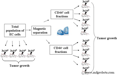
比较分析总群体和分离的CD44 + 致瘤能力的实验设计 和 CD44 – EC分数
在第一组实验中,我们评估了总群体和分离的 CD44 + 的致瘤能力 和 CD44 – 以用于 EC 启动的标准剂量将它们施用于动物时的 EC 分数 (3 × 10 6 0.3 毫升生理盐水中的细胞)。
动物被分为以下几组(n =10):
-
第 1.1 组 - 施用 EC 细胞总数(3 × 10 6 细胞/动物)
-
组 2.1 – CD44 + 的管理 EC细胞的比例(3 × 10 6 细胞/动物)
-
第 3.1 组–管理 CD44 – 部分EC细胞(3 × 10 6 细胞/动物)
各实验组接种后7天,计算动物PC总细胞数,评估细胞表型特征(如上所述),CD44 high /CD117 + EC 细胞比例确定为 CD44 high CD117 + 的百分比 细胞 [22]。总 EC 群体和分离的 CD44 + 细胞的增殖潜力 和 CD44 – 基于以下数据估计分数:培养期间细胞群过剩的多重因子(MF),M =N/N0;和时间加倍(TD),TD =(log22)* t /[log2 (N /N 0)],其中 t 为细胞培养时间(h),N 是 t 处的单元格数 时间; N 0为初始单元格编号[23]。
在第二组实验中,估计了总群体和分离的 CD44 + 的给药细胞的最小剂量 和 CD44 – EC 组分,诱导肿瘤生长。总细胞悬液和分离的 CD44 + 和 CD44 – EC组分以3 × 10 6 的剂量腹腔注射给小鼠 , 3 × 10 5 , 3 × 10 4 , 和 3 × 10 3 每只小鼠的细胞在 0.3 ml 盐水中,并在 PC 中培养 7 天。
本组实验中使用的动物分为以下几组(n =10):
-
第 1.1 组 - 施用 EC 细胞总数(3 × 10 6 细胞动物)
-
第 1.2 组 - 施用 EC 细胞总数(3 × 10 5 细胞/动物)
-
第 1.3 组 - 施用 EC 细胞总数(3 × 10 4 细胞/动物)
-
第 1.4 组 - 施用 EC 细胞总数(3 × 10 3 细胞/动物)
-
组 2.1 – CD44 + 的管理 EC细胞的比例(3 × 10 6 细胞/动物)
-
第 2.2 组 – CD44 + 的管理 EC 细胞的比例 (3 × 10 5 细胞/动物)
-
第 2.3 组 – CD44 + 的管理 EC 细胞的比例 (3 × 10 4 细胞/动物)
-
第 2.4 组 – CD44 + 的管理 EC 细胞的比例 (3 × 10 3 细胞/动物)
-
第 3.1 组–管理 CD44 – 部分EC细胞(3 × 10 6 细胞/动物)
-
第 3.2 组–管理 CD44 – 部分EC细胞(3 × 10 5 细胞/动物)
-
第 3.3 组–管理 CD44 – 部分EC细胞(3 × 10 4 细胞/动物)
-
第 3.4 组–管理 CD44 – 分数 EC 细胞 (3 × 10 3 细胞/动物)
各实验组接种EC后7天测定PC总细胞数和腹水发育动物总细胞数。
纳米复合物的合成
在闪烁材料研究所合成了包含浓度为 1.30 克/升的球形纳米粒子 (NP)(直径为 2-3 纳米)和浓度为 0.55 克/升的绵羊胆固醇(“Acros Organics”,比利时)的混合纳米复合物乌克兰国家科学院(哈尔科夫)的报告 [18]。基于稀土元素正钒酸盐的纳米颗粒 GdYVO4:Eu 3+ 如 [24] 所述制备浓度为 1.30 g/l 的球形。已使用“Cellu Sep H1”3.5 KDa 膜通过透析纯化了基于原钒酸盐的胶体水溶液中的杂质。
在混合纳米复合物中,由于范德华力和疏水相互作用,带负电荷的 NPs 位于胆固醇颗粒的外围。 NPs 通过静电相互作用稳定纳米复合物。合成的纳米复合物的尺寸不超过 100 纳米。此外,NPs 具有抗氧化性能,不会被氧化。这一事实有助于提高胆固醇水分散体对活性氧物质的抵抗力。杂化纳米复合物的结构示意图见图2。
<图片>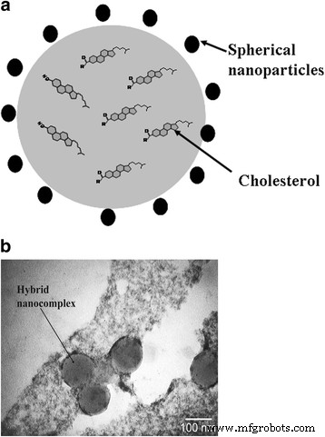
混合纳米复合物:a 示意图和b 碳网络上胆固醇水溶液制备的杂化纳米复合物的透射电子显微镜显微照片
为了在体外研究期间记录混合纳米复合物在细胞中的积累,可以将疏水性荧光染料 1,1'-dioctadecyl-3,3,3',3'-tetramethylindocarbocyanine perchlorate (DiI) 添加到胆固醇水分散体中,允许在局部发光光谱中通过单体的比率评估复合物整合到细胞膜中的动力学 - “J-聚集体”发光带 [25]。我们早期的研究表明,混合纳米复合物能够整合到不超过 10% 的总 EC 细胞群中,并且几乎可以整合到所有分离的 CD44 + 具有最高致癌潜力的部分。这允许在该修饰中使用混合纳米复合物(NPs + 胆固醇 + DiI)作为识别癌细胞中纳米复合物局部积累的方法 [26, 27]。
用纳米材料预处理 EC 细胞
具有混合纳米复合物或 NP 的 EC 细胞的总悬浮液在室温下在 5% 葡萄糖溶液(“Infusion”CJSC,Kyiv)中孵育 3 小时。先前发现这样的孵育时间是纳米复合物与细胞结合的最佳时间[26]。
测试了以下用纳米材料预处理的EC细胞变体:
-
选项 1–900 μl EC 细胞 (1 × 10 7 ) 加入 100 μl 球形纳米颗粒 (1.3 g/l)。
-
选项 2–900 μl EC 细胞 (1 × 10 7 ) 加入 100 μl 混合复合物(球形 NPs (1.3 g/l) + 胆固醇 (0.55 g/l))。
对照是EC总群体的细胞,其在没有用纳米复合材料处理的情况下在5%葡萄糖溶液中温育。每个实验组的动物数量不少于20。
孵育后,所有测试组的EC细胞通过离心(10分钟,300g)用盐水(1:1)洗涤3次。
通过腹腔注射3 × 10 6 评估纳米材料预处理后EC发展的强度 0.3 ml 生理盐水中的细胞。各研究组接种EC细胞后7天,测定:
-
腹腔内EC细胞总数(TN)。
-
根据公式Ri =(TN(c) – TN(e))的EC生长抑制率(Ri):TN(c) × 100%,其中TN(c)––PC中EAC细胞的总数对照组,TN(e)——实验组PC中EAC细胞总数。
-
用公式Rg(e) =Rg(c) - Ri计算EC的生长率(Rg),其中Rg(e)——实验组动物肿瘤的生长率; Rg (c)——对照组肿瘤生长率,Ri——实验组动物EC生长抑制率;对照组EC生长抑制率为100%,此时EC生长无抑制作用。
-
CD44 高 /CD117 + 比率(CD44 高 的比率 CD117 + 的百分比 细胞)。
-
在腹腔注射未处理和用所有类型的纳米复合材料 EC 细胞处理后,评估动物存活至第 20 天。
使用非参数 Mann-Whitney U 进行统计处理 在 Statistica 6.0 软件中进行测试。 P 处的差异被认为具有统计学意义 <0.05.
结果
获得的结果表明存在异质性的 EC 细胞群,其表面带有 CD44、CD24、Sca-1 标记物,以及可归因于微环境的辅助调节元件 (CD117) 的那些。表 1 中显示了具有这些特征的细胞在总 EC 库(组 1.1)中的浓度,并且与之前关于 EC 亚群组成的发现完全一致 [28]。几乎在所有 EC 细胞中鉴定了 Sca-1 结构,使其成为这种肿瘤类型的通用标志物。
就 CSC 的表型鉴定而言,信息最丰富的是 CD44 分子表达,它本身或与其他表面标志物结合用于从包括 EC 在内的各种肿瘤中分离该细胞群。根据经典观念,乳腺癌发展过程中肿瘤细胞的分化伴随着CD-44受体表达的降低,随着CD-44受体的逐渐消失和表达CD24标志物的细胞的出现[3]。
CSCs 在 EC 过程中发挥作用的候选者可能是 CD44 高 的细胞 表型是 CD44 + 的一部分 CD24 – - 人口。这种关于 EC 依赖于 CD44 + 亚群功能活性的假设 在评价CD44 + 诱导的肿瘤生长强度时检测细胞 和 CD44 – 派系和 EC 总人口。表1表明CD44 + 细胞固有的最高的肿瘤诱导活性 分数。实际上,在管理 3x10 6 之后 CD44 + 细胞(组2.1),PC中细胞绝对数比EC细胞总数(组1.1)高23倍,比CD44 – 高105倍 给予部分(第 3.1 组)。
因此,发现了发展中肿瘤的不仅数量上而且质量上的组成的变化。 CD44 + 的分数 形成以CD44 + 为主的腹水 细胞,即 CD44 high , CD44 + CD24 – , 和 CD44 + CD24 + 细胞。此外,CD44的浓度 高 与 1.1 组相比,细胞数高 2 倍,3.1 组高 16 倍。 CD44的分数 – 相比之下,形成了包含更多成熟细胞的肿瘤,即CD44 - CD24 + 表型。 3.1组细胞亚群组成的重新分布显然决定了PC中细胞的最小绝对含量。
重要的是已经确定的事实,即在具有 CD117 + 的亚群的 EC 细胞中存在 标记。 CD117 分子是一种跨膜酪氨酸激酶受体。在正常情况下,它被相应的配体激活,即干细胞生长因子 (SCGF) [29]。在肿瘤病理学中,c-KIT 受体的配体依赖性激活发生,这通常(高达 92% 的病例)是 c-kit 癌基因突变的结果,或者是由该受体功能的调节机制紊乱引起的[30]。
考虑CD117 + 细胞作为肿瘤微环境的细胞 肿瘤生长强度依赖于 CD117 + 细胞及其浓度与 CD44 high 的关系 细胞是逻辑。如表 1 所示,当通过在 PC 中引入总细胞群(组 1.1.)来启动 EC 时,形成了 34.80 ± 1.27 × 10 7 CD44 高 的细胞 /CD117 + 0.02个相对单位。
CD44的致瘤潜力 – 分数低 4 倍(组 3.1),表现为 CD44 high /CD117 + 如果与组 1.1 相比,比例相同(4 倍)。 CD44 高 的这种变化 /CD117 + 指数主要是由于CD44 high 下降 CD117 + 含量降低背景的浓度(8.5倍) 细胞(2次)也是如此。
评估腹水生长强度时,由 CD44 + 产生 部分,注意到 PC 中细胞总数的显着增加(如果与组 1.1 相比,几乎是 24 倍)。同样重要的是CD44的两倍 high CD117的浓度和缺乏 + 细胞。 CD44的初始素材 + 部分(培养前)根据数据的流式细胞术分析,CD44 高 的含量 细胞比 EC 细胞总数高 15 倍(数据未提供)。
综上所述,我们可能会争辩说,具有高 CD44 标记(CD44 high ),而CD117最重要的功能之一 + 亚群是 CD44 high 致瘤活性的调节(“抑制”) 细胞。 CD117 + 的缺失 细胞(2.1 组)似乎使整个 CD44 池的增殖和分化潜能成倍增加 + 细胞,导致PC中细胞总数显着增加。
EC 细胞总池和 CD44 + 增殖潜力的分析 派赞成这种解释。结果表明,在2.1组PC中培养的总体群体中的倍增因子(MF)。如果与组 1.1 相比,在 7 天内增加了几乎 24 倍。这伴随着细胞时间从第 1.1 组的 24.47 ± 2.75 小时增加一倍减少至第 2.1 组的 14.70 ± 1.35,这可能表征了从 CD44 + 生长的腹水细胞群 分数,作为更积极增殖的一个(表 1)。
证明CD44 + 的特殊作用 细胞在启动和维持肿瘤时,即使以最小剂量给予 EC,比较评估分离的 CD44 + 的致瘤能力是有意义的 和 CD44 – 以不同浓度给药时的分数。发现引入3 × 10 6 后 在总 EC 群体的细胞中,在 100% 的动物中观察到肿瘤生长 (10/10)(表 2)。细胞给药剂量减少10倍(3 × 10 5 ) resulted in a proportional decrease in absolute number of cells in the PC, tumor developed only in 50% of animals (Table 2). Reducing the administered dose of total EC population of cells down to 3 × 10 4 did not lead to tumor formation in the PC.
Initiations of EC by introducing of CD44 + cells at concentrations of 3 × 10 6 and 3 × 10 5 cells per animal resulted in almost 100% tumor development for both cases. Herewith, tumorigenic potential of CD44 + fraction exceeded that of total population of EC cells administered in the same doses (in 23 and 21 times, respectively). Moreover, introduction of 3 × 10 4 cells of CD44 + fraction caused a tumor formation in 33% of animals, while total population of EC cells used in the same dose, did not cause the formation of ascites. With the introduction of 3 × 10 3 cells of CD44 + fraction, no animals with the developed EC have been identified.
Fraction of CD44 – cells just in a dose of 3 × 10 6 was capable of forming tumors in 50% of animals, the number of cells in the PC in this case was 4.5 times less than when introducing the total population and in 105.9 times less than when inducing by CD44 + 分数。 Thus, the results of this part of research suggest that CSCs are mainly present in the pool of cells with CD44 + 表型。 This emphasizes the importance of this subpopulation of cells in initiation and development of EC.
As noted above, identification and inactivation of CSCs is a major theoretical and practical issue of oncology. On this basis, the next task of our study was to investigate the impact of hybrid nanocomplexes designed at the Institute for Scintillation Materials of National Academy of Sciences of Ukraine on the tumorigenic activity of EC cells.
As Table 3 demonstrates an incubation of EC cells with only NPs as a component of hybrid nanocomplexes (option 1) decreased the concentration of CD44 high virtually twice if compared to the control and 5 times the content CD44 + CD24 – cells in ascites formed in vivo. The number in it of more differentiated CD44 + CD24 + , CD44 – CD24 + cells remained practically unchanged if compared to the control. In this group, there was established reduction of CD117 + cells (35%) at a slightly changed content of Sca-1 + 亚群。 Based on the data, the inhibition rate of EC growth (59.41 ± 3.45%) in variant 1 was accompanied by a twofold decrease in the concentrations of CD44 high cells in comparison with the control that was also reflected in the reduction of CD44 high /CD117 + ratio (Table. 3).
Pretreatment of EC cells with hybrid nanocomplexes (option 2) reduced almost 10 times the concentration of CD44 high and CD44 + CD24 – cells in the developed ascites if compared to the control (Table 3). It should be noted that the concentration of more differentiated CD44 + CD24 + and CD44 – CD24 + cells after this treatment increased slightly if compared to the control. The redistribution pattern of EC subpopulation composition in this option was accompanied with a pronounced enhancement of tumor growth inhibition compared to option 1 (74.70 ± 4.38 and 59.41 ± 3.45%, respectively, P < 0.05) that underlined the importance of cholesterol as a targeted compound of antitumor therapy. Pretreatment with hybrid nanocomplexes (option 2) led to maximal reduction there was found a maximum reduction of CD44 high /CD117 + ratio (10 times) as compared with option 1, that again confirmed a specific role of ratio of these cell subpopulations in the EC growth.
For all the types of EC pretreatment, the reduction of CD44 high /CD117 + ratio was accompanied by a decrease in tumor growth rate and increased survival of animals to day 20 of EC development (Fig. 3).

Tumor growth rate of EC, survival of animals and CD44 high /CD117 + ratio after incubation with nanocomplexes. Note:differences are statistically significant as compared with administration of the control (*), option 1 (**) (P < 0.05)
Discussion
One of the tasks of current oncology is elucidation of the mechanisms of initiation and development of malignant neoplasms. Mandatory participants in these events are the CSCs and so-called accessory-regulatory cells of tumor microenvironment. The variety of functional and structural characteristics of the CSCs in the development of different types of tumors determines the need for their further study. This is facilitated by the expansion of experimental model systems. One of them is the transplantable line of tumor cells of EC.
The elucidation of the peculiarities of this experimental model development, the subpopulation composition of tumor and tumorigenic potential of individual cell populations within the general pool of the EC cells will facilitate the development of new approaches to cancer therapy.
Using the method of phenotypic evaluation of progenitor cells of various levels of differentiation in the tumor focus makes it possible the identifying the stages, dynamics of development and invasiveness of the process. The established fact of heterogeneity of the EC subpopulation composition is important and there has been emphasized the value of CD44 + subpopulation in maintaining the growth of this type of tumor.
The most important role in implementing a tumorigenesis is played by an expression rate of the molecule. Indeed, in contrast to leukocytes for adhesion of those normally a low expression rate of CD44 receptor is required, triggering and self-maintenance in CSCs are implemented its much greater density on a cell surface [31].
It is known that CD44-glycoprotein is a hyaluronic acid (HA) receptor, a main component of extracellular matrix. The emerging set of HA-CD44 activates many receptor tyrosine kinases, resulting in activation of PI3K/Akt/ mTOR way [32, 33], which plays the role of a single universal signal transmission mechanism to the translation apparatus and is responsible for the integration of proliferative stimuli.
Among two known CD44-isoforms in normal hematopoietic cells its standard isoform (CD44s) is predominantly expressed [34]. In most malignant tissues there were detected both CD44s and variable isoforms of CD44- molecule (CD44v), resulting from alternative splicing of exons 6-15. Namely alternative splicing leads to a lengthening of CD44-extracellular domain, promoting its greater interaction with HA and tumor metastasis [35]. Due to that the role of CD44 high cells in triggering and maintaining the tumorogenesis is clear. It was previously found that a minor subpopulation of CD44 high cells had a high proliferative potential and played a critical role in EC developing [20].
In this paper, a special role of CD44 + -cells of the EC in initiation and maintenance of the tumor process in the EC under administration even in minimal doses has been shown. CD44 + cells were able to form a tumor even at a cell concentration of 100 times lower (10 4 cells/ mouse) if compared with the introduction of a total EC population (10 6 cells / mouse). The belonging of tumor cells to the CD44 + fraction was also confirmed by the fact that the EC initiation by the fraction of CD44-cells even at a dose of 10 6 cells / mouse caused the formation of a tumor only in 50% of cases, with an absolute number of cells in the PC 5 times less than in under introduction of a similar amount of the total population of EC and more than 100 times less than after the introduced CD44 + -fraction.
This is in accordance with the data of Shipitsin M et al. has shown that CD44 + and CD24 + cells in breast cancer development there are cell populations with different genetic profiles [36]. The research performed by Shipitsin M CD24 + cells have been noted to be more differentiated, while more progenitor-like functions are inherent to CD44 + 细胞。 The research performed by Shipitsin M CD24 + cells have been noted to be more differentiated, while more progenitor-like functions are inherent to CD44 + 细胞。 The authors suggest that CD24 + cells can be derived from CD44 + cells [36]. Fillmore C. and Kuperwasser C. supposed that CD24 + population was mainly characterized by less differentiated basal type of breast cancer, and CD44 + cells caused the development of luminal form of breast cancer, being more differentiated type of tumor [37].
Analyzing the patterns of tumor development, the classic hypothesis of «seed and soil» looks very actual [38], which postulated that an appropriate microenvironment (soil) is required for optimal growth of tumor cells (CSCs).
Most often the carcinoma-associated fibroblasts (CAFs) act as a tumor stroma in breast cancer and pancreatic cancer [39]. It has been shown that the CAFs, derived from invasive forms of human breast carcinomas, activated much stronger the growth of human breast cancer cell line MCF-7-Ras when administered to immunodeficient mice if compared with normal fibroblasts [9]. This function is implemented by the microenvironment cells due to the secretion by them of cytokines, chemokines and growth factors [10, 40].
Although so far the phenotypic identification of the microenvironment cells for various types of tumor has remained a subject of debate, most often used for this purpose the surface markers of primitive hematopoietic and endothelial cells, including c-kit (CD 117), CD133, VE-cadherin, VEGFR-2 and endoglin are used [41]. In this experimental model the most probable candidate to the role of tumor microenvironment cells is CD 117 + .
It is known that the c-KIT receptor (CD117 + ) is highly expressed in normal epithelium of the breast and progressively decreases with the development of breast carcinoma in situ and is almost completely lost in invasive breast cancer [42, 43]. Some authors proposed this kind of change in the expression rate of this marker as a possible test to assess the effectiveness of antitumor therapy [44].
Previously, after analysis of the significance of the content ratios for different subpopulations of EC cells when maintaining tumor growth, we proposed to use the CD44 high /CD117 + ratio as a prognostic criterion of tumor development [22].
Adequacy of using this index is confirmed in this study using the applied nanocomposites as therapeutic agents when treating the EC. The inhibition rate of EC growth (59.41 ± 3.45%) when treated with spherical NPs (option 1) was accompanied by a 2-fold decrease if compared to the control in the CD44 high -cell concentration, which was reflected in the reduced CD44 high / CD117 + 指数。 The maximum decrease in the CD44 high /CD117 + index (10 times if compared to option 1) was established using the hybrid nanocomplexes for a pre-treatment of EC cells. Thus, many cells of a total pool of EC, but primarily those with the phenotype CD44 high and CD117 + , can be the target of the effect of the studied nanocomplexes (both direct and indirect). A significant decrease in their concentrations in the growing pool of EC after pretreatment with hybrid nanocomplexes clearly coincides with a reduced intensity of tumor growth.
Judging by the decrease in the amount of CD44 high as the most potent CSCs forming the entire subsequent series of advanced tumor cells, the main component in manifestation of antitumor effect of the synthesized hybrid nanocomplexes is spherical NPs. Introduction of cholesterol having affinity to tumor cell membranes into composition of hybrid nanocomplexes enhanced an inhibitory activity of NPs. Similar data were obtained by Betker J.L. et al. after analysis of the structure and functioning principles of the membranes of tumor cells. The authors concluded that the incorporation of cholesterol into membranes of tumor cells could be a prerequisite for a targeted delivery of liposomes with therapeutic agents directly into a cell.
Thus, the importance of cooperative interactions of cells with different phenotypic signs in maintaining the EC growth has been proven. The cells with the CD44 high phenotype being the part of the population of CD44 + CD24 – can be considered as CSCs in this model system. The use of new forms of nanocomposites that are capable to bind to CSCs and induce tumor destruction as the EC is a promising direction the treatment of oncopathology.
Conclusions
- 1.
On the base of the findings of phenotypic assessment and functional potential studies, the Ehrlich carcinoma is a heterogeneous population of tumor cells of varying differentiation extent referred to high and less potent tumor-inducing precursors, as well as the cells composing their microenvironment.
- 2.
A high (tenfold) tumorigenic activity of the EC CD44 + cells if compared to CD44 – cells was proven. In this pair of comparison, the CD44 + cells had a higher potential of generating in PC of CD44 high , CD44 + CD24 – , CD44 + CD24 + cell subpopulations, highlighting the presence of CSCs in a pool of CD44 + cells.
- 3.
There was found an ability of the synthesized nanocomplexes based on rare earth orthovanadates and cholesterol to inhibit the growth of CD44 + cell pool (CD44 high , CD44 + CD24 – , CD44 + CD24 + ) that was accompanied by a reduced intensity of EC growth (by 75%) and increased survival of the animal with tumors (in 3.5 times) in comparison with the control.
- 4.
It has been shown that the reduction in tumor growth rate after pretreatment with hybrid nanocomplexes was accompanied with a change in the composition of EC subpopulation that was reflected in a decrease in the CD44 high /CD117 + 比率。 This ratio can be offered as one of diagnostic and prognostic tests of the severity and extent of oncology inactivation.
缩写
- BC:
-
Breast cancer
- CAFs:
-
Carcinoma-associated fibroblasts
- CSCs:
-
Cancer stem cells
- DiI:
-
1,1′-Dioctadecyl-3,3,3′,3′-tetramethylindocarbocyanine perchlorate
- EC:
-
Ehrlich carcinoma
- HA:
-
Hyaluronic acid
- MF:
-
Multiplicity factor
- NOD/SCID mice:
-
Nonobese diabetic-severe combined immunodeficiency mice
- NPs:
-
纳米粒子
- PC:
-
Peritoneal cavity
- Rg:
-
Growth rate of EC
- Ri:
-
Inhibition rate of EC growth
- SCID mice:
-
Severe combined immunodeficiency mice
- TD:
-
Time doubling
纳米材料