

使用新型均匀磁场改善磁性聚乙烯亚胺纳米颗粒向 MG-63 成骨细胞的磁转染
摘要
本研究旨在通过将新型均匀磁场与低分子量聚乙烯亚胺修饰的超顺磁性氧化铁纳米粒子 (PEI-SPIO-NPs) 结合使用来改善 MG-63 成骨细胞的磁感染。 PEI-SPIO-NPs 的优良特性,如大小、zeta 电位、pDNA 结合和保护能力,被确定适用于基因传递。新型均匀磁场使聚乙烯亚胺修饰的超顺磁性氧化铁纳米粒子/pDNA 复合物(PEI-SPIO-NPs/pDNA 复合物)能够快速均匀地分布在 MG-63 细胞表面,避免局部转染并减少膜的破坏通过带正电荷的 PEI-SPIO-NPs 的集中,从而增加转染过程中磁性基因载体的有效覆盖,提高磁转染效率。这种创新的均匀磁场可用于确定 PEI-SPIO-NPs 与 pDNA 之间的最佳用量,以及筛选均质条件下磁性基因载体的最佳配方设计。最重要的是,新的均匀磁场有助于将 PEI-SPIO-NPs/pDNA 转染到成骨细胞中,从而为将治疗基因靶向递送至骨肉瘤组织提供了一种新方法,也为其他肿瘤的治疗提供了参考。
背景
骨肉瘤是最常见的恶性骨肿瘤,主要影响儿童和青少年。由于传统疗法提供的改善有限,因此必须采用新的治疗策略 [1,2,3]。基因治疗的出现促使研究人员评估其在骨肉瘤中的应用 [4,5,6]。安全有效的基因传递系统对于基因治疗至关重要。基因传递系统可分为病毒基因传递系统和非病毒基因传递系统。病毒基因递送系统已被证明在多种原代细胞和细胞系中具有高转染效率。病毒基因传递系统与安全问题密切相关,例如引发炎症反应和基因突变 [7],促使研究人员将研究转向非病毒基因传递系统。然而,大多数非病毒基因转染技术在转染骨肉瘤细胞系方面并不是特别有效[8, 9]。
在过去的十年中,基因枪 [10]、超声波 [11] 和电穿孔 [12] 等各种物理方法已被有效地用于改善转染。然而,这些物理方法也会引起细胞损伤 [13]。由于磁场一般不会对细胞造成损伤,因此磁辅助转染技术引起了研究人员的关注。马等人。通过将携带绿色荧光蛋白 (GFP) 的重组腺相关病毒 (rAAV) 与磁性纳米颗粒连接起来,在基因转染实验中引入磁性纳米颗粒,从而在体外和体内实现靶向特异性增强的转染。与纯腺病毒相比,磁性腺病毒纳米粒子在相同转染率条件下可将携带 GFP 的 rAAV 的剂量降低至 1% [14]。舍勒等人。创造了术语“磁转染”来表示使用磁性颗粒的磁介导转染 [15]。随后,普朗克等人。描述了使用非病毒载体的磁性转染技术 [16]。为了进一步提高非病毒载体的磁转染效率,Fouriki 等人。调整振荡磁场的频率和幅度,以对磁性纳米粒子产生动态机械刺激效应,从而增加与靶细胞接触的可能性[17]。口头等。通过反向旋转磁铁的六边形轴来控制磁场的速度和方向,进一步提高了转染效率并降低了基因载体的细胞毒性[18]。在另一项研究中,Vainauska 等人。使用通过旋转圆柱形永磁体产生的动态梯度磁场来靶向递送磁性基因载体[19]。
随着磁场从静态和单一演变为动态和复杂,磁转染的效率显着提高。然而,关注磁场均匀性和有效范围的研究是有限的。据我们所知,大多数磁体装置无法在特定的三维空间中形成均匀的磁场,从而导致磁性纳米粒子的分布不均匀,只能实现磁场的局部转染效果,无法解决由此产生的细胞毒性问题。不均匀的磁场也阻碍了磁转染试剂的比较,降低了转染效率。
为了解决这些问题,我们的研究小组与重庆大学电气工程学院合作开发了一种新型磁场发生器[20, 21]。磁场发生器可在一定高度形成均匀磁场,加入的磁性纳米粒子迅速均匀分布在培养板底部。均匀磁场不仅便于比较研究各种转染试剂的剂量与转染效率之间的关系,还可用于评估细胞对磁性纳米颗粒的摄取。
在目前的设计中,我们旨在建立一个可靠的非病毒基因传递系统,在骨肉瘤细胞系中具有高磁转染效率。选择线性 Mw20000 聚乙烯亚胺 (PEI-20000) 来制备磁性纳米粒子,因为之前的几项研究已经证实,线性 PEI 递送系统在体内具有比分支 PEI 递送系统更好的转染效率 [22, 23]。评价了聚乙烯亚胺改性的超顺磁性氧化铁纳米颗粒 (PEI-SPIO-NPs) 的性能。开发了一种新型磁场发生器以形成均匀磁场,用于将 PEI-SPIO-NPs/pDNA 复合物转染到 MG-63 骨肉瘤细胞系中。系统研究了不同磁场对磁感的影响。
方法
材料和试剂
超顺磁性氧化铁纳米粒子 (SPIO-NPs)、聚乙烯亚胺盐酸盐 (PEI) 和 Hoechst-33,324 购自 Sigma-Aldrich Co. (St Louis, MO, USA)。 PolyMag-200(商业磁转染试剂)购自北京首席东方科技有限公司(Qwbio)(中国北京)。 1-乙基-3-[3-(二甲氨基)丙基]碳二亚胺(EDC)和N -羟基琥珀酰亚胺(NHS)购自成都西雅试剂有限公司(中国四川)。 PolyMag-200 和 96 孔细胞培养磁性板购自 Chemicell GmbH(德国柏林)。 Dulbecco 改良 Eagle 培养基 (DMEM) 和青霉素-链霉素和胎牛血清 (FBS) 购自 Invitrogen-Gibco (CA, USA)。罗丹明 B 异硫氰酸酯购自 Aladdin Ind., Co.(中国上海)。 LysoTracker Green DND-26 购自 Shanghai Wei Jin Biological Technology Co., Ltd.(中国上海)。 CCK-8试剂盒购自七海生物科技有限公司(中国上海),Endo-free Plasmid Maxi Kit-25购自OMEGA(美国GA)。磷酸盐缓冲液(PBS)和其他试剂在我们实验室制备。所有溶剂和化学品均为分析纯。
PEI-SPIO-NPs 的合成
如前所述制备 PEI-SPIO-NPs [24]。简而言之,将 0.1 g EDC 和 0.5 g NHS 添加到 15 mL 羧基修饰的 SPIO-NPs 水溶液(5 mg/mL,pH 调至 5)中,并在室温下搅拌溶液 4-6 小时以激活氧化铁纳米颗粒的羧基。接着,加入相同体积的聚乙烯亚胺盐酸盐水溶液(20mg/mL)反应数小时。使用浸没在蒸馏水中 2 天的渗析膜 (MWCO 20,000) 渗析所得结合的 PEI-SPIO-NPs 溶液,以去除任何未结合的 PEI 分子和中间体。将等分的纳米颗粒溶液冷冻干燥并储存。
PEI-SPIO-NPs/pDNA 复合物的合成和表征
PEI-SPIO-NPs 的合成如上所述,PEI-SPIO-NPs 和质粒 DNA (pDNA) 在 37°C 下分别预热 10 分钟,然后以不同的 N/P 比混合在一起以制备PEI-SPIO-NPs/pDNA 复合物。 PEI-SPIO-NPs/pDNA 复合物的形态通过透射电子显微镜(TEM,CM100,Philips,Netherlands)测量,流体动力学直径通过动态光散射(DLS,Nicomp 380,PSS,FL,USA)测量.使用 EV-11 振动样品磁力计(PPMS-9,Quantum Design,San Carlos,CA,USA)在 300 K 下检查它们的 SPION 和 PEI-SPIO-NP 的磁特性。测量值归一化为铁的重量每个样品,并通过电感耦合等离子体发射光谱仪(ICP-OES)验证测量结果。
通过使用动态光散射仪(Malvern,Southborough,MA,USA)在磷酸盐缓冲盐水(PBS,pH =7.4)中测定 zeta 电位来测量表面电荷。通过将 PEI-SPIO-NPs 溶液添加到 pDNA 溶液中,以 2.5 至 25 的各种 N/P 摩尔比制备 PEI-SPIO-NPs/pDNA 复合物,并将最终 pDNA 浓度调整为 30 μg/mL。
使用原子力显微镜(IPC-208B,重庆大学,重庆,中国)检测 PEI-SPIO-NPs/pDNA 复合物的成分。将用作观察载体的小片状金属用丙酮洗涤并干燥。然后,将几滴反义探针样品放在金属上并风干。在700 nm × 700 nm和1000 × 1000像素的大尺度扫描区域内观察样品的表面形貌。在9 nm × 9 nm和800 × 800像素的小范围扫描区域内观察样品的分子结构和微观结构。将原始图像数据传输至计算机,使用 G2DR 软件进行 3D 重建[25]。每组重复测量三次。
通过凝胶电泳分析与 PEI-SPIO-NPs 结合后 pDNA 的迁移率。 PEI-SPIO-NPs/pDNA 的 N/P 比为 1/5、1、5、10、15、20、25 和 30,其中 DNA 的含量保持在 3 μg。在 37°C 下孵育 15 分钟后,通过 1% 琼脂糖凝胶电泳(90 V,30 分钟)分析 10 μL 混合溶液。裸pDNA作为对照。
评估了 PEI-SPIO-NPs 对 pDNA 对 DNase I 的保护作用。将各种 N/P 比的 PEI-SPIO-NPs/pDNA 复合物和裸 pDNA (3 μg) 在 5% CO2 半湿润环境中与 DNase I (4 U) 在 DNase/Mg 中的 DNase I (4 U) 下在 37°C 下孵育 30 分钟 2+ 由 50 mM Tris-HCl (pH=7.6) 和 10 mM MgCl2 组成的消化缓冲液。然后通过添加 EDTA 溶液 (pH =8.0) 灭活 DNase I,直到最终浓度为 2.5 mM。然后将样品在 65°C 下孵育 15 分钟,并加入 10 μL 的 1 mg/mL 肝素钠以从 PEI-SPIO-NPs/pDNA 复合物中释放 pDNA [26]。 pDNA 的完整性通过 1% 琼脂糖凝胶电泳(90 V,30 分钟)进行评估。
磁场
新型磁场发生器(图 4)是我们团队与重庆大学电气工程学院合作开发的 [20, 21]。我们使用 3D CAD 图像来显示均匀磁场的磁铁排列。显示了感兴趣区域中磁场的 XOY 平面分布和 3D 分布。观察PEI-SPION-NPs在不同磁场中的分布,以非均匀磁场(96孔Nd-Fe-B永磁板)为对照。
细胞培养
人骨肉瘤细胞系MG-63(原美国典型培养物保藏中心)购自重庆市干细胞治疗工程研究中心(中国重庆)。将细胞维持在补充有 10% FBS、100 U/mL 青霉素和 100 mg/mL 链霉素的 DMEM 中,并在 37°C、5% CO2 和 95% 相对湿度下培养。
体外细胞毒性
使用 CCK-8 检测试剂盒确定不同纳米粒子有或没有 pDNA 以及磁化或未磁化的纳米粒子对 MG-63 骨肉瘤细胞的体外细胞毒作用。将细胞以4 × 10 3 的密度接种于96孔培养板中 每孔一个细胞,加入不同的纳米颗粒并孵育 24、48、72 和 96 小时。在预设时间点将 CCK-8 溶液(10% 体积的培养基)加入每个孔中,并将细胞再培养 2 小时。使用荧光酶标仪在 450 nm 波长下评估细胞的光密度,吸光度值反映高细胞活力;高活力的观察表明纳米颗粒诱导低细胞毒性作用。 PolyMag-200/pDNA 和 PolyMag-200 用作阳性对照。为了研究不同磁场对细胞的毒性作用,我们选择了PEI-SPIO-NPs(不含pDNA)作为基因载体,并考察了不同时间点不同磁场干预下细胞的光密度。
共聚焦显微镜分析
通过共聚焦激光扫描显微镜观察 PEI-SPIO-NPs/pDNA 复合物或聚乙烯亚胺纳米颗粒 (PEI-NPs) 的吸收。 MG-63骨肉瘤细胞以3 × 10 5 的密度接种于24mm玻璃皿中 每口井。将罗丹明 B 异硫氰酸酯 (RBITC) 标记的 PEI-SPIO-NPs/pDNA (3 μg) 复合物加入细胞中,并在均匀磁场或非均匀磁场的作用下孵育 30 分钟;在没有磁场干预的情况下,将 RBITC 标记的 PEI-NPs/pDNA 复合物添加到细胞中。然后立即用 PBS (0.01 M) 洗涤细胞两次以去除任何游离的 RBITC 标记的 PEI-SPIO-NPs/pDNA 复合物或 RBITC 标记的 PEI-NPs/pDNA 复合物,并将处理过的细胞孵育 6-24 小时.转染后 6、12 和 24 小时去除培养基,细胞核用 Hoechst 33342 染色 5 分钟。去除 Hoechst 33342 残留物后,将细胞在含有 0.5 mM LysoTracker Green DND-26 的预热培养基中孵育 1 小时,然后用预热的 PBS 洗涤两次。细胞在共聚焦激光扫描显微镜(LSM 700,Carl Zeiss,Oberkochen,Germany)下成像,物镜× 40 以可视化具有以下激发 (Ex) 和发射 (Em) 波长 [GFP (Ex:488 nm) 的荧光染料; Em:530 nm), Hoechst (Ex:350 nm; Em:460 nm), LysoTracker Green (Ex:443 nm; Em:505 nm), and RBITC (Ex:554 nm; Em:576 nm)] [27 ].
流式细胞术分析
MG-63细胞接种于12孔板(1 × 10 5 细胞每孔)24 小时,然后用 PBS 冲洗两次,并在转染前用 0.8 mL Opti-MEM 培养基在 37°C 下预孵育 1 小时。将含有 3 μg pDNA 的 PEI-SPIO-NPs/pDNA 复合物(N/P =10)或 PEI-NPs/pDNA 复合物(N/P =10)加入到每孔中,并将细胞培养板置于均匀的或非均匀磁场 20 分钟。 PolyMag-200/pDNA 用作阳性对照。 4 小时后,将细胞用 1 mL PBS (0.01 M) 冲洗 3 次以去除任何游离的 PEI-SPIO-NPs/pDNA 复合物或 PEI-NPs/pDNA 复合物,并将细胞再培养 24 小时。孵育后,收集细胞并通过流式细胞术(BD FACS Canto II,BD Biosciences,San Jose,CA,USA)评估转染效率,该部分实验重复3次。
统计分析
一式三份获得定量数据 (n =5) 并表示为平均值 ± 标准偏差。统计分析使用单向方差分析进行多重比较和学生的 t 组间比较检验。一个 P 值 <0.05 被认为具有统计学意义。
结果与讨论
磁化原理
磁转染被定义为与超顺磁性纳米颗粒相关的载体,并通过应用磁梯度场在靶细胞上积累 [15]。图 1 展示了 PEI-SPIO-NPs/pDNA 复合物的成分和形态以及磁转染的主要原理。缓慢的载体积累和因此在靶组织中的低载体浓度已被确定为有效基因传递的简单但强大的障碍 [28]。磁转染的主要功效增强机制似乎是完整的载体剂量在靶细胞上的快速沉淀,这样高达 100% 的这些细胞将在几分钟内将载体颗粒结合到它们的表面。因此,磁转染是一种合适的工具,可以克服载体缓慢积累的强大障碍,从而克服靶组织中载体浓度低的问题。 PEI-SPIO-NPs/pDNA 复合物是随机的,在没有磁场的情况下,它在目标细胞中的沉积是有限且耗时的。然而,PEI-SPIO-NPs/pDNA复合物在磁场存在的情况下可以在较短的时间内广泛均匀地接触靶细胞,这可能会增加细胞单位面积内吞的机会,从而提高PEI-SPIO-NPs/pDNA复合物的利用率,提高转染效率。
<图片>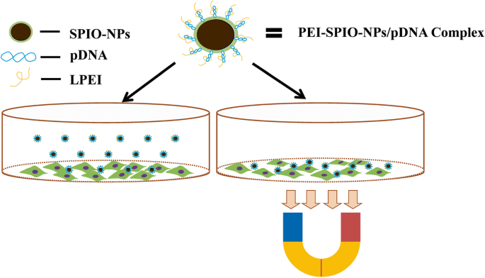
使用 PEI-SPIO-NPs/pDNA 复合物的磁转染概述。内衬聚乙烯亚胺 (LPEI) 和超顺磁性氧化铁纳米粒子 (SPIO-NPs) 通过脱水缩合反应相关联。为此,SPIO-NPs 涂有 LPEI,即 LPEI 与 SPIO-NPs 的表面紧密结合,它们一起形成 PEI-SPIO-NPs。如果 PEI-SPIO-NPs 与裸 pDNA 混合,带负电的 pDNA 会通过静电吸附与带正电的 PEI-SPIO-NPs 结合。在存在磁场的情况下,将细胞与 PEI-SPIO-NPs/pDNA 复合物一起孵育,该磁场将复合物吸引向细胞。磁转染的结果是基本上所有细胞都与载体接触,并且高比例的细胞被快速转染
PEI-SPIO-NPs/pDNA 复合物的表征
图 2a 表明 PEI-SPIO-NPs 大致呈球形,PEI-SPIO-NPs/pDNA 复合物呈聚集状态,均具有良好的分散性,正负电荷之间的吸引力使 PEI-SPIO-NPs/pDNA 复合物能够获得一个小尺寸。 PEI-SPIO-NPs/pDNA 复合物的滞后环如图 2b 所示。通过原子吸收光谱仪测量的 PEI-SPION-NPs 中氧化铁的重量百分比为 20.33(± 2.87)%。 PEI-SPIO-NPs/pDNA 复合物的饱和磁化强度为 21.5 (± 1.6) emu/g 铁。尽管与未修饰的超顺磁性氧化铁纳米粒子相比磁性有所降低,但 PEI-SPIO-NPs/pDNA 复合物表现出优异的磁响应性,这是高效磁转染所必需的。
<图片>
PEI-SPIO-NPs/pDNA 复合物的特征。 一 PEI-SPIO-NPs 和 PEI-SPIO-NPs/pDNA 复合物的 TEM。 b SPIO-NPs 和 PEI-SPIO-NPs/pDNA 复合物的滞后环。 c PEI-SPIO-NPs/pDNA 复合物在各种 N/P 比下的 Zeta 电位和流体动力学直径。 d AFM 灰度图像和 PEI-SPIO-NPs/pDNA 复合物的分子结构。红点表示SPIO化学基团的位置,绿点表示氮原子,黄点表示碳原子,磷原子由白圈黑点表示。 e (a) PEI-SPIO-NPs/pDNA 复合物和 e 的尺寸分布 (b) PEI-SPIO-NPs/pDNA 复合物的 zeta 电位,由 Malvern Zetasizer 动态光散射仪测量。结果表示为平均值 ± SD (n =5)
流体动力学直径和zeta电位被认为是基因载体不可或缺的参数。图 2c 显示 PEI-SPIO-NPs/pDNA 复合物的流体动力学直径为 200 (± 21.7) nm,N/P 比(NH2-PEI 中的基团/PO4-pDNA 中的基团)为 2.5 和 175 (± 16.4) ) nm 当 N/P 比为 5 时,表明在电位从负向正转变的过程中发生了尺寸的快速变化。此后,PEI-SPIO-NPs/pDNA复合物之间的排斥力随着N/P比的升高而增加,表明纳米颗粒的尺寸增加。
我们根据原子的排列分析了化学键的类型,并进一步推测了 PEI-SPIO-NPs/pDNA 复合物的分子结构。 PEI-SPIO-NPs/pDNA 复合物的分子结构通过原子力显微镜(AFM,图 2d)间接公开,这表明超顺磁性氧化铁纳米颗粒的羧基通过酰胺键与 PEI 的伯胺结合。此外,pDNA 通过核苷酸链的磷酸基团和 PEI 的胺基团之间的静电结合被包裹在 PEI 分子链中,形成 PEI-SPIO-NPs/pDNA 复合物。在AFM灰度图中,红点代表铁原子的位置,绿点代表氮原子,黄点代表碳原子,白圈黑点代表磷原子。
如图 2e 所示,使用动态光散射仪检测 PEI-SPIO-NPs/pDNA 复合物的潜力。在 pH =7 时,超顺磁性氧化铁纳米粒子 (SPIO-NPs) 的电位为 - 6.6 (± 1.1) mV,这表明其表面存在羧基。 PEI-SPIO-NPs 的电位为 + 18.2 (± 1.5) mV。用 SPIO-NPs 修饰的 PEI 的电位转化为带正电荷的表面,可用作与带负电荷的 pDNA 结合的基因载体。我们评估了 PEI-SPIO-NPs/pDNA 复合物在不同 N/P 比下表面电荷的变化。 PEI-SPIO-NPs/pDNA 复合物即使在 2.5 的低 N/P 比率下也显示出负表面电荷,随着 N/P 比率增加到 5,它逐渐转化为正表面电荷。 N/P 比率的这种增加在 PEI-SPIO-NPs/pDNA 复合物中导致复合物的正表面电荷增加。在 N/P 比为 10 时,PEI-SPIO-NPs/pDNA 复合物的电位 (N/P =10) 为 + 11.9 (± 1.2) mV,表面电荷达到平台。带正电的复合物与带负电的细胞膜接触,使细胞能够吸收复合纳米颗粒 [29]。 PEI-SPIO-NPs 不仅可以作为带负电荷的 pDNA 的基因载体,还可以为靶向药物递送提供一定程度的磁性,成为用于磁共振成像 (MRI) 对比成像的造影剂 [30, 31] .
基因载体的一个基本要求是转染载体必须有效地与核酸形成稳定的复合物。为了评估其结合能力,将类似体积的 PEI-SPIO-NPs 溶液和 pDNA 溶液以不同的 N/P 比混合并涡旋。然后通过琼脂糖凝胶电泳分析 10 μL 混合物的等分试样(图 3a)。与裸 pDNA 相比,质粒的迁移在 N/P 比为 5 时被完全阻断,表明 PEI-SPIO-NPs 完全浓缩了 pDNA [32]。
<图片>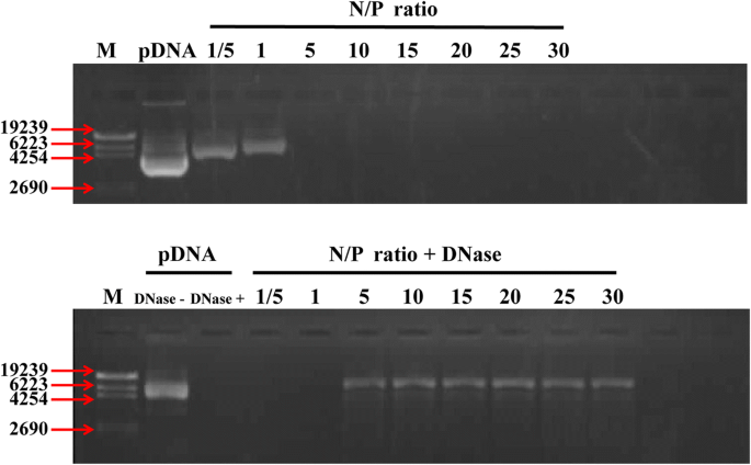
PEI-SPIO-NPs 的 pDNA 结合试验和 pDNA 保护试验。 一 PEI-SPIO-NPs/pDNA 复合物在不同 N/P 比下的琼脂糖凝胶电泳。 b DNase-I 处理后 PEI-SPIO-NPs/pDNA 的电泳迁移率分析。将各种 N/P 比和 pDNA (3 μg) 的 PEI-SPIO-NP 与 DNase-I (4 U) 在 DNase/Mg 中在 5% CO2 半湿润环境中在 37°C 下孵育 30 分钟2+ 由 50 mM Tris-HCl (pH =7.6) 和 10 mM MgCl2 组成的消化缓冲液。然后通过添加 EDTA 溶液 (pH =8) 灭活 DNase I,直到最终浓度为 2.5 mM。然后将样品在 65°C 下孵育 15 分钟,并添加 10 μL 的 1 mg/mL 肝素钠以从 PEI-SPIO-NPs/pDNA 中释放 pDNA。 pDNA 的完整性通过 1% 琼脂糖凝胶电泳(90 V,30 分钟)进行评估
PEI-SPIO-NPs 作为潜在基因载体的另一个特点是它们可以保护 pDNA 不被核酸酶降解,从而有利于转染。图 3b 显示裸 pDNA 被 DNase-I 显着降解。 N/P 摩尔比 <5 的 PEI-SPIO-NPs/pDNA 复合物被完全消化,而从 PEI-SPIO-NPs/pDNA 复合物(N/P =5:1)释放的 pDNA 保持完整。这些 DNase-I 保护试验的结果表明 PEI-SPIO-NPs 有效地保护 pDNA 免受 DNase-I 消化,从而暗示其在基因治疗中的潜在应用。
如上所示,当 N/P <5 时,PEI-SPIO-NPs 不能保护 pDNAs 免受核酸酶降解。当 N/P> 10 时,PEI-SPIO-NPs/pDNA 复合物的大小增加,这不适合载体运输 [33, 34]。 PEI-SPIO-NPs/pDNA复合物的尺寸最小,在N/P =5时表现出稳定性。此外,当N/P> 10时,PEI-SPIO-NPs的zeta电位没有显着增加。因此,为保证PEI-SPIO-NPs/pDNA的稳定性,后续实验选择N/P比为10。
磁场的产生
新型 Halbach 磁场发生器(图 4a、b)由我们小组与重庆大学电气工程学院合作开发 [20, 21]。新型磁场发生器由九个相同的长方体永磁模块和两个无源匀场片组成(图4c),在水平面产生高度均匀的磁场。
<图片>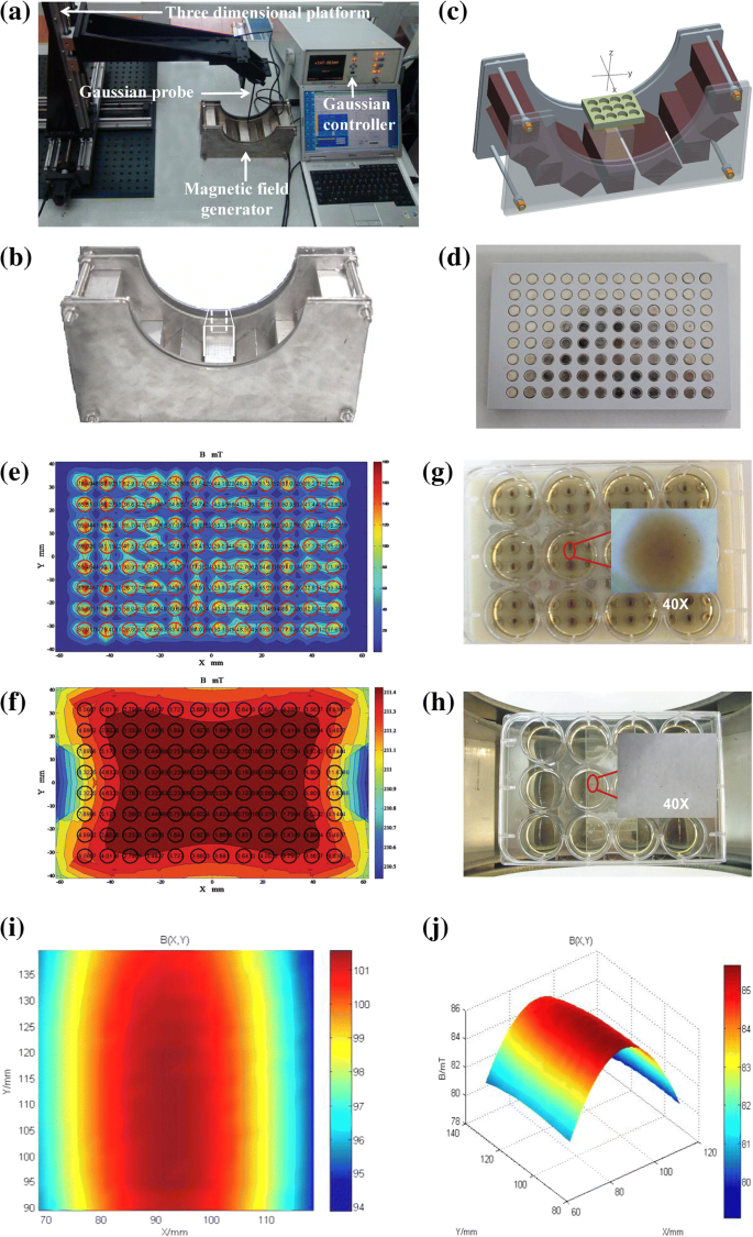
磁场发生器及其磁场均匀性和分布在不同磁场中的 PEI-SPIO-NPs。 一 用 Graussmeter 测量磁场均匀性。 b 均匀磁场发生器。 c 均匀磁场发生器磁铁排列的 3D 图像(其中每个磁铁的尺寸为 40 × 40 × 200 毫米 3 )。 d 96 孔磁场发生器。 e 96井磁场发生器的磁场均匀性。 f 均匀磁场发生器的磁场均匀性。 g PEI-SPIO-NPs 在 96 孔磁场中的分布。 h PEI-SPIO-NPs 在均匀磁场中的分布。 我 均匀磁场 (50 mm × 50 mm) 和 j 的 XOY 平面分布 均匀磁场3D分布
优化的磁体结构可以在YOZ平面内50mm × 50mm区域的水平方向产生平坦分布的磁场。梯度沿垂直方向分布,梯度为 2 mT/mm。磁场均匀分布在 50 mm × 50 mm 的 XOY 区域内,均匀性为 1.3 × 10 -3 在0.0739 T的磁场中,放置细胞培养板的冠状面和矢状面的磁场强度分别为0.0632 T和0.07 T。96孔细胞培养磁板中各孔的均匀性差异约为80%(图 4d、e)。然而,新型Halbach磁场中每口井的均匀度差异小于2‰,因此两个磁场之间的均匀度差异> 100倍(图4f)。在这种情况下,我们将96孔细胞培养磁板设置为非均匀磁场,新的Halbach磁场是一个相对均匀的磁场,作为后续研究的实验工具。
PEI-SPIO-NPs 的分布受到磁场的显着影响。 PEI-SPIO-NPs 由于重力而逐渐下沉,并且在没有磁场的情况下随机分布。在施加磁场后,PEI-SPIO-NPs 迅速下沉到板的底部。此外,分布也可能在不同的磁场中发生显着变化。在传统的非均匀磁场(96 孔细胞培养磁板)中,PEI-SPIO-NPs 聚集成团块或条带,并沿磁力线分布(图 4g)。然而,PEI-SPIO-NPs 均匀分布在由新型磁场发生器感应的均匀磁场中(图 4h)。从磁场的XOY平面分布和3D分布(图4i,j)可以看出,图中红色区域为相对均匀的磁场,均匀度约为千分之二(Z =10 mm),可以看出目标区域的磁场梯度约为 1.3 t/m (130 G/cm)。 The magnetic field generated in the designated area could obtain a good flat property in the horizontal direction by adjusting the arrangement of the magnet module and changing the magnetization direction of the magnet module.
Assessment of In Vitro Cytotoxicity
We used CCK-8 kits to evaluate the effects of magnetization, magnetic field, and pDNA on the cytotoxicity of nanoparticles. Figure 5a shows that with the prolongation of culture time, the absorbance values increased in all the groups except for the PolyMag-200/pDNA groups. The absorbance values of PEI-SPIO-NPs/pDNA group were higher than that of PEI-NPs/pDNA group and PolyMag-200/pDNA group (P < 0.05) and lower than that of the control group (P > 0.05), suggesting that the cytotoxicity of PEI-SPIO-NPs/pDNA complexes is lower than unmagnetized PEI-NPs/pDNA complexes and PolyMag-200/pDNA complexes. Figure 5b shows the negative effects on the absorbance values caused by the uniform and non-uniform magnetic fields. The negative effect caused by the uniform magnetic field was smaller than that caused by the non-uniform magnetic field (P <0.05)。 As shown in Fig. 5c, the absorbance values of nanoparticles without pDNA is significantly lower than that of nanoparticles with pDNA (P < 0.05), which meant nanoparticles added with pDNA have low cytotoxicity effect to Mg-63 osteoblasts.

Comparison of cytotoxicity effects of the uniform magnetic field and non-uniform magnetic field, PEI-SPIO-NPs/pDNA complexes, PolyMag-200/pDNA complexes and PEI-NPs/pDNA complexes, and different nanoparticles with pDNA and without pDNA on MG-63 osteoblasts. Cytotoxicity is detected by the CCK-8 test kit, the absorbance was measured at a wavelength of 450 nm, which reflects the cell viability, and the high viability is indicative of the low cytotoxicity. The results are expressed as the mean ± SD (n = 5, one-way ANOVA, *P <0.05, **P <0.01)。 一 The cytotoxicity effects of PEI-SPIO-NPs/pDNA complexes, PolyMag-200/pDNA complexes and PEI-NPs/pDNA complexes on MG-63 osteoblasts. b The cytotoxicity effects of the uniform magnetic field and non-uniform magnetic field on MG-63 osteoblasts and c the cytotoxicity effects of different nanoparticles with pDNA and without pDNA
Although PEI is an efficient transfection reagent, its cytotoxicity is strongly and positively correlated with its transfection efficiency and the mechanism of cytotoxicity caused by PEI is not very clear [35]. The present study found that during transfection, PEI increases the permeability of the cell membrane and damages the integrity of the mitochondrial and nuclear membranes [36, 37]. Sonawane et al. [38] reported that PEI25K promotes the release of mitochondrial protons and inhibits the electron transport chain in a dose- and time-dependent manner, indicating that PEI induces cell apoptosis. Another study showed that PEI induces cell autophagy, which is closely related to cytotoxicity [39]. The cytotoxicity of magnetized PEI decreased, and this might be attributable to the surface carboxyl groups of superparamagnetic iron oxide nanoparticles, which are negatively charged, thus partly neutralizing the positive charge of the PEIs and decreasing the chances of incurring irreversible damage. The non-uniform magnetic field induced PEI-SPIO-NPs/pDNA complexes to gather into masses or bands, and be distributed along with the lines of magnetic force (Fig. 4b), thereby causing severe damage to parts of the cell membrane and even cell death due to the excessively high number of positive charges [40]. In contrast, PEI-SPIO-NPs/pDNA complexes were uniformly distributed across the uniform magnetic field, thereby decreasing the accumulation of positive charges and reducing the cytotoxicity.
Nanoparticles added with pDNA have low cytotoxicity effect to Mg-63 osteoblasts than nanoparticles without pDNA; this may be attributed to the negatively charged pDNA partially neutralize the surface positive potential of the cationic nanoparticles at the beginning of magnetofection progress. Moghimi SM et al. concluded that PEI-induced cellular toxicity could been defined as a two-stage process, with the first stage taking place within 30 min of PEI uptake [41]. Stage-one toxicity has been defined as necrosis that is based on compromise of cell membrane integrity mediated by PEI binding to negatively charged plasma membrane proteoglycans, with highly cationic NPs being extremely cytotoxic [42]. After internalized by MG-63 osteoblast, different nanoparticles exhibits similar cytotoxic mechanisms, internalization leads to proton buffering, osmotic pressure, and eventual lysis of lysosomal membranes, releasing hydrolytic enzymes and other lysosomal constituents into the cytoplasm [43].
Cellular Uptake of PEI-SPIO-NPs/pDNA Complexes
To better understand the intracellular distribution of PEI-SPIO-NPs/pDNA complexes or PEI-NPs/pDNA complexes and the relationship between intracellular uptake of complexes and transfection efficiency, a laser scanning confocal microscope was used to trace the magnetized RBITC-PEI-SPIO-NPs/pDNA complexes and non-magnetized RBITC-PEI-NPs/pDNA complexes during transfection of MG-63 cells. LysoTracker Green D26 is a specific marker for lysosomes, and Hoechst 33342 specifically stains nuclei. Overlapping green and red fluorescence, which yields yellow fluorescence, represents colocalization and indicates entrapment of polyplexes in lysosome.
Figure 6 shows that extensive co-localization was observed at 6 h post-transfection, which indicates that most of the complexes were entrapped in endosomes. The RBITC-PEI-SPIO-NPs/pDNA + uniform magnetic field group showed a higher frequency of co-localization than the RBITC-PEI-SPIO-NPs/pDNA + non-uniform magnetic field group and the RBITC-PEI-NPs/pDNA group (arrow). At 12 h post-transfection, most of the red fluorescence had already translocated from the green fluorescence of lysosomes to the surrounding blue fluorescence of nuclei, and some green fluorescence of gene expression was visible in the cytoplasm, which indicated that most complexes escaped the endosomes via the proton sponge effect [44, 45] (arrow). The fluorescent signals of the RBITC-PEI-SPIO-NPs/pDNA + uniform magnetic field group were more distinct than those of the RBITC-SPIO-PEI-NPs/pDNA + non-uniform magnetic field group and the RBITC-PEI-NPs/pDNA group. It was interesting that the cytoplasm emitted green fluorescence in the RBITC-PEI-SPIO-NPs/pDNA + uniform magnetic field group, which indicated expression of GFP, thereby illustrating the proton sponge effect that facilitates gene expression. At 12 h post-transfection, some nuclei emitted red fluorescence in the RBITC-PEI-SPIO-NPs/pDNA + uniform magnetic field group, indicating that complexes had been transported into nuclei, which may be attributable to the mechanical effect of magnetofection (arrow). At 24 h post-transfection, the green fluorescence in the cytoplasm showed maximal levels, the RBITC-PEI-SPIO-NPs/pDNA + uniform magnetic field group showed more intense green fluorescence than the RBITC-PEI-SPIO-NPs/pDNA + non-uniform magnetic field and the PBITC-PEI-NPs group (arrow). The affiliated magnetic field enabled the magnetic nanoparticles to attach rapidly to the cell membrane and accelerate intracellular uptake of magnetic nanoparticles. By contrast, up to 100% of these cells will have vector particles bound to their surfaces within a few minutes in the presence of the novel uniform magnetic field; more vectors adherence leads to a greater probability of cellular uptake, and transfection efficiency is positively correlated with uptake capability [46].

Intracellular tracking of PEI-SPIO-NPs/pDNA complexes in the presence of uniform magnetic field (uniform MF) or non-uniform magnetic field (non-uniform MF) and PEI-NPs/pDNA complexes without magnetic field (No MF) at 6, 12, and 24 h post-transfection to MG-63 osteoblasts. Confocal images were obtained from three channels and overlaid:red indicates RBITC-PEI-SPIO-NPs/pDNA complexes (excitation:554 nm; emission:576 nm), green represents lysosomes stained with Lysotracker-DND26 and the homogeneous green in the cytoplasm implies GFP expression (excitation:443 nm; emission:505 nm), blue signifies the nuclei stained by the Hochest 33342. (excitation:359 nm; emission:461 nm)
In Vitro Transfection
Because PEI-SPIO-NPs are endowed with promising attributes such as pDNA condensation ability and high cell viability, we next used it as a gene carrier to determine the role of novel uniform and non-uniform magnetic fields on transfection efficiency. To evaluate the effectiveness of magnetofection, we selected the MG-63 osteosarcoma cell line as target cells and used GFP-pDNA as the reporter gene.
GFP expression was observed by inverted fluorescence microscopy at 24 h post-transfection. Figure 7a shows that the PEI-SPIO-NPs/pDNA group yielded significantly higher transfection efficiencies than the PEI-NPs/pDNA group in the presence of the uniform or non-uniform magnetic field, which is obvious in the uniform magnetic field group (P < 0.05), the transfection efficiency of the uniform magnetic field group was 42.1%, which is roughly two times higher than that of the non-uniform magnetic field group. Although PolyMag-200/pDNA showed high transfection efficiency and has been confirmed to transfect most adherent cell lines, it is also highly cytotoxic. The cells were collected for flow cytometry at 48 h post-transfection (Fig. 7b), and the statistical analysis results were in agreement with the findings from fluorescence microscopy (Fig. 7c).

MG-63 osteoblasts transfected with PEI-SPIO-NPs/pDNA or PEI-NPs/pDNA under the condition of no magnetic field, non-uniform magnetic field, or uniform magnetic field. GFP-pDNA was used for reporter gene in combination with PEI-NPs or PEI-SPIO-NPs (N/P = 10), and the cells were exposed to the non-uniform or uniform magnetic fields for 20 min. At 24-h post-transfection, cell images were captured under an inverted fluorescent microscope. PolyMag-200 comprise commercial magnetic transfection reagents that were used as positive control, naked pDNA was used as the negative control. 一 At × 40 magnification in an inverted fluorescent microscope. b Transfection efficiency was calculated by flow cytometer and c statistical analysis of the transfection efficiency of PEI-SPIO-NPs/pDNA group, PolyMag-200/pDNA group and PEI-NPs/pDNA group. The results are expressed as the mean ± SD (n = 5, one-way ANOVA, *P <0.05, **P < 0.01)
The mechanism underlying the increase in transfection efficiency by magnetofection is currently unclear. Previous studies have demonstrated that magnetofection does not involve magnetic nanoparticles being pulled directly into the cells by the magnetic field, magnetic nanoparticles enter cells via endocytosis and remain intact upon cellular uptake [47]. The observed significant increase in transfection efficiency may be due to the synergistic effect of accelerated sedimentation and fast internalization [48, 49]. The number of magnetic complexes that enter each cell apparently influences pDNA content. Although genes must still escape endosomes before transcription, transfection efficiency is positively correlated with uptake capability [50], branched PEIs showed a higher uptake rate. However, once inside the cell, lysosomal escape becomes the key to transfection, especially with higher N/P ratios, and linear PEIs showed higher rates of lysosomal escape [51,52,53]. Because of the magnetic force, PEI-SPIO-NPs can quickly come in close contact with cell membranes, which to some extent, increases the uptake capability of complexes. Once the complexes are inside the cell, PEIs provide a structural advantage and facilitate timely release of pDNA, which is eventually expressed by the cells.
结论
The novel magnetic field generator has been developed to induced a uniform magnetic field, in which the PEI-SPIO-NPs/pDNA complexes are rapidly and uniformly distributed on the surface of MG-63 osteoblasts, thereby averting local transfection and decreasing disruption of the membrane caused by centralization of positively charged PEI-SPIO-NPs/pDNA complexes, ultimately resulting in an increase in the effective coverage of the magnetic gene carriers during transfection, and improving magnetofection efficiency. This innovative uniform magnetic field could be used to determine the optimal amount of PEI-SPIO-NPs and pDNA, and screen for the optimal formulation design of a magnetic gene carrier under homogeneous conditions. Most importantly, the novel uniform magnetic field facilitates the transfection of PEI-SPIO-NPs/pDNA complexes into osteoblasts, which provides a novel approach for the targeted delivery of therapeutic genes to osteosarcoma tissues and serves as a reference for the treatment of other tumors. However, a series of comprehensive studies are warranted to establish the therapeutic potential of PEI-SPIO-NPs that are integrated into a novel uniform magnetic field in combating osteosarcoma.
缩写
- 原子力显微镜:
-
原子力显微镜
- CLSM:
-
Confocal laser scanning microscopy
- DLS:
-
动态光散射
- DMEM:
-
Dulbecco改良Eagle培养基
- EDC:
-
1-Ethyl-3-[3-(dimethylamino) propyl] carbodiimide
- FBS:
-
胎牛血清
- GFP:
-
Green fluorescent protein
- ICP-OES:
-
Inductively coupled plasma optical emission spectrometer
- MF:
-
Magnetic field
- MG-63:
-
Human osteoblasts
- MRI:
-
Magnetic resonance imaging
- MWCO:
-
Molecular weight cut off
- NHS:
-
N -hydroxy succinimide
- PBS:
-
磷酸盐缓冲盐水
- pDNA:
-
Plasmid DNA
- PEI:
-
Polyethylenimine
- PEI-NPs:
-
Polyethylenimine nanoparticles
- PEI-SPIO-NPs:
-
Polyethylenimine modified superparamagnetic iron oxide nanoparticles
- RBITC:
-
Rhodamine B isothiocyanate
- SPIO-NPs:
-
Superparamagnetic iron oxide nanoparticles
- TEM:
-
透射电子显微镜
- VSM:
-
Vibrating sample magnetometer
纳米材料