

Sonodynamic N2O 微泡增强型高强度聚焦超声治疗的研究
摘要
高强度聚焦超声(HIFU)是一种具有代表性的非侵入性癌症治疗方法,但其治疗效果低和对周围正常组织有损伤的风险阻碍了其进一步的临床开发和应用。声动力疗法 (SDT) 在超声治疗期间通过声敏剂产生的活性氧分子杀死肿瘤细胞。 SDT 可以像微泡一样增强 HIFU 的功效。在这项工作中,我们通过改进的机械振荡方法开发了纳米级 N2O 微泡 (N2O-mbs)。这些微泡显示出良好的生物相容性和肿瘤细胞结合。通过检测活性氧的产生,在细胞外和细胞内检测到 N2O-mbs 的声敏性。评估了这些声动力学微泡对肿瘤细胞的毒性作用和对 HIFU 治疗的协同作用。显着的细胞凋亡是由超声照射下 N2O-mbs 产生的活性氧引起的。 N2O-mbs 联合 HIFU 增加了体外肿瘤细胞的坏死和凋亡,以及体外牛肝靶区的凝固坏死体积和回声强度。这些声动力学微泡也已被证明可以有效地抑制体内肿瘤的生长。 N2O-mbs由于具有微气泡和非凡的声敏性等优点,对HIFU的治疗和消融效果有显着影响。这一发现表明N2O-mbs可能是一种新型的超声辅助剂,可用于促进HIFU肿瘤热消融。
介绍
恶性肿瘤是威胁人类生命和健康的主要疾病之一[1]。高强度聚焦超声(HIFU)是一种用于肿瘤无创局部消融的新技术。 HIFU已成功用于治疗肝脏、肾脏、胰腺、前列腺、乳腺、骨骼和子宫的良性和恶性实体瘤[2, 3]。但HIFU治疗过程中超声能量衰减、靶区能量积累减弱、非靶健康组织损伤等固有局限性降低了其治疗效果[4, 5]。 HIFU照射形成的焦场很小,通常为毫米级。消融大肿瘤需要很长时间。 HIFU照射时间的延长会使体温升高,甚至达到39.2℃。随着体内肿瘤深度的增加,HIFU消融的能量也需要增加[6]。这些会增加对患者造成身体伤害的风险,并降低 HIFU 治疗的效率和安全性。因此,近年来研究人员一直致力于寻找提高HIFU治疗效率的方法[7,8,9]。微泡可以协同提高 HIFU 治疗效果 [7, 10]。微泡中的气体属于高声阻抗材料。当HIFU照射肿瘤组织区域时,微泡增加了超声波在目标区域的散射和反射[11, 12]。微泡通过增加靶区 HIFU 能量的积累来提高肿瘤靶区的温度,增强 HIFU 治疗的热效应[13]。此外,微泡作为外源性空化核也降低了组织空化阈值,增强了HIFU在通过血液循环进入肿瘤组织时的空化效应[14]。近年来,研究表明声动力疗法(SDT)可以协同提高HIFU治疗效果[15, 16]。声敏剂和超声波之间的相互作用会产生活性氧 (ROS),包括单线态氧和羟基自由基,它们通过细胞凋亡和坏死杀死肿瘤细胞并抑制肿瘤生长 [17]。近年来,SDT在非侵入性肿瘤治疗领域取得了很大进展。与传统的单一疗法相比,它能够减少药物剂量和 HIFU 照射功率。为了充分发挥两者的优势,本研究将微泡与声动力效应结合起来,进一步提高HIFU的疗效。声敏剂是 SDT 的重要组成部分 [18, 19]。目前研究的传统声敏剂大多来自光敏剂。 N2O 是一种光敏剂 [20, 21],在紫外线照射条件下会产生有毒物质。在这项研究中,还初步探讨了 N2O 的声敏性。我们以前发现在 HIFU 手术中应用 N2O 吸入麻醉会加重组织的损伤程度,包括增加腹部和皮肤充血的厚度[22]。 N2O与HIFU联合利用对组织损伤起到协同作用。但是,N2O 在 HIFU 治疗中的生物学效应尚不清楚。 N2O 是一种相对稳定的小分子气体,在细胞内没有生物转化和组织特异性。可广泛应用于组织,对机体无毒副作用[23, 24]。因此,它作为声敏剂具有诱人的前景。在这项研究中,使用脂质包裹的 N2O 和 C3F8 制备了纳米级微气泡 (N2O-mbs)。 N2O的用量减少,其体内作用范围也有效地限制在肿瘤组织区域。考察了 N2O 的声动力学效应及其对 HIFU 疗效的协同作用。
材料和方法
材料
1,2-Dihexadecanoyl-rac-glycero-3-phosphocholine (DPPC) 和 1,2-distearoyl-sn-glycero-3-phosphoethanolamine-N -[甲氧基(聚乙二醇)-2000] (DSPE-mPEG2000) 购自 Avanti Polar Lipids (Alabaster, AL, USA)。甘油和琼脂糖获自 Sigma Aldrich (St. Louis, MO, USA)。 N2O 和 C3F8 购自重庆瑞新燃气有限公司(中国)。 4',6-二脒基-2-苯基吲哚 (DAPI), 1,1'-双十八烷基-3,3,3',3'-四甲基吲哚羰花青高氯酸盐 (DiI), 2',7'-二氯荧光素二乙酸酯 (DCFH-DA) ,3-氨基,4-氨基甲基-2',7'-二荧光素,二乙酸酯(DAF-FM DA)和CCK-8购自Beyotime Biotechnology(中国重庆)。 Calcein-AM (CAM) 和碘化丙啶 (PI) 购自 Santa Cruz Biotechnology (TX, USA)。膜联蛋白 V-FITC/PI 购自南京凯根生物技术公司(中国南京)。单线态氧传感器绿色 (SOSG) 购自 Invitrogen (NY, USA)。
N2O-mbs 和 C3F8-mbs 的制备
N2O-mbs是通过旋转膜结合机械振荡法制备的。我们以DPPC和DSPE-PEG2000为壳材料,N2O为核心。简而言之,将 5 mg DPPC、2 mg DSPE-PEG2000 和 10 mL 氯仿同时倒入圆底烧瓶中,混合并通过超声波充分溶解。然后,通过旋转蒸发器(60 分钟,55°C,100 r/min)形成脂质膜。接下来,加入 2 mL 10% 甘油以水合脂质膜并制备悬浮液。将其中的 500 μL 添加到胶囊管中。胶囊管用橡胶帽盖住。通过气体置换装置用 N2O 和 C3F8 置换管中的空气。然后,悬浮液通过银汞胶囊混合器(YJT-2,上海医疗器械有限公司,中国)机械振荡 150 秒。最后,将所得悬浮液进行差速离心以获得目标 N2O-mbs,并以 300 rpm 离心 5 分钟,然后以 800 rpm 离心 5 分钟。 N2O-mbs重悬于PBS中,4℃保存备用。
C3F8-mbs 是使用相同的机械振荡方法制造的,如前所述 [25]。 DiI 标记的 N2O-mbs 可以通过在自旋膜形成过程中添加 DiI 来制备。值得一提的是,N2O 分子量低,不稳定。微泡中的 N2O 容易溢出。因此,微泡的半衰期减少。 C3F8 是一种高分子惰性气体,广泛用于超声微泡造影剂的研究。 C3F8 可以增加微泡的稳定性。因此,在本研究中,N2O和C3F8(2:1)混合作为N2O-mbs核心以增加其稳定性。
N2O-mbs 的制造和性能检测
在明场光学显微镜下观察 N2O-mbs 的形态和尺寸分布。 N2O-mbs 和 C3F8-mbs 的浓度是根据计算指南用球度计计算的。 N2O-mbs 的平均粒径和 zeta 电位由动态激光散射系统(Malvern Instruments,Malvern,UK)测定。 N2O-mbs 的结构和形态通过透射电子显微镜(TEM,Hitachi H-7600,Hitachi Ltd.,Tokyo,Japan)表征。制备的 N2O-mbs 储存在 4°C。为了观察N2O-mbs的稳定性,在第一天、第二天和第三天记录形态和浓度。同时测定了微泡的平均粒径和zeta电位。
细胞培养和动物模型
人乳腺癌细胞(MDA-MB-231;重庆金喜鹊科技发展有限公司)在复苏后在实验室传代不到3个月。使用含有 10% 胎牛血清、50 μg/L 链霉素和 50 μg/L 青霉素的 DMEM,将细胞维持在 37 °C 的 5% CO2 培养箱中。当细胞达到80%汇合时,用于实验。
所有动物实验均按照重庆医科大学动物伦理委员会的指导方针进行。一定数量的雌性裸鼠(4-6 周龄;体重 15-20 g)购自重庆医科大学动物实验中心(中国重庆)。对于实体瘤的建立,MDA-MB-231 细胞(1 × 10 6 将悬浮在生理盐水(100 μL)中的细胞/mL)皮下注射到每只小鼠的右后腹。当肿瘤体积达到150 mm左右时,所有荷瘤小鼠用于治疗实验 3 .
N2O-mb 的细胞内摄取和生物相容性评价
将生长期MDA-MB-231细胞以约1×10 5 的密度接种 每个共聚焦激光扫描显微镜 (CLSM) 烧瓶中的细胞数为 24 小时。接下来,100 μL (1 × 10 5 气泡/mL)的 DiI 标记的 N2O-mbs 完全培养基稀释溶液被添加到共聚焦盘中。将培养皿用微孔板密封剂密封并倒置储存在培养箱中,保持 N2O-mbs 与细胞接触。共孵育 2 小时和 4 小时后,用 PBS 轻轻洗涤细胞 3 次,用 4% 多聚甲醛 (PFA) 固定 20 分钟,然后用 DAPI 染色 20 分钟。最后,通过CLSM(Nikon A1,Japan)观察N2O-mbs的细胞靶向行为。
N2O-mbs 的细胞毒性通过标准 CCK-8 测定进行评估。 MDA-MB-231 细胞接种于 96 孔板 (5 × 10 4 细胞/孔)24 小时。接下来,100 μL 不同浓度(1 × 10 4 /mL, 1 × 10 5 /mL 和 1 × 10 6 /mL) 的 N2O-mb。孵育 24 h 后,每孔加入 CCK-8 溶液(10 μL)。然后,将 MDA-MB-231 细胞在 37°C 下再培养 2 小时。最后,用Bio-Tek酶标仪(Bio-Tek Instruments, Thermo Fisher Scientific, Winooski, VT, USA)在450 nm处测量各孔的吸光度。
将10只体重约20g的健康雌性裸鼠随机分为两组。 N2O-mbs (n =5)组静脉注射N2O-mbs(200 μL,1×10 5 /mL) 和对照组 (n =5) 注射生理盐水 (200 μL)。 21天后,所有小鼠的主要器官(心、肝、脾、肺和肾)均被切除并用苏木精-伊红(H&E)染色。
N2O-mbs 的体外 SDT 效率
氧自由基检测方法基于之前的报告[26]。简而言之,将 10 μL SOSG (500 μM) 与 2 mL 样品溶液混合。处理分组如下:C组(空白对照组),单独加入PBS; N2O-mbs 组,100 μL N2O-mbs (1 × 10 5 /mL) 加入 2 mL PBS 溶液中; C3F8-mbs 组,100 μL C3F8-mbs (1 × 10 5 /mL) 加入 2 mL PBS 溶液中; LIFU组,PBS照射超声(2 W/cm 2 ;重庆医学超声影像研究所,重庆,中国)100 s; LIFU-N2O-mbs组,加入N2O-mbs的PBS超声辐照; LIFU-C3F8-mbs 组,加入 C3F8-mbs 的 PBS 进行超声辐照。实验组治疗参数设为2 W/cm 2 和100 s通过对实验结果的分析比较和查阅文献[27, 28]。用于治疗目的的超声参数设置如下:频率,650 kHz;焦距,12.5 毫米;脉冲波模式,50%占空比;和脉冲持续时间,1 s。单线态氧的产生( 1 O2) 通过荧光光谱法 (Cary Eclipse, Agilent Technologies) 通过测量荧光强度 (λ 激发/λ 发射 =504 nm/525 nm)。超声强度和持续时间的选择与体外细胞实验一致。
使用 SOSG 法检测细胞外活性氧 (ROS) 的产生后,使用基于 DCFH-DA 的 ROS 检测试剂盒检测细胞内 ROS 的产生。 MDA-MB-231 细胞以大约 1 × 10 5 的密度接种 每个 CLSM 烧瓶的细胞数。然后,将它们随机分为六组:对照组(未处理),仅 N2O-mbs 组(100 μL,1 × 10 5 气泡/毫升),仅 C3F8-mbs 组(100 μL,1 × 10 5 气泡/毫升),仅 LIFU 组(2 W/cm 2 100 秒),LIFU-C3F8-mbs 组,LIFU-N2O-mbs 组。在孵育 24 小时以允许细胞附着后,每组都接受上述相应的实验干预。然后,在各组对应的培养皿中加入等量的DCFH-DA(30 μM),培养皿避光孵育20 min。然后用PBS轻轻冲洗细胞3次以去除未进入细胞的DCFH探针。最后,通过 CLSM 确定细胞内 ROS 的产生。为了进一步验证 N2O-mbs 的 SDT 效率,如上所述,将细胞接种在 6 孔板中,培养 24 小时并随机分为六组。然后对各组进行相应的处理。将各组细胞消化制备成适当浓度的细胞悬液,置于CLSM培养皿中。最后,在用 CAM 和 PI 染料共染色后,通过 CLSM 扫描细胞以区分死(红色)和活(绿色)细胞。通过流式细胞术和膜联蛋白 V-FITC/PI 染色检测细胞凋亡。处理1 h后,离心各组细胞,收集细胞。 Annexin V-FITC/PI共染色后,进行流式细胞术(Becton-Dickinson, Franklin Lakes, NJ)检测。
N2O-mbs 的体外声动力学效应增强 HIFU 治疗效果
HIFU 系统(JC200,中国重庆 HIFU 科技有限公司)由诊断超声单元、治疗超声单元和中央处理系统组成,如前所述 [29]。用于治疗目的的 HIFU 参数设置如下:工作频率,0.94 MHz;焦距,140 毫米;直径,220 毫米;和实时诊断传感器频率,3.5 MH。集成换能器浸没在脱气水中。为了评估 N2O-mbs 对 HIFU 处理的增强作用,将 MDA-MB-231 细胞离心,5 × 10 5 /mL,并分为四组:对照组(未处理),仅 HIFU 组(125 W,5 s),仅 HIFU-N2O-mbs 组,仅 HIFU-C3F8-mbs 组。接下来,对每组进行不同的各自处理。将处理过的细胞离心并用PBS洗涤,用Annexin V-FITC/PI双染色并收集用于流式细胞术。使用一氧化氮 (NO) 探针 DAF-FM 检测各处理组上清液中的 NO。所有实验均进行 3 次。 TEM 用于鉴定处理细胞的超微结构形态。每次处理后,处理过的细胞立即用 2% OsO4 固定,在一系列分级酒精中脱水,然后平铺在 Epon 812(Electron Microscopy Sciences,Fort Washington,PA)中。制备超薄切片(100 nm),醋酸双氧铀和柠檬酸铅染色,电镜下观察。
N2O-mbs 的体外和体内声动力学效应增强 HIFU 消融功效
新鲜牛肝组织购自当地屠宰场,屠宰后 12 小时内使用。将结缔组织少、血管少的新鲜离体牛肝切成 10 cm × 5 cm × 5 cm 切片,37 °C 脱气 1 h。牛肝分为三组:C3F8-mbs组、N2O-mbs组和PBS组。首先,将新鲜分离的牛肝放入装有脱气水的容器中。然后,200 μL (1 × 10 5 以图 6a 所示的方式将 PBS 中的微泡(气泡/毫升)直接注射到牛肝中。在诊断超声换能器的引导下,HIFU 焦点位于注射部位。然后,使用治疗换能器在牛肝注射部位以不同功率(100 W、125 W、150 W)进行 5 s 消融。最后,根据测得的最大长度、宽度和深度(mm),通过下式计算牛肝组织中靶区的凝固坏死体积:V (mm 3 ) =π/6 × 长 × 宽 × 深。并在超声诊断图像上记录各组HIFU消融前后靶区的灰度值。
20只荷瘤小鼠随机分为四组(n =每组 5 个),包括对照组(未进行任何处理)、HIFU-PBS 组、HIFU-C3F8-mbs 组和 HIFU-N2O-mbs 组。小鼠接受200 μL C3F8-mbs组、N2O-mbs和PBS的静脉注射后,应用HIFU(125 W,5 s)消融肿瘤。实体瘤的 HIFU 消融过程类似于离体离体牛肝的消融过程。首先,小鼠用 1% 戊巴比妥 (8 mL/kg) 麻醉。将小鼠的肿瘤部位接种到装有脱气水的容器中。在诊断超声换能器的引导下,HIFU 焦点定位在肿瘤组织上。每隔3天记录小鼠体重和肿瘤体积。最后,治疗后21天处死所有小鼠,解剖小鼠肿瘤组织,送H&E、增殖细胞核抗原(PCNA)染色、末端脱氧核苷酸转移酶dUTP缺口末端标记(TUNEL)。
统计分析
所有数据均使用 GraphPad Prism(第 7 版,GraphPad Software Inc.,La Jolla,CA,USA)呈现和分析。数据表示为平均值±SD。每个实验至少重复 3 次。统计分析通过单向方差分析与 Bonferroni 后检验(比较所有组)或 Dunnett 后检验(将所有组与对照组进行比较)进行。 p 的值 <0.05 被认为具有统计学意义。
结果
N2O-mbs 的特征
通过改进的机械振荡方法制备 N2O-mbs 会产生高度稳定的乳白色悬浮液(图 1a)。在室温下储存 72 小时后,N2O-mbs 的粒径和形态没有显着变化。在光学显微镜下,观察到 N2O-mbs 具有均匀的尺寸、良好的分散性和高稳定性(图 1b)。 N2O-mbs的浓度为5.12±0.45×10 8 气泡/毫升。如 TEM 图像(图 1c)所示,N2O-mbs 的尺寸分布均匀,平均尺寸约为 450 nm。 N2O-mb 动态光散射结果显示流体动力学直径为 458.1 ± 17.26 nm(多分散指数 =0.368;图 1d),这与 TEM 结果一致。 N2O-mbs 的电位为 − 16.2 ± 0.35 mV(图 1e)(附加文件 1:图 S1)。
<图片>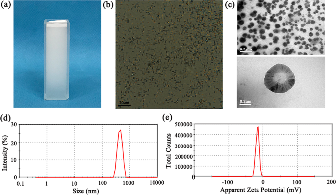
一 分散在去离子水中的 N2O-mbs 的照片; b 明场光学显微镜下的 N2O-mbs 图像; c N2O-mbs 的透射电子显微镜图像; d N2O-mbs 的尺寸分布; e N2O-mbs的表观zeta电位
N2O-mbs 的细胞内摄取和生物相容性
N2O-mbs 在体外对细胞的细胞毒性通过 CCK-8 方法进行研究。我们将对照组的细胞活力定义为 100%。孵育 24 小时后,细胞活力结果显示对 MDA-MB-231 细胞没有显着的细胞毒性。即使在高 N2O-mb 浓度下,细胞活力仍保持在 95% 以上(图 2a)。分析了 N2O-mbs 在体内对健康裸鼠的细胞毒性。如图 2b 所示,主要器官(心脏、肝脏、脾脏、肺和肾脏)的组织病理学分析表明,N2O-mbs 不会对小鼠造成显着的毒性损伤。这些结果表明N2O-mbs具有较高的生物相容性。
<图片>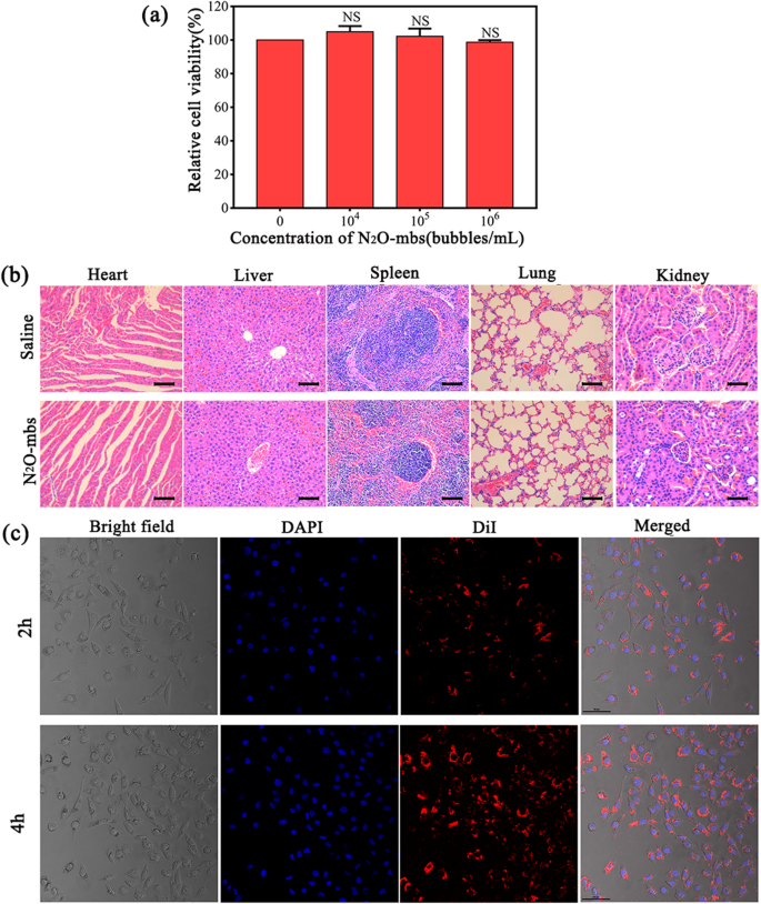
一 CCK-8 法测定细胞活力; b 接受或未接受 N2O-mbs 的健康雌性裸鼠主要器官(心脏、肝脏、脾脏、肺和肾脏)的 H&E 染色; c 来自 a 的图像 d 显示明视野中的细胞,DAPI染色的细胞核,DiI染色的N2O-mbs,以及三张荧光图像的合并结果;比例尺 =50 μm [NS 与对照组相比无显着性(0气泡/mL)]
N2O-mbs 在延长的孵育时间(2 小时和 4 小时)内的细胞内吸收通过 CLSM 进行可视化。细胞与 DiI 标记的 N2O-mbs 孵育导致癌细胞膜和细胞质中大量 N2O-mbs 的积累。如图 2c 所示,红色荧光 N2O-mbs 与蓝色荧光细胞具有良好的共定位性。这一发现表明 N2O-mbs 被有效地内化到 MDA-MB-231 癌细胞中。
N2O-mbs 体外声动力学效应
SOSG是一种常用的单线态氧检测探针,对氧自由基产生的检测具有高灵敏度和可靠性。为了探索 N2O-mbs 作为声敏剂的潜力,使用 SOSG 检测体外 ROS 的产生 [30, 31]。 SOSG 与 1 结合 O2 形成 SOSG-EP,其特点是荧光强度增加。如图 3a 所示,超声辐照含有 N2O-mbs 的 SOSG 溶液后,荧光强度值显着增加;与其余各组相比,氧自由基的产生显着增加(p <0.01).
<图片>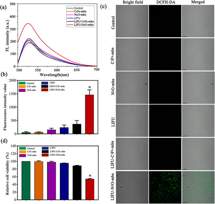
一 LIFU 辐照 SOSG 与 N2O-mbs 的荧光光谱; b 通过CCK8测定确定细胞活力; c 细胞内 ROS 生成的共聚焦图像(绿色荧光表示用 DCFH-DA 染色的 ROS 呈阳性染色); d Image J计算的各组平均荧光强度值(DCFH-DA)(数据以平均值±SD表示,n =每组 5 个,*p <0.05,与LIFU-C3F8-mbs组相比)
除了检测 N2O-mbs 超声照射后水溶液中 ROS 的产生外,还使用 DCFH-DA ROS 测定试剂盒测定 N2O-mbs 产生的细胞内 ROS。如图 3b 所示,阳性对照组表现出最强的荧光强度。次强的绿色荧光信号仅出现在 LIFU-N2O-mbs 组中,揭示了在 N2O-mbs 和 LIFU 照射同时存在的情况下随之产生的 ROS。然而,在任何其余组中均未检测到明显的荧光(图 3c)。对照组的细胞活力定义为100%。 CCK-8 测定结果显示 LIFU-N2O-mbs 组中大约 46% 的细胞死亡(图 3d)。 N2O-mbs 与 LIFU 组合的细胞毒性通过 CLSM 观察处理后的细胞死亡来评估。 CAM 用于用绿色荧光染色活细胞。 PI 用于用红色荧光染色死细胞。如图 4a 所示,LIFU-N2O-mbs 组中有许多死细胞。此外,流式细胞术结果显示,MDA-MB-231细胞组的凋亡率明显高于其他组(p <0.05) 在结合 N2O-mbs 与 LIFU 处理后(图 4b)。 C3F8-mbs 与 LIFU 联合治疗未引起明显的细胞死亡或凋亡。结果进一步表明,SDT 发生了广泛的细胞凋亡和坏死。流式细胞术结果与 CLSM 一致。这些结果表明N2O-mbs联合LIFU在体外诱导肿瘤细胞凋亡(附加文件2:图S2;附加文件3:图S3)。
<图片>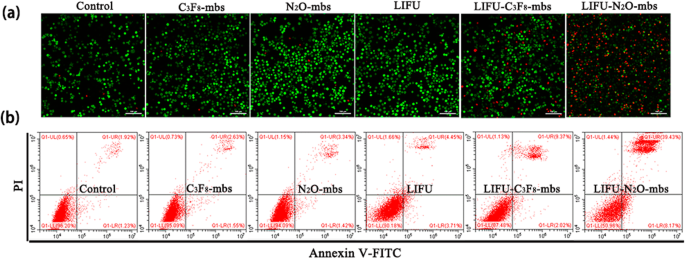
一 肿瘤细胞凋亡和坏死的流式细胞术分析; b CAM/PI 共染色的 MDA-MB-231 细胞经过各种处理后的共聚焦图像。比例尺为 100 μm
体外 HIFU 在癌细胞中的协同效应评估
流式细胞术结果如图 5a 所示。 N2O-mbs 和 C3F8-mbs 均增加了 HIFU 消融后 MDA-MB-231 细胞的死亡率。此外,与 HIFU-C3F8-mbs 相比,N2O-mbs 显着增加 MDA-MB-231 细胞凋亡 (p <0.05;图 5b)。 NO检测结果显示HIFU-N2O-mbs组上清液的荧光强度也显着高于其余各组(p <0.05)(图 5c)。通过 TEM 观察到细胞质和细胞器的变化(图 5d)。对照组(未进行任何处理)中的细胞显示出完整的细胞形态。 HIFU组细胞质中发现大量液泡状物质。但细胞仍然具有完整的细胞膜和核膜。然而,HIFU-N2O-mbs 组和 HIFU-C3F8-mbs 组均表现出细胞膜完整性的丧失和细胞结构的完全分解。值得一提的是,TEM观察只是各组治疗后细胞死亡和凋亡过程中的时刻。 N2O-mbs作为外源性微泡对HIFU处理具有明显的协同作用。在 HIFU-N2O-mbs 组中,N2O-mbs 的声动力学效应可以促进更多的细胞凋亡。结果已在流式细胞术检测和体内实验中得到验证(附加文件 4:图 S4;附加文件 5:图 S5)
<图片>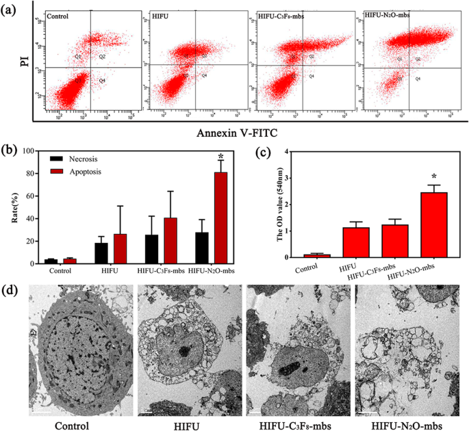
一 Annexin V-FITC/PI结合测定结果; b 各组平均细胞凋亡率; c HIFU辐照N2O-mbs产生NO的定量分析; d 细胞的透射电子显微镜图像。比例尺为 100 μm。 (数据显示为平均值±SD,n =每组 5 个,*p <0.05,与对照相比,HIFU , HIFU–C3F8-mbs 组)
N2O-mbs 作为 HIFU 疗法的增效剂
在对分离的牛肝进行 HIFU 消融后,目标区域出现灰白色凝固性坏死区域(图 6b)。 HIFU前后目标区域的平均回波强度变化如图6c所示。与PBS组相比,HIFU治疗后C3F8-mbs组和N2O-mbs组的靶回波强度值显着增加(p <0.05) 并且随着 HIFU 消融强度的增加而增加。后两组的凝固性坏死体积和靶区B超图像回波强度均随着HIFU功率的增加而增加(图6d、e)。
<图片>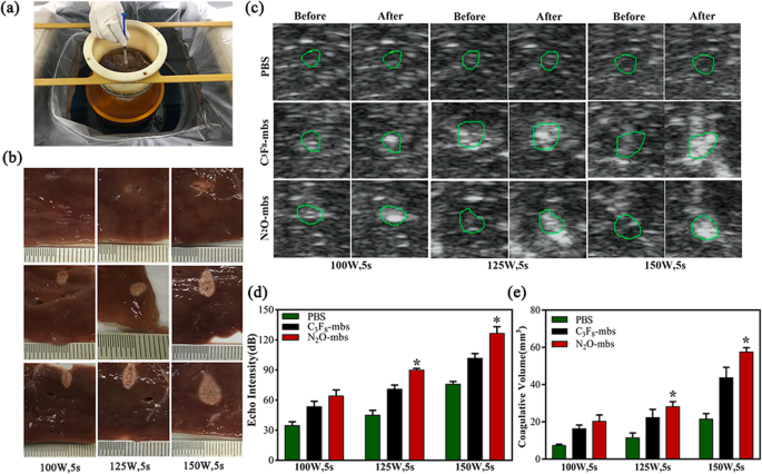
一 脱气牛肝离体 HIFU 消融示意图; b HIFU 消融后切除的牛肝目标区域的照片; c 在 100 W、125 W 和 150 W 下对牛肝脏进行 HIFU 照射 5 s 前后的实时超声图像; d 回波强度定量分析; e HIFU 消融后牛肝目标区域凝固性坏死体积的定量分析。 (数据显示为平均值±SD,n =每组 5 个,*p <0.05,与100 W、5 s、125 W、5 s组相比)
在体内进一步验证了 N2O-mbs 对 HIFU 治疗的协同作用。 As shown in Fig. 7a, the digital photos of mice and corresponding tumor tissue were recorded 21 days after different treatment. Tumor growth was more effectively inhibited after N2O-mbs combined with HIFU treatment. The synergistic effect of N2O-mbs on HIFU treatment was also evaluated by H&E, PCNA, and TUNEL staining (Fig. 7b), which demonstrated that N2O-mbs combined with HIFU irradiation could cause larger scale of tumor cell necrosis as compared with HIFU–C3F8-mbs group. The cell morphology changes exhibited in HIFU–N2O-mbs were more obvious than other groups, including karyopyknosis, karyorrhexis and karyolysis, indicating the substantial necrosis of cancer cells.

一 Digital photos of tumor-bearing mice and ex vivo tumors 21 days after different treatments; b H&E, PCNA and TUNEL staining of tumor sections after various treatments; c the corresponding calculated tumor volume of each group after 21 days of different treatments; d body weight of tumor-bearing mice for each group. The scale bars in HE staining are 100 μm; the scale bars in TUNEL and PCNA staining are 50 μm. (The data were shown as mean ± SD, n =5 per group, *p <0.05, compared with HIFU-C3F8-mbs group)
In addition, the tumor volume of HIFU–N2O-mbs exhibited a more significant inhibited tendency than HIFU–C3F8-mbs within the 21 days (Fig. 7c, d).
讨论
HIFU, as a new method of oncotherapy, has been clinically recognized and has achieved notable progress in the treatment of many solid tumors [32]. The thermal effect, cavitation effect, mechanical effect and acoustic chemical effect play significant roles in killing tumor cells and causing irreversible damage to the target area tissue during HIFU ablation [33]. HIFU has limited impact on surrounding normal tissues and is essentially a non-invasive tumor treatment. However, the energy carried by the ultrasound is attenuated due to increased transmission distance, absorption by the blood flow in the target area and the blockage of bones or gas in the body during the HIFU treatment, so that the energy transferred to the target area is reduced. However, prolonging the ablation time and increasing the treatment power increases the risk of normal tissue damage, such as skin burns, neurovascular damage and other complications [34]. Therefore, it is necessary to increase the efficacy of HIFU. Many studies have confirmed that both microbubbles and SDT can improve the efficacy of HIFU [15, 16, 35]. The N2O-loaded microbubbles prepared in this study have a smaller particle size, but similar characteristics as ultrasonic microbubble contrast agents. Studies have shown that nano/microbubbles have the same imaging capability as microbubbles in vitro, but have greater capability than microbubbles in vivo [36]. So, N2O-mbs not only could be used for ultrasound guidance and localization for HIFU treatment, but also could be used as exogenous microbubbles to enhance the efficacy of HIFU (Additional file 6:Figure S6). At the same time, the sonodynamic effect of N2O-mbs could effectively improve the efficacy of HIFU. The generation of ROS after ultrasonic excitation is one of the most important factors for sonosensitizers [16]. To verify the sonosensitivity of N2O-mbs, the production of ROS in both extracellular and intracellular environments was detected in this study. The consistent result of both tests showed that N2O-mbs can generate ROS in both aqueous solution and intracellularly. Studies have also shown that gases (N2 and O2) trapped in cavitation nuclei in solution can undergo cleavage reactions and produce N and O free radicals [37, 38], as follows:
$$ {N}_2O\to {N}_2+{O}_2\kern0.50em {O}_2\to 2\bullet O\ \ \ {N}_2\to 2N.N.+ HO.\to NO+H. NO+ HO.\to HNO $$The cavitation effect and the advantage of microbubble packaging reduce the difficulty of the N2O reaction [39]. When incubated with cells, N2O-mbs exhibit good biocompatibility. The high selectivity, high affinity and high drug loading of nanoparticles [40], as well as the active phagocytosis by cells, promote efficient N2O-mbs binding to cancer cells. The nanobubbles display increased penetration, stability and ease of cell entry or aggregation around the cells [36]. In addition, the sonoporation effect of ultrasound can effectively promote the transfer of microbubbles into cells [41].
In contrast to the traditional microbubble contrast agent C3F8-mbs, N2O-mbs have a greater inhibitory effect on cell growth after ultrasonic irradiation, which is characterized by increased intracellular ROS, decreased cell viability, and apoptosis and necrosis. These results preliminarily suggested the sonodynamic toxic effect of N2O-mbs.
In the HIFU synergy experiment, the treatment parameters in the experimental group were set to 125 W for 5 s by analyzing and comparing the experimental results and consulting the literature, which validated a low HIFU power could induce sufficient ablation effect and reduce adverse effects. The results showed that N2O-mbs increased the necrosis and advanced apoptosis of more tumor cells. N2O-mbs acted as a nanoscale microbubble synergizer and its sonodynamic effect in response to HIFU may be the reason for the different experiment results. As a microbubble, N2O-mbs increased the deposition of HIFU energy in the target area and increased the temperature of the target area and increased the thermal effect of HIFU treatment [11]. The generation of the cavitation effect is closely related to the cavitation threshold and cavitation nucleus concentration [42]. N2O-mbs were cavitation nucleus that reduce the cavitation threshold of HIFU and facilitate the cavitation effect. Therefore, N2O-mbs effectively promoted cell necrosis. As a nanoscale microbubble synergizer, N2O-mbs were lysed and generate free radicals and nitrogen oxide in response to ultrasound. Studies have confirmed that ROS are important factors in apoptosis induction [43]. It is worth noting that many kinds of free radicals can cause damage to cells. In addition to the generation of oxygen radicals, N2O can generate other types of free radicals in cleavage reactions, such as nitrogen free radicals, which contribute to the killing of tumor cells. A small amount of nitric oxide (NO) production was detected in the cell supernatant after HIFU treatment, further indicating that the N2O cleavage reaction occurred. An et al. [44] reported that NO may have an anti-tumor function, the mechanism of which involves NO attacking suspected iron enzymes that cause cells to fail to grow and divide when they are trapped. In addition, unstable NO can interact with oxygen molecules to form hydroxyl radicals (HO · ) and NO2. Hydroxyl radicals are extremely cytotoxic and promote tumor cell apoptosis. NO is also an endothelium-derived relaxing factor. The generation of NO may be the reason for hyperaemia and abnormal swelling in patients during HIFU operation. In an excised bovine liver experiment, we carefully compared the echo intensity of B-mode ultrasound images, pathological changes and coagulative necrotic volume of the target area after HIFU ablation in the presence of N2O-mbs, C3F8-mbs and PBS. All of these results were consistent. Compared with PBS, N2O-mbs effectively enhanced the ablation effect of HIFU. This synergistic effect was also better demonstrated in the ablation of solid tumor in vivo, manifested by tumor cell apoptosis increased, tumor volume reduced and tumor growth inhibition. This finding further indicates that N2O-mbs may be a novel auxiliary agent for ultrasound that can be used to promote HIFU thermal tumor ablation. The synergistic effect of N2O-mbs in HIFU and the synergistic mechanism provide new ideas and theoretical practice for HIFU synergist research. But, the other related mechanisms of the synergy should be further explored. The effect of tumor ablation by HIFU combined with N2O-mbs should be further addressed.
结论
In summary, we have developed N2O-mb nanoparticles. The characterization and biocompatibility of the N2O-mbs were determined. The sonosensitivity of N2O-mbs was examined by detecting the production of ROS in aqueous solution and intracellularly. Compared with C3F8-mbs, N2O-mbs generated ROS and promoted the apoptosis and necrosis of tumor cells, which exhibited extraordinary sonosensitivity. In the HIFU synergy experiments, the sonodynamic effects of N2O-mbs significantly increased the apoptosis and necrosis of tumor cells in vitro, and increased the coagulative necrotic volume and echo intensity in the ex vivo isolated bovine liver target area, and effectively inhibited tumor growth in vivo. These results suggested that N2O-mbs could be a new kind of sonosensitizer and a novel auxiliary agent for ultrasound therapy that can be used for the ablation of tumors by HIFU.
数据和材料的可用性
本手稿中的结论是基于本文提供和展示的所有数据。
缩写
- N2O-mbs:
-
N2O microbubbles
- C3F8-mbs:
-
C3F8 microbubbles
- HIFU:
-
high-intensity focused ultrasound
- LIFU:
-
low-intensity focused ultrasound
- SOSG:
-
singlet oxygen sensor green
- SDT:
-
sonodynamic therapy
- DPPC:
-
1,2-dihexadecanoyl-rac-glycero-3-phosphocholine
- DSPE-mPEG2000:
-
1,2-distearoyl-sn-glycero-3phosphoethanolamine-N -[methoxy(polyethylene glycol)-2000
- DAPI:
-
4′,6-diamidino-2-phenylindole
- DiI:
-
1,1′-dioctadecyl-3,3,3′,3′-tetramethylindocarbocyanine perchlorate
- DCFH-DA:
-
2′,7′-dichlorofluorescin diacetate
- DAF-FM:
-
3-amino,4-aminomethyl-2′,7′-difluorescein, diacetate
- CAM:
-
Calcein-AM
- CCK-8:
-
Cell Counting Kit-8
- CLSM:
-
confocal laser scanning microscopy
- TEM:
-
transmission electron microscope
- DMEM:
-
Dulbecco改良Eagle培养基
- FBS:
-
fetal bovine serum
- PBS:
-
phosphate-buffered saline
- DI:
-
deionized water
- ROI:
-
regions of interest
- ROS:
-
reactive oxygen species
- NO:
-
nitric oxide
纳米材料
- 案例研究:本田方式
- 将活性氧种类的毒性调整到高级肿瘤治疗中
- 环境响应金属-有机框架作为肿瘤治疗的给药系统
- 青蒿琥酯的纳米颗粒递送通过激活线粒体介导的细胞凋亡来增强抗肿瘤效率
- 在 Au 纳米颗粒修饰的玻碳电极上增强光电化学检测尿酸
- 新型纳米粒子增强蠕虫状胶束系统的研究
- 硼烯稳定性和STM图像的第一性原理研究
- TiO2-水纳米流体在螺旋槽管中的流动和传热特性的实验研究
- Pt/Nd:SrTiO3结中多级电阻开关记忆和记忆状态相关光电压的研究
- 5-氨基乙酰丙酸-角鲨烯纳米组件用于肿瘤光检测和治疗:体外研究
- 研究纳米粒子聚集/附聚对聚合物纳米复合材料杨氏模量影响的两步法
- 天然和合成纳米材料的电化学、生物医学和热特性的比较研究