

HDAC1 介导的 MicroRNA-124-5p 调节 NPY 以影响抑郁症大鼠的学习和记忆能力
摘要
以抑郁症组蛋白去乙酰化酶 (HDAC) 为中心的研究已经过度进行,但对 HDAC1 的研究并不多。其中,本研究旨在揭示HDAC1/microRNA(miR)-124-5p/神经肽Y(NPY)轴在抑郁症中的作用机制。 Sprague Dawley 大鼠通过慢性不可预测的轻度应激刺激建立抑郁模型。向抑郁的大鼠注射抑制的 HDAC1 或抑制的 miR-124-5p,以探索它们在体重、学习和记忆能力、血清中的氧化应激和炎症以及海马组织中神经递质表达中的作用。测试了海马中的 MiR-124-5p、HDAC1 和 NPY 表达。还证实了 miR-124-5p、HDAC1 和 NPY 表达的相互作用。在抑郁大鼠的海马中发现较高的 miR-124-5p 和 HDAC1 和较低的 NPY 表达水平。抑制 miR-124-5p 或抑制 HDAC1 减弱学习和记忆能力并增加抑郁大鼠的体重。敲除 miR-124-5p 或抑制 HDAC1 可抑制氧化应激和炎症,并促进抑郁大鼠的神经递质表达。 HDAC1 介导 miR-124-5p 来调节 NPY。 NPY 的敲除消除了被抑制的 miR-124-5p 对抑郁大鼠的保护作用。我们的研究表明,抑制miR-124-5p或HDAC1均可上调NPY以改善抑郁小鼠的记忆和学习能力,这可能更新现有的抑郁症知识,为抑郁症的治疗提供新的参考。
介绍
抑郁症是指一种严重的致残性精神病,在全球范围内造成严重的经济负担和社会后果[1]。抑郁症的特点是生理、认知和行为变化,威胁患者的整体健康[2]。随着氯胺酮作为速效抗抑郁药的应用以及能够选择性地适应神经元亚型人群活动的设备的改进,抑郁症的治疗方法得到了发展[3]。此外,碳点等多功能纳米结构适用于神经系统疾病 [4]。然而,抑郁症治疗不当会导致表现不佳、行为障碍、身体疾病、药物滥用甚至自杀[5]。因此,迫切需要发现治疗抑郁症的创新药物。
先前有记录表明,患有抑郁症和抗抑郁药的血浆中 microRNA (miR)-124 水平升高 [6]。在慢性不可预测的轻度压力(CUMS)诱导的抑郁症期间,海马中的 miR-124 表达呈现动态变化,表明抑郁症不同阶段的各种病理变化[7]。此外,miR-124 是改善重度抑郁症中抑郁样行为的候选药物 [8]。此外,抑制 miR-124 可作为前额叶皮层的抗抑郁剂,这反映在小鼠的抑郁样行为减弱 [9]。组蛋白去乙酰化酶 1 (HDAC1) 是 miR-124 [10] 的介质,可形成用于转录调控和表观遗传修饰的多蛋白复合物 [11]。据我们所知,HDAC1 活性受损可促进重度抑郁症的恢复能力 [12]。此外,抑制大脑中的 HDAC1 可以改善情绪障碍和其他神经可塑性改变的大脑疾病 [13]。 HDAC 抑制剂可以减轻焦虑样和饮酒行为,并可以提高神经肽 Y (NPY) 的表达 [14]。此外,HDAC 活性受损可导致杏仁核中央核和杏仁核内侧核中 NPY 表达增加 [15]。 NPY 是一种下丘脑促食欲神经肽,足以预防焦虑、社交障碍和抑郁症状 [16]。除此之外,已证明 NPY 及其受体对脂多糖诱导的抑郁症大鼠模型具有抗炎和抗抑郁作用 [17]。综上所述,HDAC1/miR-124-5p/NPY 轴在抑郁症中的联合相互作用是不明确的。因此,本研究旨在揭示该轴的作用机制,以研究抑郁症的治疗候选者。
材料和方法
道德声明
本研究所涉及的动物实验均符合哈尔滨医科大学第一附属医院实验动物伦理要求。优化实验以改善实验动物饲养条件,减少使用动物数量,减轻动物痛苦。
实验动物
无特定病原体(SPF)级的 Sprague-Dawley(SD)雄性大鼠,体重 200-220 g,由哈尔滨医科大学第一附属医院动物研究中心(中国哈尔滨)提供。将大鼠饲养在 SPF 级环境(22-24°C,60-64% 湿度,12 小时光照和黑暗循环)中。除造模期间及其他特定时间点外,大鼠自由饮水和进食。
大鼠分组与模型建立
造模大鼠随机分为8组(n =10):正常组(未进行任何处理的大鼠)、CUMS组(CUMS诱导的抑郁大鼠)、sh阴性对照(NC)组(注射sh-HDAC1慢病毒NC的CUMS诱导的抑郁大鼠)、sh-HDAC1组(注射sh-HDAC1慢病毒的CUMS诱导抑郁大鼠)、抗miR-NC组(注射抗miR-124-5p慢病毒NC的CUMS诱导抑郁大鼠)、抗miR-124-5p组(CUMS-抗miR-124-5p慢病毒注射诱导抑郁大鼠),抗miR-124-5p + sh-NC组(CUMS诱导抑郁大鼠注射抗miR-124-5p慢病毒和sh-NPY慢病毒NC) 、抗miR-124-5p + sh-NPY组(注射抗miR-124-5p慢病毒和sh-NPY慢病毒的CUMS诱导抑郁大鼠)。
CUMS诱导的抑郁大鼠模型通过Willner的方法建立[18]。正常组大鼠喂食加水,不给予任何刺激,其他7组大鼠在独立笼子中接受为期35天的CUMS。这些大鼠通过在容器(水深 30 厘米)中在 4°C 的冰水中游泳 5 分钟,昼夜颠倒(大鼠白天在黑暗中呆 12 小时,并在由白炽灯夜间 12 小时)、燕尾夹夹尾 30 秒、摇晃 1 分钟、禁食和禁水 24 小时、30 分钟超声波刺激(50 赫兹)、湿垫和空调在 17℃。每天用其中一种刺激物随机刺激大鼠,施加相同刺激物的次数不超过5次。
大鼠用 2% 戊巴比妥钠 (50 mg/kg) 麻醉,双侧海马(前后径 =4.8 mm,中外侧距离 =± 2.5 mm,后腔与前囟门之间的距离 =- 3.5 mm)注射 15 mm 病毒。 μL/min 由带有 26-G 针头的 Hamilton 微型注射器制成。针头在注射部位保持 5 分钟以防止回流。用骨蜡封住头骨,缝合切口,7 d 后恢复大鼠[19]。 sh-HDAC1 慢病毒及其 NC,抗 miR-124-5p 慢病毒及其 NC,以及 sh-NPY 及其 NC(滴度:10 8 TU/mL) 购自 GenePharma Co. Ltd.(中国上海)。
行为功能测试
分别于CUMS建模前一天(第0天)、CUMS建模后(第36天)和慢病毒干扰后(第52天)称重。
旷场试验(OFT)可以检测大鼠在新环境中的自主和探索行为,常用于评估抑郁行为。该测试在黑色木制露天箱(100 cm × 100 cm × 50 cm)中进行,视频跟踪系统放置在露天箱上方以记录大鼠活动。开箱底部用白线分成25个格子(20 cm × 20 cm)。将大鼠以预先定义的随机顺序放置在中央网格中,并通过网格的数量(大鼠进入四肢或双前肢一后肢进入网格)和饲养发生率(大鼠抬起前肢并直立站立后肢)由视频跟踪系统记录。该测试是在安静昏暗的室内环境中进行的。每次试验结束后,用75%酒精对露天箱进行清洗,去除异味。
快感缺失是抑郁症的重要症状之一。蔗糖偏好试验(SPT)可以评估大鼠的抑郁样行为。 SPT前,每只大鼠给予2瓶糖水(1%蔗糖,w/w)24小时,并给予1瓶无菌水和1瓶糖水24小时。之后,大鼠的蔗糖偏好百分比(SPP)检测如下:禁食23小时和禁水后,允许大鼠饮用1瓶无菌水和1瓶糖水(1%蔗糖)30分钟。 1 小时后,将两个瓶子的位置颠倒,大鼠可以在这两个瓶子中再自由活动 30 分钟。在这 1 小时内,测量这两瓶无菌水和糖水(1% 蔗糖)的消耗量 (mL) 并计算 SPP。 SPP =糖水消耗量/(糖水消耗量 + 无菌水消耗量) × 100%。
Morris水迷宫试验(MWM)被广泛应用于评估大鼠的空间学习记忆能力。 MWM 测试在圆形水箱(直径 150 cm,高度 60 cm)中进行,水深为 40 cm。水温保持在22 ± 1℃,用无毒易清洗的染料将水染成黑色。水箱和水面被划分为4个象限(SE、SW、NW和NE象限),每个象限内壁装饰有相应的颜色鲜艳和特殊形状的图标。目标平台为圆形透明平台(直径 11 cm),位于 NE 象限中心,浸入水面以下 1.5 cm。水箱中央上方安装了一个视频跟踪系统,用于记录游泳速度、路径以及老鼠进入目标平台并在象限内游泳所花费的时间。将大鼠从4个象限随机放入水中,在60 s内探索并登上目标平台。将大鼠登上平台的时间记录为逃逸潜伏期。如果大鼠未能在 60 s 内登上目标平台,则对其进行引导,并将逃逸潜伏期记录为 60 s。每次试验后,让大鼠在目标平台上停留15 s,每天训练4次,共5天。最终的逃逸潜伏期为3~5日逃逸潜伏期的平均值。在MWM测试的第6天,进行了太空探索测试。将目标平台从水箱中取出,大鼠从SW象限进入水中。将大鼠60 s内在NE象限内游泳的时间记录为空间探索时间。
本实验时间线如图1所示。
<图片>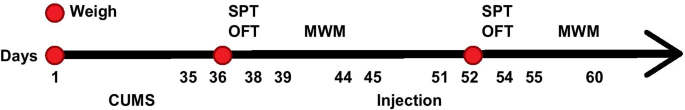
本研究的年表
标本集
在最后一次 MWM 测试后一天,将大鼠安乐死并腹腔注射 2% 戊巴比妥钠 (50 mg/kg)。用注射器打开胸腔取心脏血,3500 r/min离心3次,离心15 min,上清液保存于-20 ℃。采血后,将导管从左心室插入主动脉,灌注生理盐水500mL冲洗全血。分离海马组织并置于 1.5 mL 离心管中,称重并储存于 - 80 °C。部分海马用4%多聚甲醛固定2 h,梯度蔗糖脱水,石蜡包埋。
氧化应激相关和炎症指数检测
复温血清以检测超氧化物歧化酶(SOD)、丙二醛(MDA)、谷胱甘肽(GSH)、白细胞介素(IL)-β、肿瘤坏死因子-α(TNF-α)和一氧化氮(NO)浓度。 SOD、MDA、GSH检测试剂盒由碧瑶生物技术研究所(中国上海)提供,IL-β、TNF-α和NO检测试剂盒由南京建诚生物工程研究所提供(中国上海)。中国南京)。
苏木精-伊红 (HE) 染色
石蜡切片脱蜡,乙醇水合,蒸馏水冲洗,苏木精染色液染色20 min。之后,切片用自来水冲洗,直到切片变蓝。然后将切片置于1%乙醇盐酸溶液中10 s,自来水冲洗,乙醇脱水,伊红染色2 min,高浓度酒精脱水,二甲苯透化。最后密封切片,在生物显微镜下观察。
神经递质表达检测
海马组织在生理盐水(100 μL/10 mg)中称重并通过超声匀浆。匀浆在 4°C 下保持 30 分钟,以 12,000 rpm (4°C) 离心 3 分钟以收集上清液。用二辛可宁酸(BCA)蛋白检测试剂盒(CWBIO,北京,中国)检测上清液的蛋白浓度,ELISA试剂盒检测去甲肾上腺素(NE)、5-羟色胺(5-HT)和多巴胺(DA)的表达水平。 NE、5-HT、DA ELISA试剂盒由北京立维平生物技术有限公司提供。
逆转录定量聚合酶链反应 (RT-qPCR)
用RNA提取试剂盒(Promega,Madison,WI,USA)从海马组织中提取RNA,紫外分光光度计检测RNA的浓度和纯度。按照逆转录试剂盒说明书(DRR047S,Takara,大连,中国),将RNA逆转录为互补DNA(cDNA)。 mRNA 通过 GoldScript 一步法 RT-PCR 试剂盒(Applied Biosystems,Carlsbad,CA,USA)逆转录成 cDNA,而 miRNA 通过 Hairpin-it™ miRNA 定量检测试剂盒(GenePharma)逆转录成 cDNA。 RT-qPCR检测试剂盒(Promega)用于检测组织中HDAC1、miR-124-5p和NPY的表达。 U6 被指示为 miR-124-5p 的内部对照,而 HDAC1 和 NPY 的 β-肌动蛋白被指示为内部对照。 PCR 引物购自 Sangon Biotech Co., Ltd.(中国上海)(表 1)。目的基因的相对表达量计算公式为2 −△△ Ct法。
蛋白质印迹分析
将海马组织在冰上切成碎片并通过放射免疫沉淀测定裂解物(北京阳光生物科技有限公司,北京,中国)裂解以提取蛋白质。 BCA法测定蛋白质浓度。将总蛋白(50 μg)与 5 × 十二烷基硫酸钠(SDS)上样缓冲液以 1:4 的比例加入,并在沸水浴中加热 5 分钟。然后,将蛋白质进行十二烷基硫酸钠聚丙烯酰胺凝胶电泳,电印迹到膜上并用5%牛奶封闭1小时。之后,用针对 HDAC1 (1:1000, Cell Signaling Technology, Beverly, MA, USA)、NPY (1:800, NeoMakers, Fremont, California, USA) 和 β-肌动蛋白 (1:1000, Abcam, Cambridge, MA, UK) 过夜,并用辣根过氧化物酶标记的二抗重新检测。膜通过增强化学发光显色,光密度通过Quantity One灰度分析软件计算。目的基因的蛋白表达量以灰度值与β-actin灰度值的比值表示。
染色质免疫沉淀 (ChIP) 检测
ChIP 检测按照 EZ-ChIP 试剂盒(Millipore,Bedford,MA,USA)的说明进行。 HEK293T 细胞与 1% 甲醛孵育 10 分钟,并用甘氨酸终止。 2000 rpm 离心 5 min,加入 SDS Lysis Buffer 进行超声处理。 4℃ 10,000 g 离心 10 min,细胞(100 μL)与 900 μL ChIP Dilution Buffer、20 μL 50 × PIC 和 60 μL ProteinA Agarose/Salmon Sperm DNA 4℃反应 1 h,允许静置10分钟。将沉淀物以 20 μL 作为输入以 700 rpm 离心 1 分钟。一管加入HDAC1抗体(1 μL)和免疫球蛋白G抗体,另一管加入无抗体。将两管中的样品温育过夜、洗脱和去交联。提取DNA后,样本进行RT-qPCR检测。
双荧光素酶报告基因检测
生物信息学网站 (https://cm.jefferson.edu/rna22/Precomputed/) 分析了 miR-124-5p 和 NPY 的结合位点。 NPY 野生型 (WT) 和 NPY 突变型 (MUT) 质粒由华月洋生物技术有限公司(中国北京)生产。结合模拟NC或miR-124-5p模拟,将质粒转染到HEK293T细胞中。采用荧光素酶检测试剂盒(BioVision)和Glomax20/20光度计(Promega)测定细胞荧光素酶活性。
统计分析
SPSS 21.0 统计软件(IBM Corp. Armonk, NY, USA)用于数据分析。结果表示为平均值 ± 标准偏差。两组比较用t检验 测试。通过单向方差分析 (ANOVA) 评估多组之间的比较,然后通过 Tukey 事后检验进行成对比较。 P 代表两侧检验和 P <0.05 被认为具有统计学意义。
结果
抑制 HDAC1 或 miR-124-5p 会增加抑郁大鼠的体重
在 CUMS 建模前 1 天(第 0 天)、停止建模后 1 天(第 36 天)和慢病毒干扰后 7 天(第 52 天)对大鼠称重。造模前各组大鼠体重未见差异(均P> 0.05)。造模后CUMS组、sh-NC组、sh-HDAC1组、anti-miR-NC组和anti-miR-124-5p组大鼠体重较正常组下降(均P <0.05)。 CUMS组、sh-NC组、sh-HDAC1组、抗miR-NC组和抗miR-124-5p组大鼠体重未见差异(均P> 0.05)。干扰后,CUMS组大鼠体重较正常组下降(均P <0.05)。 CUMS组、sh-NC组和抗miR-NC组大鼠体重未见差异(均P> 0.05)。与NC组相比,sh-HDAC1组和anti-miR-124-5p组大鼠体重增加(均P <0.05),表明抑制HDAC1或miR-124-5p会增加抑郁大鼠的体重(图2a)。
<图片>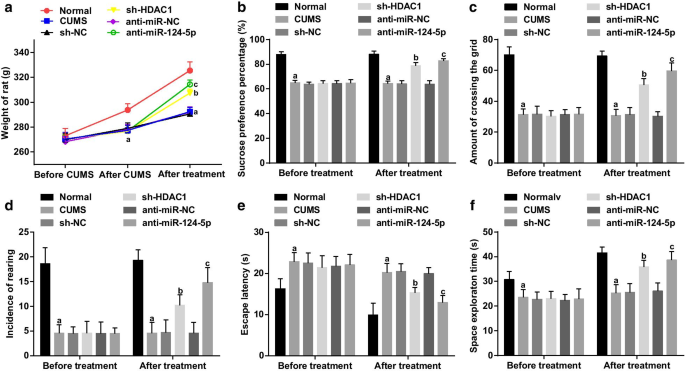
抑制 HDAC1 或 miR-124-5p 会增加体重并改善抑郁大鼠的学习和记忆能力。 一 HDAC1或miR-124-5p抑制对抑郁大鼠体重的影响; b HDAC1或miR-124-5p抑制前后的SPP; c 用HDAC1或miR-124-5p抑制前后在OFT中穿过网格的频率; d 在用 HDAC1 或 miR-124-5p 抑制之前和之后在 OFT 中饲养的发生率; e 用 HDAC1 或 miR-124-5p 抑制前后 MWM 测试中的逃逸潜伏期; f 用HDAC1或miR-124-5p抑制前后MWM测试中的空间探索时间; n =10; P 与正常组比较<0.05; b P 与sh-NC组相比<0.05; c P 与抗miR-NC组相比<0.05。多组间比较采用单向方差分析,成对比较采用 Tukey 事后检验
抑制 HDAC1 或 miR-124-5p 可改善抑郁大鼠的学习和记忆能力
应用SPT、OFT和MWM测试检测SPP、过网频率和饲养发生率,以及逃逸潜伏期和空间探索时间。概述(图2b-f)在干扰前,与正常组相比,SPP,穿越网格的频率,饲养发生率和空间探索时间减少,而CUMS组的逃逸潜伏期延长,sh-NC组、sh-HDAC1组、抗miR-NC组和抗miR-124-5p组(均P <0.05),表明大鼠出现抑郁样行为。 CUMS组、sh-NC组、sh-HDAC1组、anti-miR-NC组和anti-miR-124的SPP、过格频率、饲养发生率、太空探索时间和逃逸潜伏期没有差异-5p 组(所有 P> 0.05)。干扰后,与sh-NC组和anti-miR-NC组相比,sh-HDAC1组和anti-miR-124-SPP、过格频率、饲养发生率和空间探索时间增加,逃逸潜伏期缩短。 5p组(所有P <0.05)。 CUMS组、sh-NC组和anti-miR-NC组的SPP、过格频率、饲养发生率、空间探索时间和逃逸潜伏期均无差异(均P> 0.05),表明沉默HDAC1或抑制miR-124-5p可以减轻大鼠抑郁样行为,提高学习记忆能力。
抑制 HDAC1 或 miR-124-5p 可减轻抑郁大鼠的病理性神经元损伤
HE染色观察海马病变(图3a),正常组大鼠海马神经元排列整齐,形态清晰,结构正常,层数致密,细胞核透明,核仁明显。 CUMS组大鼠神经元萎缩,数量减少,排列松散,染色质分布不均,层变薄。 sh-NC和anti-miR-NC组大鼠表现出与CUMS组相同的情况。与 NC 组相比,sh-HDAC1 和抗 miR-124-5p 组的大鼠神经元依次增加并减轻损伤。表明抑制HDAC1或抑制miR-124-5p可减轻抑郁大鼠的海马病变。
<图片>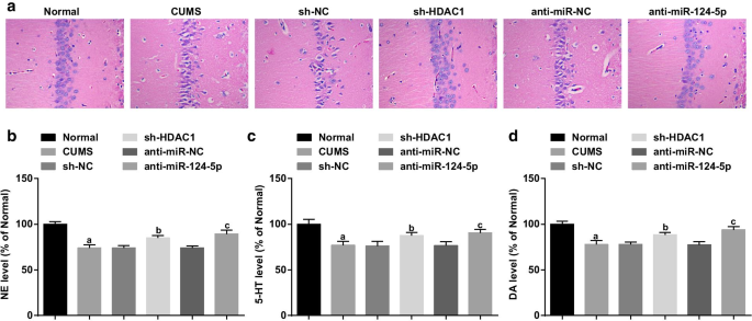
抑制 HDAC1 或 miR-124-5p 可减轻病理性海马体损伤并上调抑郁大鼠的神经递质表达。 一 抑郁大鼠海马病变HE染色; b 海马组织中NE表达的ELISA; c 海马组织中5-HT表达的ELISA; d 海马组织中DA表达的ELISA; n =6; P 与正常组比较<0.05; b P 与sh-NC组相比<0.05; c P 与抗miR-NC组相比<0.05。多组间比较采用单向方差分析,成对比较采用 Tukey 事后检验
抑制 HDAC1 或 miR-124-5p 上调抑郁大鼠的神经递质表达
抑郁症与脑神经递质紊乱有关。因此,通过ELISA测量大鼠海马中DA、NE和5-HT神经递质的水平。结果表明(图3b-d)与正常组相比,CUMS组、sh-NC组、sh-HDAC1组、抗miR组大鼠DA、NE和5-HT水平降低-NC组和抗miR-124-5p组(均P <0.05)。 CUMS组、sh-NC组和anti-miR-NC组神经递质表达无差异(均P> 0.05)。相对于sh-NC和anti-miR-NC组,sh-HDAC1和anti-miR-124-5p组大鼠的DA、NE和5-HT水平升高(均P <0.05),提示HDAC1的下调或miR-124-5p的降低可以上调抑郁大鼠海马中DA、NE和5-HT的水平。
抑制 HDAC1 或 miR-124-5p 可抑制抑郁大鼠的氧化应激和炎症
测量血清中氧化应激相关和炎症因子的表达。与正常组相比,SOD和GSH活性受损,而其他抑郁模型组MDA、IL-1β、TNF-α和NO水平升高(均P <0.05)。 CUMS组、sh-NC组和抗miR-NC组的SOD、GSH活性、MDA、IL-1β、TNF-α、NO水平无差异(均P> 0.05)。与 sh-NC 组和抗 miR-NC 组相比,观察到 SOD 和 GSH 活性增加,并观察到 sh-NC 组中 MDA、IL-1β、TNF-α 和 NO 水平降低。 HDAC1组和抗miR-124-5p组(均P <0.05)(图4a-f),表明HDAC1的消耗或miR-124-5p的下调可以减轻抑郁大鼠的氧化应激和炎症。
<图片>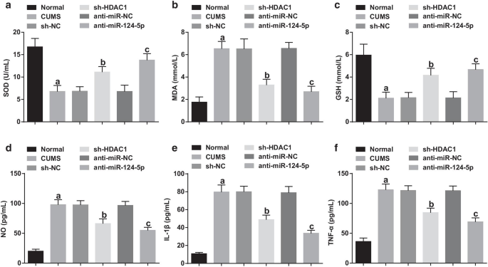
抑制 HDAC1 或 miR-124-5p 可抑制抑郁大鼠的氧化应激和炎症。 一 –c 抑制HDAC1或miR-124-5p对抑郁大鼠血清SOD、MDA和GSH浓度的影响; d –f 抑制HDAC1或miR-124-5p对抑郁大鼠血清IL-1β、TNF-α和NO浓度的影响; n =10; P 与正常组比较<0.05; b P 与sh-NC组相比<0.05; c P 与抗miR-NC组相比<0.05。多组间比较采用单向方差分析,成对比较采用 Tukey 事后检验
HDAC1 介导 miR-124-5p 以调节 NPY
RT-qPCR和蛋白质印迹分析用于检测海马中的HDAC1、miR-124-5p和NPY。概述(图5a,b)与正常组相比,CUMS组中HDAC1和miR-124-5p增加而NPY减少(所有P <0.05)。 HDAC1、miR-124-5p 和 NPY 表达在 CUMS 组和 sh-NC 组中没有变化(所有 P> 0.05)。相对于sh-NC组,sh-HDAC1组表现为HDAC1和miR-124-5p降低,NPY表达水平升高(均P <0.05)。以上结果提示慢病毒干扰成功,miR-124-5p与HDAC1表达呈正相关。
<图片>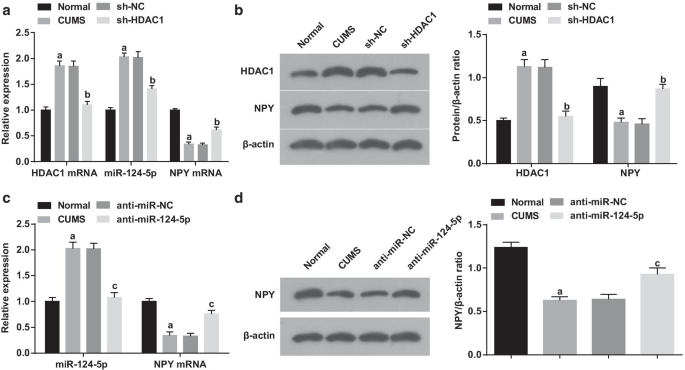
HDAC1 介导 miR-124-5p 以调节 NPY。 一 RT-qPCR抑制HDAC1后海马组织中HDAC1、miR-124-5p和NPY mRNA的表达(n =6); b 通过蛋白质印迹分析抑制HDAC1后海马组织中HDAC1和NPY蛋白的表达(n =6); c RT-qPCR抑制miR-124-5p后海马组织中MiR-124-5p和NPY mRNA的表达(n =6); d Western印迹分析抑制miR-124-5p后海马组织中NPY蛋白的表达(n =6); P 与正常组比较<0.05; c P <0.05 与 sh-NC 组相比。多组间比较采用单向方差分析,成对比较采用 Tukey 事后检验
此外,在 miR-124-5p 抑制后检测到 miR-124-5p 和 NPY 表达,结果(图 5c、d)表明与正常组相比,miR-124-5p 升高和 NPY 降低出现在CUMS 组(均 P <0.05)。相反,与抗miR-NC组相比,抗miR-124-5p组趋向于降低miR-124-5p和升高NPY(均P <0.05)。 CUMS组和抗miR-NC组的miR-124-5p和NPY表达未见差异(均P> 0.05)。结果表明慢病毒干扰成功,NPY与miR-124-5p呈负相关。
ChIP检测是为了检测HDAC1是否能与miR-124-5p启动子结合,结果表明(图6a、b)HDAD1仅与miR-124-5p启动子相关(P <0.001),表明HDAC1可以直接调控miR-124-5p。
<图片>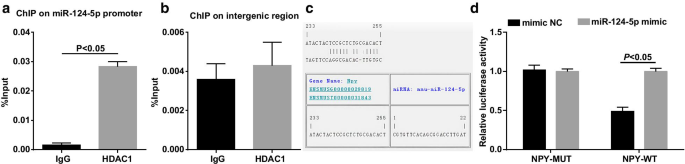
HDAC1 与 miR-124-5p 的启动子结合。 一 通过 ChIP 测定法测定 HEK293T 细胞中 HDAC1 和 miR-124-5p 启动子区域的结合丰度 (n =3); b 通过 ChIP 测定,HEK293T 细胞中 HDAC1 和无关基因间区域的结合丰度 (n =3); c MiR-124-5p和NPY结合位点生物信息软件网站; d 双荧光素酶报告基因检测miR-124-5p与NPY的靶向关系(n =3);两组比较用t检验 测试
通过RNA22工具和双荧光素酶报告基因测定预测并验证了miR-124-5p和NPY之间的靶向关系(图6c、d)。对于与 NPY-3'UTR-WT 和模拟物 NC 共转染的细胞,共转染 NPY-3'UTR-WT 和 miR-124-5p 模拟物的细胞显示出荧光素酶活性受损(P <0.05)。 No difference was recognized in the luciferase activity in the cells co-transfected with NPY-3′UTR-MUT and mimic NC, and the cells co-transfected with NPY-3′UTR-MUT and miR-124-5p mimic (P> 0.05).
Knockdown of NPY Abolishes the Protective Effects of Inhibited miR-124-5p on Depressed Rats
Spontaneous depletion of NPY and miR-124-5p was programmed to explore their interplay in depressed rats. It was exhibited that (Fig. 7a, b) lower NPY expression level was noticed in the anti-miR-124-5p + sh-NPY group versus the anti-miR-124-5p + sh-NC group (P <0.05)。 Body weight and behavioral function tests illustrated that (Fig. 7c–f) by comparison with the anti-miR-124-5p + sh-NC group, the rats in the anti-miR-124-5p + sh-NPY group presented reduced weight, SPP, frequency of crossing the grid, incidence of rearing and space exploration time with increased escape latency (all P <0.05)。 HE staining of the hippocampal lesions pictured that (Fig. 8a) in comparison with the anti-miR-124-5p + sh-NC group, the number of hippocampal neurons was reduced, and hippocampal neurons were darkly stained, sparsely and disorderly arranged in an irregular shape with reduced cell layers in the anti-miR-124-5p + sh-NPY group. Moreover, versus the anti-miR-124-5p + sh-NC group, the rats in the anti-miR-124-5p + sh-NPY group was accompanied by reduced DA, NE and 5-HT (all P < 0.05) (Fig. 8b). Besides, impaired SOD and GSH activities, and increased MDA, IL-1β, TNF-α and NO levels existed in the rats of the anti-miR-124-5p + sh-NPY group versus the anti-miR-124-5p + sh-NC group (all P < 0.05) (Fig. 8c, d). Collectively, knockdown of NPY abolished the protective effects of repressed miR-124-5p on depressed rat.

Knockdown of NPY abolishes the effects of inhibited miR-124-5p on body weight and behavioral function of depressed rats. 一 MiR-124-5p and NPY mRNA expression in hippocampal tissues after inhibition of both miR-124-5p and NPY by RT-qPCR (n = 6); b NPY protein expression in hippocampal tissues after inhibition of both miR-124-5p and NPY by western blot analysis (n = 6); c Body weight of depressed rats after inhibition of both miR-124-5p and NPY (n =10); d SPP of depressed rats after inhibition of both miR-124-5p and NPY (n =10); e Frequency of crossing the grid and incidence of rearing after inhibition of both miR-124-5p and NPY (n =10); f Escape latency and space exploration time after inhibition of both miR-124-5p and NPY (n =10); Comparisons between two groups were tested by t 测试

Knockdown of NPY abolishes the effects of inhibited miR-124-5p on hippocampal damages of depressed rats. 一 HE staining of hippocampal lesions of rats after inhibition of both miR-124-5p and NPY (n = 6); b ELISA of NE, 5-HT and DA expression in hippocampal tissues of rats after inhibition of both miR-124-5p and NPY (n = 6); C. Effect of inhibited NPY and suppressed miR-124-5p on SOD, MDA and GSH concentration in serum (n =10); D. Effect of inhibited NPY and suppressed miR-124-5p on IL-1β, TNF-α and NO concentration in serum (n =10)。 Comparisons between two groups were tested by t 测试
Discussion
Depression is a type of psychiatric disorder comprising a variety of conditions with diverse symptoms [20]. Though emerging studies have implied the role of miRNAs in depression, the precise action of miR-124-5p has been rarely investigated. Hence, the present study is projected for better comprehension of the mechanism of miR-124-5p/HDAC1/NPY axis in depression with the major outcome elaborating that silencing of either HDAC1 or miR-124-5p up-regulated NPY to improve memory and learning abilities of depressed rats.
To begin with, this study discovered HDAC1 expression in hippocampal tissues with the findings suggesting the elevation in the HDAC1 expression. Subsequently, with the purpose to decipher the functional roles of HDAC1 in depressed rats, loss-of-function assays were performed and it was disclosed that knockdown of HDAC1 increased the body weight, improved learning and memory abilities, attenuated pathological damage, up-regulated neurotransmitter expression, and suppressed oxidative stress and inflammation in depressed rats. Currently, a study has implied an increment in the expression of HDAC1 in depressive-like and anxiety-like phenotypes resulted by stress-offspring [21]. Moreover, the expression of HDAC1 is documented to up-regulate in penumbra in photothrombotic stroke [22]. Besides that, the incremental HDAC1 mRNA expression is found in granule and pyramidal cells in temporal lobe epilepsy [23]. As to the functional role of HDAC1, it has been reported that virus-mediated overexpression of neuronal HDAC1 in the hippocampus of mice imposes influences on loss of contextual fear memory in particular [24]. Mechanically, HDAC inhibitors reverse cognitive deficits found in neurodegenerative diseases and age-related memory loss [25]. Actually, it is accepted that the HDAC1 suppression by tianeptinaline has advanced neuroplasticity and reinforced memory [26]. As mentioned in a prior study, it is concluded that repression of HDAC1 inhibits the pathogenic processes that lead to motor neuron degeneration in mitochondrial diseases [27]. Experimentally, the silencing of HDAC1 by 5-thienyl-substituted 2-aminobenzamide-type is partially involved in the prevention of neuronal cell death in Parkinson's disease models [28]. Further supported by those researches, the protective effects of silenced HDAC1 have been witnessed in brain diseases, including but not limited to depression.
Then, our study discovered a targeting relationship between HDAC1 and miR-124-5p, which was supported by a prior study which suggests miR-124 transcription is in the charge of EVI1, acting by connection with the deacetylase HDAC1 [29]. miR-124-5p was the overexpressed gene in depressed rats and knockdown of miR-124-5p had the similar functions of silenced HDAC1 in depressed rats. In fact, there is a study indicating that the expression of miR-124 in the hippocampus is up-regulated from 5 to 6 weeks in depression-like behavior phenotypes [7]. Another study has identified the increase in the miR-124 expression in female with cocaine use disorder [30]. Also, it is previously described that miR-124-3p is highly expressed in stressed rodents in major depressive disorder [31]. Regarding to the effects, knockdown of miR-124 in the prefrontal cortex is reported to attenuate depression-like behavior of mice [9]. Besides that, miR-124 knockdown is believed to serve as an antidepressant agent of chronic corticosterone-induced gypenosides in mice [32]. Drawn from a prior study, knockdown of miR-124 can result in improved behavioral susceptibility to a milder stress paradigm [33]. It is reported that suppression of miR-124 by lentivirus transfection in the hippocampus can protect ketamine-induced neurodegeneration in vivo and in vitro [34]. Anyway, miR-124-5p suppression is an active actor to attenuate depression.
Furthermore, NPY expression was verified to be regulated by HDAC1 and miR-124-5p. The deteriorated deficits associated with HDAC2 in histone acetylation may be related to the decreased expression of NPY and can used to control anxiety-like and drinking behaviors [14]. NPY was down-regulated in depressed rats and up-regulation of NPY promoted learning and memory ability recovery in depressed rats. In the development of depressive-like behaviors, the rat models are manifested with reduced NPY expression [17]. Academically, the expression of NPY is evidenced to decline in mice with depression [35]. Consistently, the above-mentioned study findings are as same as the previous literature to some extent.
The novel findings of study suggested that inhibited miR-124-5p or suppressed HDAC1 attenuated learning and memory abilities and increased body weight of depressed rats. In addition, knockdown of miR-124-5p or inhibition of HDAC1 suppressed oxidative stress and inflammation and promoted neurotransmitter expression of depressed rats. Moreover, HDAC1 mediated miR-124-5p to regulate NPY. In the rescue experiments, knockdown of NPY abolished the protective effects of inhibited miR-124-5p on depressed rats.
Conclusion
In summary, this study highlighted the effect of HDAC1/miR-124-5p/NPY axis in depression with the major findings suggesting inhibited miR-124-5p or suppressed HDAC1 attenuated learning and memory abilities, increased body weight, suppressed oxidative stress and inflammation, as well as promoted neurotransmitter expression in depressed rats. HDAC1/miR-124-5p/NPY axis may provide a reference to treat neurological disorder, which may also update the existed knowledge of depression. However, further studies are still required for thorough comprehension of the complex mechanism of HDAC1/miR-124-5p/NPY axis in depression.
数据和材料的可用性
不适用。
缩写
- miR-124-5p:
-
MicroRNA-124-5p
- HDAC1:
-
Histone deacetylase 1
- NPY:
-
Neuropeptide Y
- CUMS:
-
Chronic unpredictable mild stress
- 标准差:
-
Sprague–Dawley
- SPF:
-
Specific pathogen-free
- NC:
-
Negative control
- OFT:
-
Open field test
- SPT:
-
Sucrose preference test
- SPP:
-
Sucrose preference percentage
- MWM:
-
Morris water maze test
- SOD:
-
Superoxide dismutase
- MDA:
-
Malondialchehyche
- GSH:
-
谷胱甘肽
- IL-β:
-
Interleukin-β
- TNF-α:
-
Tumor necrosis factor-α
- NO:
-
Nitric oxide
- ELISA:
-
酶联免疫吸附试验
- HE:
-
Hematoxylin–eosin
- BCA:
-
Bicinchoninic acid
- NE:
-
Norepinephrine
- 5-HT:
-
Serotonin
- DA:
-
多巴胺
- RT-qPCR:
-
Reverse transcription quantitative polymerase chain reaction
- cDNA:
-
Complementary DNA
- SDS:
-
十二烷基硫酸钠
- ChIP:
-
Chromatin immunoprecipitation
- UTR:
-
Untranslated region
- MUT:
-
Mutant type
- WT:
-
Wild type
- ANOVA:
-
One-way analysis of variance
纳米材料
- 使用 tinyML 和自动化机器学习构建有效的物联网应用
- ST:在低成本 SO-8 封装中具有丰富模拟和 DMA 的 8 位 MCU
- 坚持不懈地领导
- 人工神经元无监督学习
- 利用机器学习提高可靠性并改善维护结果
- 具有高级农业化学活性的智能纳米材料和纳米复合材料
- 具有自整流开关特性的 TaO/HfO x 和 TaO/AlO x RRAM 的保持模型
- 涂有 CuS 纳米粒子的有色导电 CuSCN 复合材料的简便合成
- RGO 和三维石墨烯网络共同修饰的高性能 TIM
- 氮化处理的基于 HfO2 的 RRAM 中的传导机制和改进的耐久性
- 电阻式随机存取存储器建模和仿真的集体研究
- 不同粒径阿维菌素纳米递送系统的制造、表征和生物活性