

毛蒿、马齿苋和夏枯草提取物的抗氧化潜力用于金纳米颗粒的生物制造和细胞毒性评估
摘要
三种水性植物提取物(Artemisia capillaris , 马齿苋 , 和 夏枯草 ) 被选择用于金纳米粒子的生物制造。研究了提取物的抗氧化活性(即自由基清除活性、总酚含量和还原能力)以及这些活性如何影响金纳米粒子的生物制造。 P。普通话 发挥最高的抗氧化活性,其次是A。毛细血管 然后P。甘蓝 . P。普通话 是生物制造过程中最有效的还原剂。由 P 生物制造的金纳米粒子。普通话 (PV-AuNPs) 具有 530 nm 的最大表面等离子体共振,具有多种形状。高分辨率 X 射线衍射分析表明 PV-AuNPs 具有面心立方结构。通过电感耦合等离子体发射光谱估计反应产率为99.3%。流体动力学尺寸确定为 45 ± 2 nm,zeta 电位为 - 13.99 mV。 PV-AuNPs 具有剂量依赖性的抗氧化活性。值得注意的是,在没有胎牛血清的情况下,PV-AuNPs 对人结直肠腺癌细胞的细胞毒性最高,而对于人胰腺导管腺癌细胞,在胎牛血清存在的情况下,观察到的细胞毒性最高。该结果表明 P.普通话 提取物是生物制备对癌细胞具有细胞毒性的金纳米颗粒的有效还原剂。
介绍
由于其广泛的应用[1],人们对金属纳米粒子的兴趣大大增加。特别是,金纳米粒子 (AuNPs) 作为药物/基因传递载体、催化剂、显像剂和化学/生物传感器具有多种有价值的应用 [2]。多种化学和物理方法,如离子溅射、反胶束和化学还原,可用于制造 AuNP。然而,这些方法因其成本和潜在毒性而受到限制。在 AuNPs 的生物制造(或绿色合成)中使用植物提取物比其他方法具有优势,这使其成为非常有前途的替代方法 [2]。生物制造过程具有成本效益且易于扩大规模。它利用环保材料,并通过结合两种材料(AuNPs 和植物提取物)的活性表现出协同活性。生物制造在合成过程中不会引入有毒的化学还原剂,这使得该过程完全绿色。此外,该特征增加了所得 AuNP 的生物相容性并增加了它们的稳定性。来自植物提取物的各种植物化学物质不仅可用作还原剂,还可用作加帽剂和稳定剂 [3]。
毛细蒿 属于蒿属 ,是菊科中最大的属之一 [4]。自古以来,A.毛细血管 由于其多种药理作用,包括抗菌、抗炎、抗氧化和保肝活性,已被广泛用作草药。以前的研究将这些影响归因于 A 的活性成分。毛细血管 ,包括毛细管蛋白、毛细管烯、scoparone 和 β-谷甾醇 [4,5,6]。 马齿苋 马齿苋科一年生多肉植物,具有多种药理作用,如抗菌、抗真菌、抗炎、抗氧化和抗溃疡活性。这些影响已被证明是由各种活性成分引起的,包括生物碱、脂肪酸、黄酮类化合物、多糖和萜类化合物 [7, 8]。 夏枯草 是唇形科的多年生草本植物。许多研究表明,P. 具有抗病毒 [9]、抗癌 [10]、免疫调节 [11]、抗氧化 [11, 12] 和降血糖活性 [13]。普通话 .促成这些作用的主要活性成分是有机酸、酚酸和三萜类化合物,包括亚麻酸、棕榈酸、顺式 和 trans- 咖啡酸、迷迭香酸、齐墩果酸和熊果酸[14]。
由于“蛋白质电晕”,纳米粒子研究的体外和体内结果之间经常存在很大差距。进入生物环境后,纳米粒子的表面会覆盖一层不同的蛋白质,称为蛋白质电晕 [15]。蛋白质电晕包覆的纳米粒子进入细胞并影响细胞摄取、细胞毒性、生物分布、排泄、细胞反应、药物释放和治疗效率 [16]。有趣的是,金属纳米粒子的物理化学性质,例如它们的形状、大小、表面电荷和表面粗糙度,在形成蛋白质冠时会深刻影响血清蛋白质的吸附 [17]。 Shannahan 及其同事报告说,在银纳米粒子 (AgNPs) 上形成蛋白质电晕介导对大鼠肺上皮细胞和大鼠主动脉内皮细胞的细胞毒性通过 清道夫受体 [18]。血清白蛋白通过降低其抗菌和细胞毒性作用来影响水凝胶包埋的胶体 AgNPs [19]。
在本报告中,我们评估了 A 的水提取物的抗氧化活性,包括自由基清除活性、总酚含量和还原能力。毛细血管 ,P。甘蓝 , 和 P。普通话 .随后,使用这三种提取物进行 AuNPs 的生物制造,以研究提取物的抗氧化活性如何影响胶体溶液的颜色和 AuNPs 的表面等离子体共振 (SPR) 带的形状。此后,生物制造的 AuNP 以每种植物的学名命名:使用 A 提取物获得的 AuNP。毛细血管 被称为 AC-AuNPs,使用 P 提取物获得的 AuNPs。甘蓝 被称为PO-AuNPs,并且使用P的提取物获得的AuNPs。普通话 被称为 PV-AuNPs。 P 的抗氧化活性。普通话 提取物最高;因此,我们通过各种光谱和显微方法对 PV-AuNPs 进行了表征,包括紫外-可见分光光度法、高分辨率透射电子显微镜 (HR-TEM)、高分辨率 X 射线衍射 (HR-XRD) 和电感耦合等离子体光发射光谱 (ICP-OES)。 PV-AuNPs 的流体动力学尺寸是通过动态光散射 (DLS) 和 zeta 电位测量的。此外,我们通过监测 2,2-diphenyl-1-picrylhydrazyl (DPPH) 自由基清除活性来检测 PV-AuNPs 的抗氧化活性。在细胞培养物中存在或不存在胎牛血清 (FBS) 的情况下,通过水溶性四唑 (WST) 测定研究对三种癌细胞的细胞毒性。使用以下癌细胞:人结直肠腺癌细胞(HT-29)、人乳腺腺癌细胞(MDA-MB-231)和人胰腺导管腺癌细胞(PANC-1)。
方法
材料
氯化钾金 (III) (KAuCl4)、丁基化羟基甲苯 (BHT)、磷酸二氢钠、磷酸氢二钠、三氯乙酸、Folin-Ciocalteu 酚试剂、碳酸钠、没食子酸、酒石酸钠钾、碳酸氢钠、硫酸,2 ,2'-azino-bis-3-ethylbenzthiazoline-6-sulphonic acid (ABTS)、过硫酸钾和 d-(+)-葡萄糖购自 Sigma-Aldrich(美国密苏里州圣路易斯)。 DPPH 购自 Alfa Aesar (Ward Hill, MA, USA)。六氰基铁酸钾 (III) 购自 Wako (Osaka, Japan)。硫酸钠、七钼酸六铵四水合物和砷酸氢二钠七水合物购自 Junsei (Tokyo, Japan)。氯化铁 (III) 购自 Duksan(韩国京畿道),五水硫酸铜购自 Deajeng(韩国京畿道)。其他试剂为分析纯,按原样使用。高糖 Dulbecco 改良 Eagle 培养基 (DMEM)、青霉素-链霉素 (10,000 U/mL)、胰蛋白酶-EDTA(0.5%,不含酚红)和磷酸盐缓冲盐水 (PBS) 购自 Gibco (Thermo Fisher Scientific,马,美国)。 FBS 来自 GE Healthcare HyClone™(维多利亚,澳大利亚)。
工具
Shimadzu UV-2600 分光光度计用于获取紫外-可见光谱并观察 SPR(Shimadzu Corporation,Kyoto,Japan)。从 HR-TEM 图像中获得有关粒度和形状的信息。为了获取 HR-TEM 图像,使用了在 200 kV 下运行的 JEM-2100F 仪器(JEOL,东京,日本)。将 PV-AuNPs 的胶体溶液加载到碳涂层铜网格(碳类型 B,300 目,Ted Pella,Redding,CA,USA)上。装载样品的网格在环境温度下风干。 Rigaku Miniflex 衍射仪(40 kV,30 mA)用于通过 CuKα 辐射源(λ =1.54056 Å) 在 10º 到 80° (2θ 规模)(理学,日本)。 FD5505 冷冻干燥机用于制备粉末样品用于 HR-XRD 分析(Il Shin Bio, Seoul, Republic of Korea)。为了估计反应产率,ICP-OES 在 Perkin Elmer Optima 8300 仪器(Waltham,MA,USA)上进行。进行离心(18,500g 力,7 小时,18°C),并收集上清液。两个样品,即胶体 PV-AuNP 溶液和上清液,进行 ICP-OES 分析。使用 Eppendorf 5424R 离心机进行离心(Eppendorf AG,Hamburg,Germany)。流体动力学尺寸和 zeta 电位由 NanoBrook 90 Plus Zeta 分析仪(Brookhaven Instruments Corporation, Holtsville, NY, USA)测量。
A 的水性提取物的制备。毛细血管 ,P。甘蓝 , 和 P。普通话
P 的干燥地上部分。普通话 和A。毛细血管 ,包括花和叶,购自 Omniherb(韩国大邱),P.甘蓝 获自 Handsherb (Youngchun,大韩民国)。通过超声处理(WUC-A22H,Daihan Scientific Co. Ltd.,大韩民国)制备每种植物地上部分的水提取物。对于每株植物,用去离子水 (1000 mL) 提取 100 g 切碎的植物 3 次。提取程序包括在 25°C 下对植物进行超声处理 3 小时。过滤水提取物,并将滤液在真空冷冻干燥机(FD8518,IlShinBioBase Co.Ltd.,大韩民国)中冻干。最后,获得每种植物的提取物粉末并在使用前储存在- 20°C。
DPPH 自由基清除活动
在 96 孔微孔板的每个孔中,将 20 μL 的每种提取物与乙醇中的 DPPH 自由基溶液(0.1 mM、180 μL)混合,得到几种最终浓度的提取物(31.25 μg/mL、62.5 μg/mL、 125 微克/毫升和 250 微克/毫升)。将板在环境温度下在黑暗中孵育 30 分钟,然后使用多检测读数器(Synergy HT,BioTek Instruments,Winooski,VT,USA)在 517 nm 处测量每个孔的吸光度。 BHT被用作标准。所有实验一式三份进行。从每个样品的吸光度中减去 AuNPs 的吸光度,以消除 AuNPs 在 517 nm 处的固有吸光度。
ABTS 激进清除活动
ABTS 自由基阳离子由 1:1 (v/v ) 7.4 mM ABTS 溶液和 2.6 mM 过硫酸钾溶液的混合物,在黑暗中在环境温度下放置 24 小时。 ABTS 自由基溶液用乙醇稀释至 730 nm 处的吸光度为 0.74,用于测量。在 96 孔微孔板的每个孔中,将乙醇中的 ABTS 自由基溶液 (180 μL) 加入 20 μL 的每种提取物(提取物的最终浓度,15.63 μg/mL、31.25 μg/mL、62.5 μg/mL、和 125 μg/mL)。在环境温度下在黑暗中孵育 10 分钟后,使用多检测读数器在 730 nm 处测量每个孔的吸光度。制备维生素 C 溶液作为标准品。所有实验一式三份进行。
降低功耗
将等体积 (500 μL) 的不同浓度提取物与 0.2 M 磷酸盐缓冲液(pH 6.6,500 μL)和 1% 六氰基高铁酸钾 (III) (500 μL) 混合。提取物的最终浓度为 52.1 μg/mL、104.2 μg/mL、208.3 μg/mL 和 416.7 μg/mL。每种混合物在 50°C 下温育。 20 分钟后,将三氯乙酸(10%,500 μL)加入混合物中,然后以 3400g 离心 力 10 分钟。然后,将氯化铁溶液(0.1%,100 μL)添加到 500 μL 的上清液中。 10 分钟后,使用多检测读数器在 700 nm 处测量反应混合物的吸光度。 BHT被用作标准。所有实验一式三份进行。
总酚含量
研究了抗氧化活性与酚类化合物含量之间的关系。将 20 微升提取物与 100 μL Folin-Ciocalteu 试剂(用去离子水稀释 10 倍)和 80 μL 碳酸钠(7.5%,w/v)混合 )。反应在黑暗中在环境温度下进行。 30 分钟后,测量 765 nm 处的吸光度,并使用由没食子酸构建的校准曲线确定总酚含量。所有测定一式三份进行。
PO-AuNPs、AC-AuNPs 和 PV-AuNPs 的生物制造
在生物制造反应之前制备两种储备溶液:氯化金 (III) 钾 (10 mM) 和提取物 (2%, w/v )。将每种提取物的原液 (2%) 离心 (18,500g 力,30 分钟,18°C)。收集上清液并用于生物制造反应。最终浓度调整为 0.5 mM 氯化钾金 (III) 和 0.05% (w/v ) 在 2 mL 玻璃小瓶中提取,用于 AuNP 的生物制造。将 Au 盐与提取物混合后,在 37°C 的干燥烘箱中孵育 5 小时。然后在300~800 nm范围内获得紫外-可见光谱。
细胞培养
三种癌细胞(HT-29、PANC-1 和 MDA-MB-231)购自韩国细胞系库(韩国首尔)。细胞在补充有 10% FBS、青霉素(100 单位/mL)和链霉素(100 μg/mL)的高葡萄糖 DMEM 中生长。细胞在培养箱中于 37°C(提供 5% CO2)培养,并在胰蛋白酶消化前保持约 80% 汇合。
细胞毒性
来自 DoGenBio(韩国京畿道)的 EZ-CYTOX 试剂盒用于 WST 测定以评估对癌细胞的细胞毒性。将细胞以5.0 × 10 3 的密度接种于96孔板中 细胞/孔。在 CO2 (5%) 气氛下,在 37°C 培养箱中培养 24 小时。孵育后,弃去培养基。然后,在存在或没有 FBS。在培养箱中在 37°C 下再培养 24 小时后,加入 EZ-CYTOX 试剂(10 μL),并将细胞在 CO2 培养箱中再培养 1 小时。使用 Cytation 混合多模式阅读器(BioTek Instruments, Winooski, VT, USA)在 450 nm 处测量吸光度。 PV-AuNPs 的固有吸光度会干扰 450 nm 处细胞毒性的测量。因此,从 450 nm 处测量的每个数据点的吸光度中减去 PV-AuNP 的背景吸光度。 PV-AuNPs 的背景吸光度是在无 WST 条件下测量的。此外,使用相同体积的去离子水代替PV-AuNPs作为对照。
统计分析
所有实验一式三份进行,结果表示为平均值 ± 标准误差(SE)。使用 ANOVA(单向方差分析)和 Tukey 多重比较检验或 Newman-Keuls 多重比较检验研究差异的显着性。使用 GraphPad 软件(GraphPad 软件 5.02 版,圣地亚哥,加利福尼亚州,美国)计算统计分析。结果被认为对于 p 的值具有统计学意义 <0.05.
结果与讨论
提取物的抗氧化活性
通过超声处理获得每种植物地上部分的水性提取物。 A的提取率。毛细血管 ,P。甘蓝 , 和 P。普通话 分别为 14.1%、39.12% 和 28.6%。最高提取率显示在 P 中。甘蓝 ,然后是 P。普通话 最后是A。毛细血管 .为了评估提取物的抗氧化活性,我们测量了自由基清除活性、还原能力和总酚含量。提取物的自由基清除活性通过 DPPH 和 ABTS 测定确定。 DPPH 被广泛用于评估酚类化合物的抗氧化活性,因为 DPPH 自由基是一种稳定的自由基,当它被抗氧化剂还原时会失去其吸收强度。 ABTS 测定测量抗氧化剂清除强氧化剂与 ABTS 反应产生的自由基的能力。评估了具有供氢特性的抗氧化剂对 ABTS 自由基吸光度的降低。水提取物以剂量依赖性方式表现出清除活性。在测试浓度(15.63 微克/毫升、31.25 微克/毫升、62.5 微克/毫升、125 微克/毫升和 250 微克/毫升)下,P 的提取物。普通话 (针对 DPPH 自由基的 IC50 50.35 ± 1.22 μg/mL;针对 ABTS 自由基的 IC50 38.6 ± 0.44 μg/mL)对 DPPH 和 ABTS 自由基表现出最有效的活性,其次是 A 的提取物。毛细血管 (对 DPPH 自由基的 IC50 156.72 ± 0.97 μg/mL;对 ABTS 自由基的 IC50 147.28 ± 2.95 μg/mL)然后是P 的提取物。甘蓝 (对 DPPH 自由基的 IC50 247.33 ± 1.57 μg/mL;对 ABTS 自由基的 IC50 305.54 ± 4.86 μg/mL)。值得注意的是,P.普通话 显示出比 BHT 更有效的 DPPH 自由基清除活性(IC50 71.37 ± 0.84 μg/mL),用作标准(表 1 和图 1a、b)。 P 的摘录。普通话 在三种提取物中显示出最高的 DPPH 和 ABTS 自由基清除活性。该结果暗示在P中可能存在更多的抗氧化化合物。普通话 比在 A 中。毛细血管 和P。甘蓝。
<图片>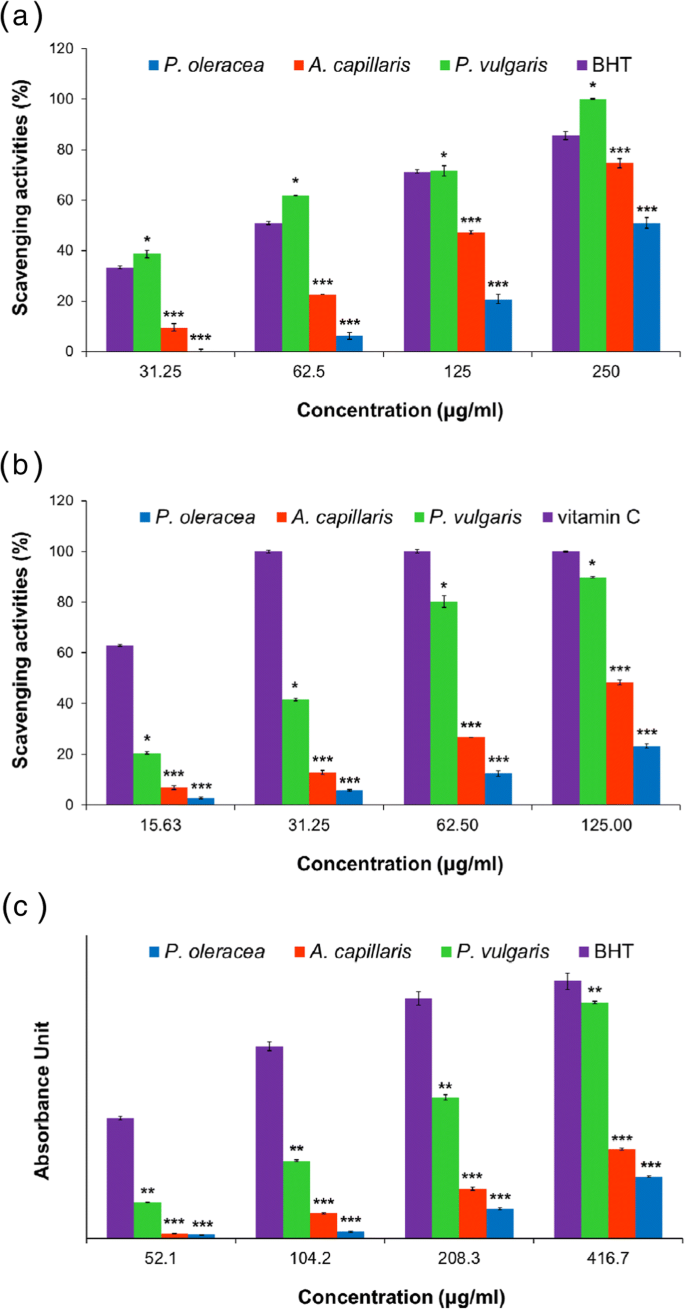
DPPH 和 ABTS 自由基清除活性和 A 水提取物的还原能力。毛细血管 ,P。甘蓝 , 和 P。普通话 . 一 DPPH 自由基清除活性。 b ABTS 自由基清除活动。 c 降低提取物的功效。数据表示为平均值 ± SEM。星号表示与标准(BHT 或维生素 C)的差异显着性:*p <0.05, **p <0.01, ***p <0.001。结果代表三个独立实验
我们还检测了提取物的抗氧化活性通过 还原力测定。在抗氧化剂、铁氰化钾 (Fe 3+ ) 转化为亚铁氰化钾 (Fe 2+ ),它与氯化铁反应形成三价铁-亚铁络合物。三价铁配合物在 700 nm 处具有最大吸光度,可用于评估抗氧化剂的还原能力。在当前的报告中,还原能力表示为在 700 nm 处吸光度为 0.7 的每种提取物的浓度 (μg/mL)。如图 1c 所示,P.普通话 (162.98 μg/mL) 的还原能力明显高于 A。毛细血管 (235.38 μg/mL) 和 P。甘蓝 (396.16 μg/mL)。如前所述,还原能力的结果还表明,P 中可能存在更多的抗氧化化合物。普通话 比在 A 中。毛细血管 和P。甘蓝。
还研究了每种提取物的总酚含量。众所周知,酚类化合物是潜在的抗氧化剂,并且由于其羟基具有清除自由基的能力。因此,植物提取物的抗氧化活性与其酚类含量密切相关。用没食子酸构建标准校准曲线,该曲线呈线性关系(y =6.0617x + 0.1293, r 2 =0.9983)。使用标准曲线,计算总酚含量并表示为每克提取物的没食子酸当量 (GAE) 毫克数。对于P,提取物的总酚含量为90.53 ± 6.81mg GAE/g。普通话 ,对于A,39.88 ± 4.55mg GAE/g。毛细血管 ,并且对于P,18.32 ± 2.76mg GAE/g。甘蓝 .总酚含量以P最高。普通话 三种提取物中。
总酚含量与提取物清除自由基的能力或其还原能力呈正相关。合起来,A. 的水提取物。毛细血管 ,P。甘蓝 , 和 P。普通话 表现出显着的抗氧化活性。特别是 P 的提取物。普通话 显示出对 DPPH 和 ABTS 自由基的最大自由基清除活性、最强的还原能力和最高的总酚含量,这意味着 P.普通话 作为 AuNPs 生物制造的还原剂,将发挥最强的活性。以下部分将讨论使用三种提取物生物制造 AuNP 的这些有趣结果。
AC-AuNPs、PO-AuNPs 和 PV-AuNPs 的生物制造
我们提出提取物的抗氧化活性强烈影响 AuNPs 的生物制造过程。由于抗氧化活性是将 Au 盐还原为 AuNPs 的主要驱动力,P.普通话 具有最高的抗氧化活性,能够成为最有效的还原剂。生物制造过程之后是紫外-可见分光光度法,因为 AuNP 在可见-近红外波长范围内具有独特的 SPR。 AuNPs 的生物制造是通过将 Au 盐与每种提取溶液混合来进行的。每种提取物都含有多种植物化学物质,可以非常有效地将 Au 盐还原为 AuNPs。
AuNPs 的独特 SPR 带导致 PO-AuNPs 的初始淡黄色溶液颜色变为深蓝色,AC-AuNPs 为荧光棕色,PV-AuNPs 为酒红色(图 2) .在 37°C 的干燥烘箱中孵育 5 小时后记录紫外-可见光谱,以观察每组 AuNP 的不同 SPR 带(图 3)。 PV-AuNP 在 530 nm 处和 AC-AuNP 在 555 nm 处观察到最大吸光度。在 PO-AuNPs 的情况下,从 500 到 700 nm 观察到一个宽的 SPR 带。这三种提取物产生了在紫外-可见光谱中具有特征 SPR 带的 AuNP。每种颜色的变化以及不同的 SPR 带都清楚地支持了每种提取物成功生物制造 AuNP。我们提出与抗氧化活性相关的三个因素(自由基清除活性、总酚含量和还原能力)影响 AuNP 生物制造。 P 的摘录。普通话 在所有三个因素中得分最高,其次是 A。毛细血管 最后由 P.甘蓝 .有趣的是,PO-AuNP 在环境温度 (25°C) 下储存 30 分钟后显示出聚集。该结果表明,PO-AuNPs 是最不稳定的,因为其抗氧化活性、总酚类化合物的量和P 的还原能力。甘蓝 是这三种提取物中最低的。相比之下,PV-AuNPs 显示出最高的吸光度和透明的酒红色溶液。 AC-AuNPs 的吸光度介于 PO-AuNPs 和 PV-AuNPs 之间。因此,基于这些观察,确定了 P 具有更高的自由基清除活性、总酚含量和还原能力。普通话 与由 A 的提取物产生的 AuNPs 相比,提取物导致合成更稳定的 AuNPs。毛细血管 和P。甘蓝 .总的来说,最大 SPR 带显示出红移和低色移,提取物的抗氧化活性降低。此外,随着抗氧化活性的降低,SPR带的形状趋于变宽。因此,PV-AuNPs 通过 HR-TEM、HR-XRD、流体动力学尺寸和 zeta 电位测量进一步表征。为了确定反应产率,进行了 ICP-OES 分析。进一步评估了FBS存在/不存在下对癌细胞的抗氧化活性和细胞毒性。
<图片>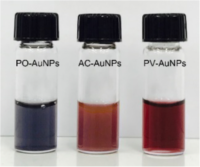
PO-AuNPs、AC-AuNPs和PV-AuNPs的数码照片
<图片>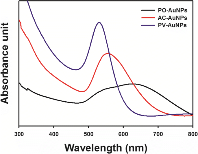
PO-AuNPs(黑线)、AC-AuNPs(红线)和PV-AuNPs(蓝线)的紫外可见光谱
PV-AuNPs 的 HR-TEM 图像
显微技术是可视化纳米粒子的最有效工具。与 HR-TEM 一起,扫描电子显微镜和原子力显微镜通常用于确定尺寸、形态、形貌和二维/三维结构。用 HR-TEM 研究了 PV-AuNPs 的尺寸和形态。如图 4 所示,观察到各种形状,包括球体、三角形、棒状、菱形和六边形(图 4a~e)。对棒状(图 4f,g)和三角形(图 4h)晶格结构的观察清楚地表明 PV-AuNP 本质上是结晶的。这一结果得到了随后的 HR-XRD 分析结果的有力支持。从 HR-TEM 图像测量每个颗粒形状的大小,所得的大小直方图如图 5 所示。所有直方图均显示高斯分布。随机选择图像中的离散纳米粒子以获得每个形状的平均尺寸。从 250 个纳米粒子中确定球体的直径为 23.8 ± 1.3 nm(图 5a)。有趣的是,产生了等边三角形;因此,我们测量了 64 个纳米粒子的高度(25.7 ± 2.2 nm,图 5b)。随机选择 27 个棒状纳米粒子来确定平均长度(28.4 ± 0.4 nm,图 5c)。杆的平均纵横比(定义为长度除以宽度)确定为 2.4。对于菱形,在图像中随机选择了 38 个菱形。如图 5d、e 所示,长对角线和短对角线的平均长度经测量分别为 22.0 ± 0.6 nm 和 15.4 ± 0.3 nm。形状多样的PV-AuNPs尺寸小于30 nm,下节将其尺寸与流体力学尺寸进行比较。
<图片>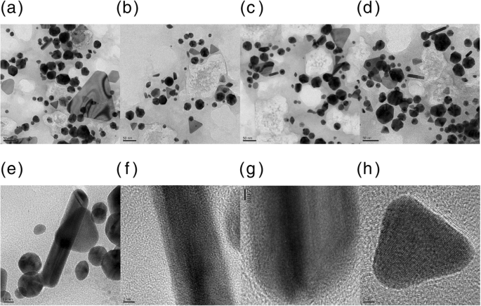
PV-AuNPs 的 HR-TEM 图像。比例尺代表a 50 纳米,b 50 纳米,c 50 纳米,d 50 纳米,e 10 纳米,f 2 纳米,g 2 纳米和 h 2 纳米
<图片>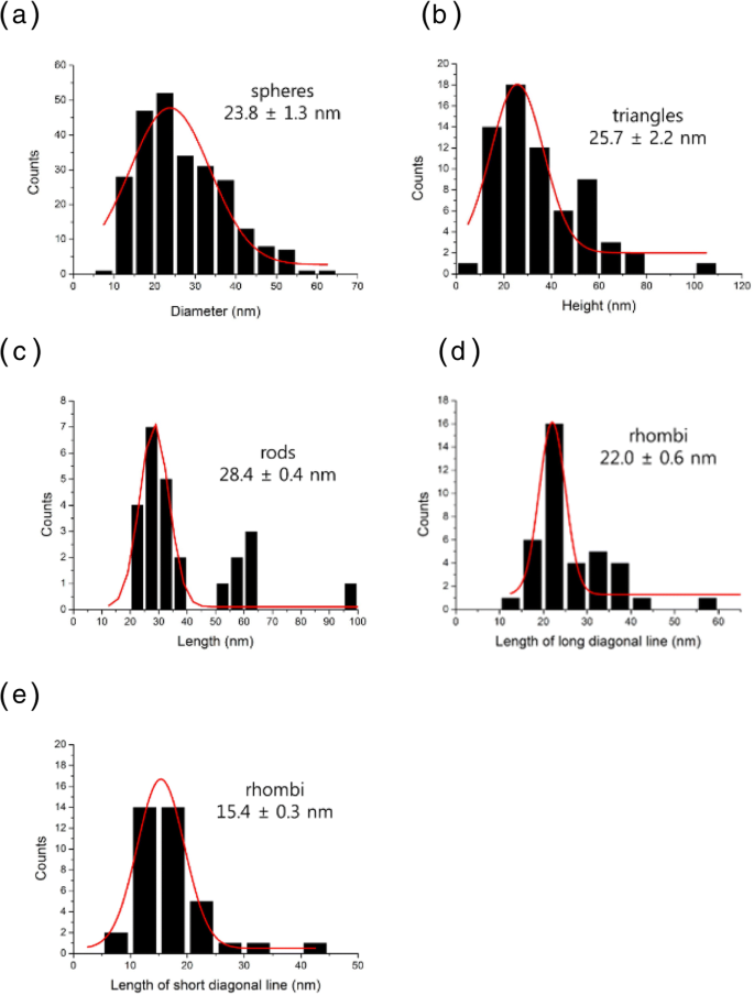
PV-AuNPs 的大小直方图。 一 球体的直径。 b 等边三角形的高。 c 杆的长度。 d 菱形的长度(长对角线)。 e 菱形的长度(短对角线)
PV-AuNPs 的流体动力学尺寸和 Zeta 电位
流体动力学尺寸和 zeta 电位是纳米粒子研究中进一步应用的重要特征。通常,从 HR-TEM 图像测量的尺寸小于流体动力学尺寸,因为流体动力学尺寸反映了结合在纳米颗粒表面的生物分子。如图 6a 所示,流体动力学尺寸确定为 45 ± 2 nm,多分散指数为 0.258,大于从 HR-TEM 图像测量的球体尺寸(23.8 ± 1.3 nm,来自 250 个纳米粒子) .该结果清楚地表明提取物中的植物化学物质与 PV-AuNP 的表面结合并使它们稳定。此外,PV-AuNPs 具有 - 13.99 mV 的负 zeta 电位(图 6b)。这些结果表明,带负电荷的植物化学物质最有可能导致负 zeta 电位。这种负 zeta 电位在 PV-AuNPs 的胶体溶液中产生排斥力,从而赋予其稳定性。因此,我们未来的工作之一将包括对P 提取物的植物化学筛选。普通话 阐明导致负 zeta 电位的确切化合物。

Hydrodynamic size and zeta potential of PV-AuNPs. 一 Hydrodynamic size. b Zeta potential
HR-XRD Analysis of the PV-AuNPs
X-ray diffraction analysis is generally employed to obtain crystallographic information about metallic nanoparticles. The characteristic diffraction patterns in terms of the Bragg reflections obtained from HR-XRD showed that the PV-AuNPs possessed a crystalline structure. The diffraction peaks (2θ values) observed at 38.1°, 44.5°, 64.8°, and 77.4° corresponded to the (111), (200), (220), and (311) planes, respectively, of a face-centered cubic structure in the PV-AuNPs (Fig. 7). The Scherrer equation (D = 0.89·λ/W·cosθ ) was used to determine the approximate size of the PV-AuNPs. We selected the most intense (111) peak for application in the equation. The definition of each term in the equation is as follows:D is the size of the PV-AuNPs determined from the (111) peak, θ is the Bragg diffraction angle of the (111) peak, λ is the X-ray wavelength that was utilized, and W is the full width at half maximum (FWHM) of the (111) peak, in radians. From the Scherrer equation, the approximate size of the PV-AuNPs was determined to be 16.7 nm. The size determined from the Scherrer equation was smaller than the sizes measured from both HR-TEM images and the hydrodynamic size. The hydrodynamic size was the largest, possibly due to the fact that phytochemicals in the extract bound to the surface of the PV-AuNPs.

HR-XRD analysis of PV-AuNPs
Reaction Yield of the PV-AuNPs
ICP-OES was used to estimate the reaction yield. First, the total concentration of Au was obtained by analyzing the PV-AuNP solution. Second, the PV-AuNPs were centrifuged and the supernatant was collected. The supernatant was analyzed to ascertain the concentration of unreacted Au in the reduction reaction. Then, the reaction yield was estimated as follows:reaction yield (%) = 100 − [Au concentration in the supernatant/Au concentration in the colloidal solution] × 100.
Based on ICP-OES analysis, the concentrations of Au in the colloidal PV-AuNP solution and the supernatant were determined to be 96.337 ppm and 0.679 ppm, respectively. Therefore, the reaction yield of the PV-AuNPs was determined to be 99.3%. From the ICP-OES results, we concluded that the reaction condition was optimal which produced a high reaction yield of PV-AuNPs.
Antioxidant Activity of the PV-AuNPs
The DPPH radical scavenging activity was examined to evaluate the antioxidant activity of the PV-AuNPs. As shown in Fig. 8, the DPPH radical scavenging activity of the PV-AuNPs was nearly constant at a concentration of less than 20.9 μg/mL (based on the Au concentration). However, as the concentration increased (41.9~334.8 μg/mL based on the Au concentration), the DPPH radical scavenging activity also increased, suggesting that the activity was dose-dependent. The IC50 of the PV-AuNPs was observed to be 165.0 μg/mL, which was equivalent to the IC50 of vitamin C (49.4 μM).

DPPH radical scavenging activity of PV-AuNPs. The concentration was expressed as Au concentration
Green-synthesized AuNPs or AgNPs have been known to possess antioxidant activity in terms of DPPH radical scavenging activity [20, 21]. Ginseng-berry-mediated AuNPs demonstrated antioxidant activity [20]. The absorption of phytochemicals in the ginseng berry extract on the surface of nanoparticles having a large surface area can be credited to the antioxidant activity of AuNPs [20]. Green-synthesized AuNPs using Angelica pubescens roots showed antioxidant activity with a dose-dependent manner [21]. Secondary metabolites in A. pubescens such as flavonoids, sesquiterpenes, and phenolic compounds are potential contributors for the free radical scavenging activity [21]. However, AgNPs demonstrated higher antioxidant activity than AuNPs in the two articles mentioned above.
Cytotoxicity of the PV-AuNPs
The cytotoxicity of the PV-AuNPs against three cancer cells from different tissue types (colon, pancreas, and breast) was evaluated to examine the tissue-specific cytotoxicity of the nanoparticles in the presence/absence of FBS (Fig. 9). When serum was present during the cell culture, various kinds of protein coronas formed. The protein corona was dependent on the characteristics of the nanoparticles, such as the size, shape, surface charge, and surface roughness [22]. The protein corona can either increase or decrease the cellular uptake and cytotoxicity of the nanoparticles [23]. However, there are controversial reports regarding whether the protein corona causes the cytotoxicity to increase or decrease. Thus, the cytotoxicity of the PV-AuNPs was evaluated in the presence and absence of FBS. In the absence of FBS, HT-29 showed the greatest cytotoxicity at the tested concentrations (29.9~478.3 μg/mL) among three cells (Fig. 9a), followed by MDA-MB-231 (Fig. 9b) and lastly by PANC-1 (Fig. 9c). However, in the presence of FBS, these cells demonstrated a different phenomenon. PANC-1 showed the highest cytotoxicity, followed by HT-29 cells. In the absence of FBS and with 478.3 μg/mL Au, both the PANC-1 and MDA-MB-231 cells did not show any significant cytotoxicity, while strong cytotoxicity was observed for the HT-29 cells (65.6% cytotoxicity). However, in the presence of FBS, cytotoxicity was observed for PANC-1 (37.5% cytotoxicity) at 478.3 μg/mL Au. In general, the PV-AuNPs surrounded by a protein corona strongly influenced the cytotoxicity against cancer cells, although the results were tissue-specific. The protein corona produced by FBS increased the cytotoxicity against PANC-1 when compared with the cytotoxicity in the absence of FBS; however, the cytotoxicity decreased in the presence of the protein corona for HT-29 when compared with the cytotoxicity without the protein corona.

In vitro cytotoxicity on cancer cells. 一 HT-29. b MDA-MB-231. c PANC-1
It has been reported that a variety of proteins bind to both positively and negatively charged AuNPs, while few proteins bind to AuNPs with a neutral charge [17, 24]. The negatively charged AuNPs showed a higher affinity and a slower release of fibrinogen protein than the positively charged AuNPs, suggesting the existence of specific binding sites for fibrinogen on the negatively charged AuNPs [17]. The PV-AuNPs possessed a negative zeta potential; thus, the binding of fibrinogen may affect the cytotoxicity against cancer cells. The investigation of the protein corona of nanoparticles is beneficial in nanomedicine research for future biomedical and clinical applications.
Conclusions
The aqueous extracts of A. capillaris , P. oleracea , and P. vulgaris exhibited antioxidant activity which was evaluated by free radical scavenging activity, total phenolic content, and reducing power. P。 vulgaris exerted the highest antioxidant activity, followed by A. capillaris 和P。 oleracea . P 的摘录。 vulgaris possessing the highest antioxidant activity was a very efficient green reducing agent for the biofabrication of AuNPs. The findings of our study demonstrate that the factors of the antioxidant activity, such as the free radical scavenging activity, reducing power, and total phenolic content, are closely correlated with the color of the colloidal solution and the shape of the SPR band of the resulting AuNPs. When the antioxidant activity was high, the resulting AuNPs showed a narrow SPR band with a strong absorbance. In contrast, a broad SPR band was observed for the PO-AuNPs, in which P. oleracea exhibited the lowest antioxidant activity among the three extracts. Furthermore, the PO-AuNPs aggregated easily after storage at ambient temperature. Consequently, the extract of P. vulgaris produced a wine-red colloidal solution of PV-AuNPs with various shapes with a maximum SPR of 530 nm. A face-centered cubic crystalline pattern of PV-AuNPs was confirmed by high-resolution X-ray diffraction analysis. The reaction yield was estimated by ICP-OES to be 99.3%. The hydrodynamic size (45 ± 2 nm) was larger than that measured from HR-TEM images suggesting that phytochemicals bound on the surface of the PV-AuNPs. Furthermore, phytochemicals with negative charges possibly attributed to a negative zeta potential of − 13.99 mV. The antioxidant activity of the PV-AuNPs was dependent on the Au concentration that was assessed based on the DPPH radical scavenging activity. Green-synthesized AuNPs using ginseng-berry and A. pubescens root extracts also showed DPPH radical scavenging activity [20, 21]. The PV-AuNPs showed cytotoxicity against HT-29, PANC-1, and MDA-MB-231 cells. Interestingly, the presence or absence of FBS dramatically affected the cytotoxicity against these cells. This phenomenon was possibly due to the protein corona that surrounded the surface of the nanoparticles.
Currently, AuNPs have diverse applications in nanomedicine as drug delivery vehicles or carriers of anticancer agents such as doxorubicin. P。 vulgaris ethylacetate fraction showed cardioprotective effects with a concentration-dependent manner on isolated rat cardiomyocytes subjected to doxorubicin-induced oxidative stress [25]. PV-AuNPs can be applied as doxorubicin delivery vehicles with a cardioprotective ability. Therefore, our subsequent work will explore the anticancer activities of the PV-AuNPs loaded with doxorubicin for future biomedical and pharmaceutical applications. Diverse plant extracts have a potential to be effective reducing agents for biofabricating AuNPs. When the biofabrication reaction is conducted, it is essential to select a plant extract that possesses a high antioxidant activity in order to produce stable AuNPs. Additionally, P. vulgaris extract has a potential to be used as a reducing agent for the green synthesis of other novel metallic nanoparticles with valuable applications in the future.
缩写
- ABTS:
-
2,2′-Azino-bis(3-ethylbenzothiazoline-6-sulfonic acid)
- AC-AuNPs:
-
Gold nanoparticles green synthesized by the extract of A. capillaris
- AgNPs:
-
银纳米粒子
- AuNPs:
-
金纳米粒子
- BHT:
-
Butylated hydroxytoluene
- DMEM:
-
Dulbecco改良Eagle培养基
- DPPH:
-
2,2-Diphenyl-1-picrylhydrazyl
- FBS:
-
胎牛血清
- GAE:
-
Gallic acid equivalent
- HR-TEM:
-
高分辨透射电子显微镜
- HR-XRD:
-
High-resolution X-ray diffraction
- HT-29:
-
Human colorectal adenocarcinoma cell
- IC50 :
-
Half maximal inhibitory concentration
- ICP-OES:
-
Inductively coupled plasma optical emission spectroscopy
- MDA-MB-231:
-
Human breast adenocarcinoma cell
- PANC-1:
-
Human pancreas ductal adenocarcinoma cell
- PO-AuNPs:
-
Gold nanoparticles green synthesized by the extract of P. oleracea
- PV-AuNPs:
-
Gold nanoparticles green synthesized by the extract of P. vulgaris
- SPR:
-
表面等离子体共振
- WST assay:
-
Water-soluble tetrazolium assay
纳米材料
- 等离子纳米粒子
- 用于化学传感器的金纳米粒子
- 用于改进诊断和治疗应用的多功能金纳米粒子:综述
- 用于癌症治疗的纳米粒子:当前的进展和挑战
- 氧化铜纳米颗粒对大肠杆菌的生物合成、表征和抗菌潜力评估
- 二氧化钛纳米颗粒对小鼠的潜在肝脏、大脑和胚胎毒性
- 用于体内 CT 成像和肾脏清除特性的新型生物相容性 Au Nanostars@PEG 纳米颗粒
- 改性超支化聚甘油作为分散剂,用于控制和稳定碳氢化合物中的金纳米粒子
- 用于光热疗法和光声成像的聚吡咯涂层铁铂纳米粒子的合成和体外性能
- 用于生物医学应用的球形共轭金-鸟蛤壳衍生的碳酸钙纳米颗粒的制造、表征和细胞毒性
- 用 6-巯基嘌呤和神经元穿透肽修饰的金纳米颗粒促进 SH-SY5Y 细胞生长
- 铜纳米粒子合成和稳定方面的环保能力:催化、抗菌、细胞毒性和抗氧化活性