

氧化锌纳米粒子的特性及其对微生物的活性
摘要
氧化锌是许多酶、防晒霜和止痛药膏的重要成分。由于宽带隙,其微晶在 UVA 和 UVB 光谱区域是非常有效的光吸收剂。氧化锌对生物功能的影响取决于其形态、粒径、暴露时间、浓度、pH 值和生物相容性。它们对枯草芽孢杆菌等微生物更有效 , 巨大芽孢杆菌 , 金黄色葡萄球菌 , 八角藤 , 大肠杆菌 , 铜绿假单胞菌 , 肺炎克雷伯菌 , 普通假单胞菌 , 白色念珠菌 , 和 黑曲霉 .作用机制归因于光激活氧化锌纳米颗粒,其通过扩散穿透细菌细胞壁。细菌细胞的扫描电镜和透射电镜图像证实,氧化锌纳米颗粒可分解细胞膜并在细胞质中与生物分子相互作用,导致细胞凋亡,导致细胞死亡。
背景
纳米技术涉及尺寸高达 100 纳米的材料的制造和应用。它们广泛用于许多过程,包括材料科学、农业、食品工业、化妆品、医疗和诊断应用 [1,2,3,4,5,6,7,8,9,10]。纳米级无机化合物由于其高表面积体积比和独特的化学和物理特性,在非常低的浓度下已显示出显着的抗菌活性 [11]。此外,这些颗粒在高温高压下也更稳定[12]。其中一些被认为是无毒的,甚至含有对人体至关重要的矿物质元素 [13]。据报道,抗菌性最强的无机材料是银、金、铜、氧化钛、氧化锌等金属纳米粒子和金属氧化物纳米粒子[14, 15]。
锌是人体必需的微量元素,没有它,许多酶如碳酸酐酶、羧肽酶和乙醇脱氢酶将失去活性,而另外两种元素镉和汞属于同一组元素,具有相同的电子构型,有毒.它对真核生物至关重要,因为它调节许多生理功能 [16, 17]。含有锌的竹盐被用作草药,通过调节 caspase-1 的活性来治疗炎症。氧化锌纳米颗粒已被证明通过抑制 NF-kB(核因子 kappa B 细胞)的激活来降低炎症细胞因子的 mRNA 表达 [18]。
在全球范围内,细菌感染被认为是严重的健康问题。新的细菌突变、抗生素耐药性、病原菌株的爆发等不断增加,因此,开发更有效的抗菌剂是时代的需要。从远古时代起,氧化锌就以其抗菌特性而闻名 [19]。它曾在法老统治时期使用过,历史记录表明,即使在公元前 2000 年,氧化锌也被用于许多治疗受伤和疖子的药膏中 [20]。它仍然用于防晒乳液中,作为补充剂、光电导材料、LED、透明晶体管、太阳能电池、存储器件 [21、22]、化妆品 [23、24] 和催化 [25]。虽然每年生产大量的 ZnO,但很少量用作药物 [26]。美国食品和药物管理局已确认 (21 CFR 182.8991) 氧化锌是安全的 [27]。它具有对生物化学物质的光催化和光氧化特性[28]。
氧化锌已被欧盟危险分类列为 N; R50-53(生态毒性)。微量的锌化合物对哺乳动物和植物具有生态毒性 [29, 30]。人体含有约 2-3 克锌,每日需要量为 10-15 毫克 [29, 31]。没有报告证明对人类有致癌性、遗传毒性和生殖毒性 [29, 32]。但吸入或摄入锌粉可能会引起锌热病,继而出现寒战、发热、咳嗽等。
氧化锌纳米颗粒的形态取决于合成过程。它们可能是纳米棒、纳米板 [33,34,35]、纳米球 [36]、纳米盒 [35]、六边形、三脚架 [37]、四脚架 [38]、纳米线、纳米管、纳米环 [39,40,41]、纳米笼和纳米花 [42, 43]。相对于同一组元素的其他纳米颗粒,氧化锌纳米颗粒对革兰氏阳性细菌的活性更高。即食食品更容易感染沙门氏菌 , 金黄色葡萄球菌 , 和 E。大肠杆菌 这对食品安全和质量构成了巨大挑战。抗微生物化合物包含在包装食品中以防止它们受到损坏。抗菌包装包含一种无毒材料,可抑制或减缓食品或包装材料中微生物的生长 [44]。供人类食用的抗菌物质必须具备以下特性。
- a)
应该是无毒的。
- b)
不应与食物或容器发生反应。
- c)
应该是好喝的或无味的。
- d)
不应该有难闻的气味。
氧化锌纳米颗粒是一种满足上述所有要求的无机金属氧化物,因此,它可以安全地用作药物、包装防腐剂和抗菌剂 [45, 46]。它很容易扩散到食物材料中,杀死微生物,防止人类生病。根据欧盟的 1935/2004/EC 和 450/2009/EC 法规,活性包装被定义为与食品接触并能够改变食品成分或周围环境的活性材料 [47]。因此,它通常用作防腐剂并掺入聚合物包装材料中以防止食品材料被微生物破坏 [48]。氧化锌纳米颗粒已被用作对伤寒沙门氏菌的抗菌物质 和S。金黄色葡萄球菌 体外。在迄今为止研究的所有金属氧化物纳米粒子中,氧化锌纳米粒子对微生物表现出最高的毒性[49]。 SEM和TEM图像也证明氧化锌纳米颗粒首先破坏细菌细胞壁,然后穿透,最后在细胞膜中积累。它们干扰微生物的代谢功能,导致它们死亡。氧化锌纳米颗粒的所有特性取决于它们的粒径、形状、浓度和接触细菌细胞的时间。此外,还研究了氧化锌纳米颗粒的生物分布研究。例如,Wang 等人。 [50] 研究了氧化锌纳米颗粒长期暴露在 3 到 35 周内对小鼠生物分布和锌代谢的影响。他们的结果表明,当小鼠在饮食中暴露于 50 和 500 毫克/千克氧化锌纳米颗粒时,它们对小鼠的毒性最小。在 5000 毫克/公斤的较高剂量下,氧化锌纳米颗粒降低了体重,但增加了胰腺、大脑和肺的重量。此外,它还增加了血清谷氨酸-丙酮酸转氨酶活性和锌代谢相关基因(如金属硫蛋白)的 mRNA 表达。生物分布研究表明,在肝脏、胰腺、肾脏和骨骼中积累了足够数量的锌。氧化锌纳米颗粒/氧化锌微粒的吸收和分布很大程度上取决于粒径。李等人。 [51] 研究了口服或腹腔注射的氧化锌纳米颗粒对 6 周龄小鼠的生物分布。在 14 天的研究中,在口服氧化锌纳米颗粒的小鼠中未检测到明显的不良反应。然而,给小鼠腹膜内注射 2.5 g/kg 体重显示锌在心脏、肝脏、脾脏、肺、肾脏和睾丸中积累。 72 小时后观察到肝脏中氧化锌纳米颗粒增加了近 9 倍。与口服喂养的小鼠相比,氧化锌纳米颗粒在肝脏、脾脏和肾脏的生物分布方面具有更好的效率。由于氧化锌纳米颗粒在低浓度下是无害的,它们会刺激人和植物中的某些酶并抑制疾病。辛格等人。 [52] 最近也综述了氧化锌纳米颗粒的生物合成及其在植物系统中的吸收、转运和生物转化。
在这篇综述中,我们试图整合所有关于氧化锌纳米颗粒作为抗菌剂的信息。还详细讨论了氧化锌纳米颗粒对多种微生物的相互作用机制。
氧化锌纳米颗粒的抗菌活性
众所周知,氧化锌纳米颗粒具有抗菌作用,并通过渗透到细胞膜中来抑制微生物的生长。氧化应激会损害脂质、碳水化合物、蛋白质和 DNA [53]。脂质过氧化显然是导致细胞膜改变并最终破坏重要细胞功能的最关键因素 [54]。它得到了大肠杆菌氧化锌纳米颗粒氧化应激机制的支持 [55]。然而,对于本体氧化锌悬浮液,已建议外部生成 H2O2 来描述抗菌性能 [56]。此外,纳米颗粒释放有毒离子的毒性也被考虑在内。由于氧化锌本质上是两性的,它与酸和碱反应生成 Zn 2+ 离子。
<图片>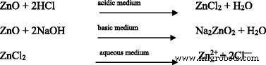
游离Zn 2+ 离子立即与蛋白质和碳水化合物等生物分子结合,细菌的所有重要功能都将停止继续。氧化锌、锌纳米颗粒和 ZnSO4·7H2O 对费氏弧菌的毒性已经过测试(表 1) .结果表明,ZnSO4·7H2O 的毒性是氧化锌纳米颗粒和氧化锌的六倍。纳米粒子实际上是分散在溶剂中,而不是溶解,因此,它们不能释放 Zn 2+ 离子。 Zn 2+ 的生物利用度 离子并不总是 100% 并且可能总是随着生理 pH 值、氧化还原电位和与之相关的阴离子(例如 Cl - )而变化 或 SO4 2− .
氧化锌在水性介质中的溶解度(1.6-5.0 毫克/升)高于氧化锌纳米颗粒(0.3-3.6 毫克/升)在相同介质中的溶解度 [57],这对藻类和甲壳类动物有毒。纳米氧化锌和块状氧化锌对 V 的毒性都比 ZnSO4 低 40-80 倍。费舍里 . ZnSO4 较高的抗菌活性与其溶解度释放 Zn 2+ 成正比 离子,由于 Zn 2+ 带正电荷,它对细菌细胞中的生物分子具有更高的迁移率和更强的亲和力 [58] 和生物分子上的负电荷。
<图片>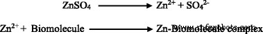
由于氧化锌及其纳米颗粒的溶解度有限,因此它们对微生物的毒性比高溶解度的 ZnSO4·7H2O 小。然而,金属氧化物纳米粒子进入细菌细胞引起毒性并不是必需的[59]。纳米颗粒与细胞壁之间的接触足以引起毒性。如果是的话,那么就需要大量的金属纳米粒子,使细菌细胞完全被包围并与环境隔绝,从而没有机会吸收营养以继续生命过程。由于纳米颗粒和金属离子比细菌细胞小,它们更有可能破坏细胞膜并抑制其生长。
许多纳米尺寸的金属氧化物,例如 ZnO、CuO、Al2O3、La2O3、Fe2O3、SnO2 和 TiO2,已被证明对 E 表现出最高的毒性。大肠杆菌 [49]。氧化锌纳米颗粒外用于治疗轻度细菌感染,但锌离子是某些病毒和人类必需的微量元素,可增加病毒整合酶的酶活性 [45, 60, 61]。当用 10 mg/L Zn 处理时,传染性胰腺坏死病毒增加 69.6% 也支持这一点 [46]。这可能是由于 Zn 离子相对于单独的 ZnO 具有更大的溶解度。 SEM 和 TEM 图像表明,氧化锌纳米颗粒会破坏细菌细胞壁 [55, 62] 并增加渗透性,然后它们在 E 中积累。大肠杆菌 防止它们的乘法 [63]。
最近,已经研究了氧化锌纳米颗粒对四种已知的革兰氏阳性和革兰氏阴性细菌的抗菌活性,即金黄色葡萄球菌 ,E。大肠杆菌 , 鼠伤寒沙门氏菌 和肺炎克雷伯菌 .观察到氧化锌纳米颗粒的生长抑制剂量为 15 μg/ml,尽管在 K 的情况下。肺炎 ,它低至 5 μg/ml [63, 64]。已经注意到随着纳米颗粒浓度的增加,微生物的生长抑制增加。当它们与最大浓度为 45 μg/ml 的氧化锌纳米颗粒一起孵育 4-5 小时时,生长受到强烈抑制。预计如果孵育时间增加,生长抑制也会增加,但作用机制没有太大改变[63]。
据报道,金属氧化物纳米颗粒首先破坏细菌细胞膜,然后渗透到其中 [64]。也有人提出释放 H2O2 可能是抗菌活性的替代方法 [65]。然而,该提议需要实验证明,因为仅存在氧化锌纳米颗粒不足以产生 H2O2。极低浓度的锌纳米颗粒或氧化锌纳米颗粒不会对人体系统产生毒性。每天需要通过食物摄入锌来执行正常的代谢功能。众所周知,氧化锌可以保护胃和肠道免受大肠杆菌的损害。大肠杆菌 [65]。胃中的pH值在2到5之间变化,因此胃中的氧化锌可以与酸反应生成Zn 2+ 离子。它们可以帮助激活有助于消化碳水化合物和酒精的羧肽酶、碳酸酐酶和乙醇脱氢酶。普雷马纳坦等人。 [66] 报道了氧化锌纳米颗粒对原核和真核细胞的毒性。氧化锌纳米颗粒对E的MIC。大肠杆菌 , 铜绿假单胞菌 , 和 S。金黄色葡萄球菌 分别为 500 和 125 μg/ml。对于氧化锌纳米颗粒的毒性,已经提出了两种作用机制,即(1)产生 ROS 和(2)诱导细胞凋亡。金属氧化物纳米颗粒诱导 ROS 产生并使细胞处于氧化应激下,从而对细胞成分(即脂质、蛋白质和 DNA)造成损害 [67,68,69]。因此,氧化锌纳米颗粒通过细胞凋亡诱导毒性。与正常细胞相比,它们对癌细胞的毒性相对更大,但无法区分它们。
最近,帕蒂等人。 [70] 已经表明氧化锌纳米颗粒破坏细菌细胞膜的完整性,降低细胞表面疏水性,并下调细菌中氧化应激抗性基因的转录。它们通过诱导 ROS 产生来增强细胞内细菌的杀灭。这些纳米颗粒破坏生物膜的形成并抑制病原体产生的溶血素毒素的溶血作用。发现氧化锌纳米颗粒的皮内给药可显着减少小鼠的皮肤感染和炎症,并改善受感染的皮肤结构。
氧化锌纳米颗粒的溶解度和浓度相关活性
纳米颗粒也已被用作载体来递送治疗剂以治疗细菌感染 [1, 9]。由于浓度高达 100 μg/ml 的氧化锌纳米颗粒对正常身体细胞无害,因此它们可用作抗生素的替代品。发现 90% 的细菌菌落在暴露于 500-1000 微克/毫升的氧化锌纳米颗粒仅 6 小时后就会死亡。即使是抗药性的S。金黄色葡萄球菌 , 耻垢分枝杆菌 , 和牛分枝杆菌 当用氧化锌纳米颗粒与低剂量的抗结核药物利福平 (0.7 μg/ml) 联合处理时,观察到它们的生长显着减少。当与 1000 μg/ml 的氧化锌纳米颗粒一起孵育 24 小时时,这些病原体被完全破坏。因此得出结论,如果重复相同的剂量,患有此类传染病的患者可能会完全治愈。还注意到,50-500 nm 范围内的氧化锌纳米颗粒对细菌生长抑制具有相同的效果。
许多研究人员已经在各种微生物和植物系统中研究了氧化锌的细胞毒性 [71,72,73,74]。氧化锌纳米颗粒的毒性取决于浓度和溶解度。已经表明,最大暴露浓度的氧化锌 (125 毫克/升) 悬浮液释放了 6.8 毫克/升的 Zn 2+ 离子。毒性是氧化锌纳米颗粒和 Zn 2+ 的综合作用 水介质中释放的离子。然而,检测到金属离子的影响很小,这表明细菌生长抑制主要是由于氧化锌纳米颗粒与微生物的相互作用。特定金属氧化物纳米颗粒的细胞毒作用具有物种敏感性,这体现在对几种细菌的生长抑制区[75]。
已表明细菌细胞的生长抑制主要发生在Zn 2+ 氧化锌纳米颗粒的细胞外溶解产生的离子 [76]。 Cho 等人。 [77] 从他们对大鼠的研究中得出结论,氧化锌纳米颗粒在中性或生物 pH 值附近保持完整,但在酸性条件 (pH 4.5) 下迅速溶解在微生物的溶酶体中,导致它们死亡。这是真的,因为在酸性条件下,氧化锌溶解,Zn 2+ 产生的离子与细菌细胞内的生物分子结合,抑制其生长。
<图片>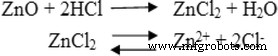
氧化锌纳米颗粒已被证明对不同的初级免疫活性细胞具有细胞毒性。转录组学分析表明,纳米颗粒具有共同的基因特征,金属硫蛋白基因的上调归因于纳米颗粒的溶解[78]。但无法确定吸收的锌是否为Zn 2+ 或氧化锌或两者兼而有之,尽管较小尺寸的氧化锌纳米粒子在血液中的浓度高于较大的纳米粒子(19 和> 100 纳米)。氧化锌纳米粒子的效率主要取决于形成Zn 2+ 的反应介质 以及它们对细胞的渗透。
蒋等人。 [79] 报道氧化锌纳米颗粒的解离导致细胞锌稳态的破坏。纳米粒子的特性及其对生物功能的影响与大块材料完全不同[80]。纳米颗粒的聚集影响巨噬细胞的细胞毒性,它们的浓度有助于调节纳米颗粒的聚集。低浓度的氧化锌纳米粒子无效,但在较高浓度(100 μg/ml)时,它们表现出不同病原体的细胞毒性。
氧化锌纳米颗粒的无意使用有时可能会对生命系统产生不利影响。已经研究了它们在人肝细胞中的细胞凋亡和遗传毒性潜力以及细胞毒性。发现当肝细胞暴露于 14-20 μg/ml 氧化锌纳米粒子 12 小时时,它们的活力会下降。它还通过氧化应激诱导 DNA 损伤。萨瓦伊等人。 [56] 已经证明 ROS 的产生与氧化锌粉末的浓度成正比。 ROS 引发线粒体膜电位降低,导致细胞凋亡 [81]。纳米颗粒的细胞摄取不是细胞毒性发生的必要条件。
氧化锌纳米颗粒的大小依赖性抗菌活性
在一项研究中,Azam 等人。 [82] 报道了对革兰氏阴性菌(大肠杆菌 和P。绿脓 ) 和革兰氏阳性 (S. 和枯草芽孢杆菌 ) 由于氧化锌纳米颗粒的粒径减小,细菌随着表面积与体积比的增加而增加。此外,在这项研究中,氧化锌纳米颗粒对 B 显示出最大(25 毫米)的细菌生长抑制作用。枯草 (图 1)。
<图片>
氧化锌纳米颗粒对革兰氏阳性和革兰氏阴性菌株的抗菌活性和/或抑菌圈,即a 大肠杆菌 , b 金黄色葡萄球菌 , c 铜绿假单胞菌 , 和 d 枯草芽孢杆菌 [82]
据报道,较小尺寸的氧化锌纳米颗粒比微米颗粒表现出更大的抗菌活性 [83]。例如,Au 55 已证明 1.4 nm 大小的纳米粒子与 DNA 的主要凹槽相互作用,这解释了其毒性 [84]。尽管报道了相互矛盾的结果,但许多工作人员表明氧化锌纳米颗粒对细菌细胞具有积极作用。然而,布雷纳等人。 [63] TEM 图像显示,10-14 nm 的氧化锌纳米颗粒被内化(当暴露于微生物时)并损坏细菌细胞膜。同样重要的是,锌/氧化锌纳米颗粒必须对人体无毒,因为它们对 5 mM 以上的 T 细胞有毒性 [85],对神经母细胞瘤细胞有 1.2 mM 以上的毒性 [86]。奈尔等人。 [87] 专门研究了氧化锌纳米颗粒对细菌和人类细胞毒性的大小影响。他们研究了氧化锌纳米颗粒对革兰氏阳性菌和革兰氏阴性菌和成骨细胞癌细胞系(MG-63)的影响。
众所周知,氧化锌纳米颗粒的抗菌活性与其大小成反比,与其浓度成正比[88]。人们还注意到,它不需要紫外线来激活;它在正常甚至漫射的阳光下起作用。细胞毒活性可能涉及 ROS 的产生和纳米颗粒在细胞质或细胞外膜上的积累。然而,H2O2 的产生及其在纳米粒子活化中的作用不容忽视。拉古帕蒂等人。 [88] 用不同的锌盐合成了氧化锌纳米颗粒,并观察到从 Zn(NO3)2 获得的纳米颗粒尺寸最小 (12 nm),表面积最大 (90.4)。作者已经表明 S. 的生长抑制。金黄色葡萄球菌 浓度为 6 mM 的氧化锌纳米颗粒取决于尺寸。细菌细胞暴露于氧化锌纳米颗粒期间的活细胞测定也表明,随着氧化锌纳米颗粒尺寸的减小,恢复的细胞数量显着减少。琼斯等人。 [89] 已经表明,8 纳米直径的氧化锌纳米颗粒抑制了硫的生长。金黄色葡萄球菌 ,E。大肠杆菌 , 和 B.枯草芽孢杆菌。 选择范围在 12 到 307 nm 之间的氧化锌纳米粒子并确认了抗菌活性与其尺寸之间的关系。它们对微生物的毒性归因于形成 Zn 2+ 当氧化锌悬浮在水中时,会从氧化锌中分离出离子,并且在一定程度上也会导致 pH 值发生轻微变化。由于 Zn 2+ 氧化锌纳米粒子几乎不释放离子,抗菌活性主要是由于氧化锌纳米粒子较小。当尺寸为 12 nm 时,它会抑制 S 的生长。金黄色葡萄球菌 ,但当尺寸超过 100 nm 时,抑制作用最小[89]。
氧化锌纳米颗粒的形状、组成和细胞毒性
由于不同的敏感性,氧化锌纳米颗粒以浓度依赖性方式和暴露的细胞类型显示出细胞毒性 [90, 91]。萨胡等人。 [90] 强调了颗粒大小和细胞对相同成分的颗粒的不同敏感性之间的细胞毒性差异。在最近的另一项研究中,Ng 等人。 [91] 检查了人肺 MRC5 细胞的浓度依赖性细胞毒性。作者通过 TEM 研究报告了氧化锌纳米颗粒被吸收和内化到人肺 MRC5 细胞中。这些颗粒以电子致密簇的形式出现在细胞的细胞质中,进一步观察到这些颗粒被囊泡包围,而在未处理的对照细胞中未发现氧化锌纳米颗粒。帕帕夫拉索普洛斯等人。 [92] 已经通过一种称为“火焰传输合成方法”的全新途径合成了氧化锌纳米颗粒四足体。与常规合成的氧化锌纳米粒子相比,四足动物具有不同的形态。它们在体外与哺乳动物成纤维细胞的相互作用表明,它们的毒性明显低于球形氧化锌纳米颗粒。四足动物呈现六方纤锌矿晶体结构,交替Zn 2+ 和 O 2− 具有三维几何形状的离子。它们阻止病毒进入活细胞,通过用紫外线辐射精确照射它们进一步增强。由于氧化锌四足动物的结构中有氧空位,单纯疱疹 病毒通过硫酸乙酰肝素附着并被拒绝进入体细胞。因此,它们在体外预防 HSV-1 和 HSV-2 感染。因此,氧化锌四足体可用作针对这些病毒感染的预防剂。氧化锌纳米颗粒的细胞毒性还取决于哺乳动物细胞的增殖速度 [66, 93]。通过控制氧化锌四足体中的氧空位也可以改变表面反应性和毒性。当它们暴露在紫外线下时,四足动物的氧空位很容易增加。或者,可以通过在富氧环境中加热来减少氧空位。因此,氧化锌四足体的独特性质可以随意改变,从而改变其抗菌效率。
动物研究表明,呼吸道接触纳米颗粒会增加肺部炎症、氧化应激等 [94]。杨等人。 [95] 研究了氧化锌纳米颗粒对原代小鼠胚胎成纤维细胞的细胞毒性、基因毒性和氧化应激。据观察,氧化锌纳米颗粒诱导的细胞毒性明显高于碳和 SiO2 纳米颗粒诱导的细胞毒性。通过测量谷胱甘肽消耗、丙二醛产生、超氧化物歧化酶抑制和 ROS 产生进一步证实了这一点。不同纳米颗粒的潜在细胞毒性作用归因于它们的形状。
聚合物涂层纳米粒子
许多细菌感染是通过接触门把手、键盘、水龙头、浴缸和电话而传播的;因此,必须用廉价的高级抗菌物质开发和涂覆此类表面,以抑制它们的生长。重要的是使用这样的抗菌物质浓度,它们可能会杀死病原体但不会伤害人类。只有当它们涂有低成本的生物相容性亲水聚合物时,才会发生这种情况。施瓦茨等人。 [96] 报道了通过混合生物相容性聚 (N -异丙基丙烯酰胺)与氧化锌纳米颗粒。复合膜的 SEM 图像显示氧化锌纳米颗粒的均匀分布。它对大肠杆菌表现出抗菌活性。大肠杆菌 在非常低的氧化锌浓度 (1.33 mM) 下。此外,发现涂层在 1 周内对哺乳动物细胞系 (N1H/3T3) 无毒。氧化锌/水凝胶纳米复合材料可安全地用作生物医学涂层,以防止人们感染细菌。
虽然氧化锌纳米粒子很稳定,但通过用不同的聚合物如聚乙烯吡咯烷酮 (PVP)、聚乙烯醇 ( 聚乙烯醇), 聚 (α, γ, l-谷氨酸) (PGA)、聚乙二醇 (PEG)、壳聚糖和葡聚糖 [97, 98]。检查了工程氧化锌纳米粒子对革兰氏阴性和革兰氏阳性病原体(即大肠杆菌)的抗菌活性。大肠杆菌 和S。金黄色葡萄球菌 并与市售氧化锌粉相比。与散装氧化锌粉末相比,聚合物涂层的球形氧化锌纳米颗粒显示出最大的细菌细胞破坏[99]。由于用聚合物包覆的纳米粒子由于溶解度低和缓释而毒性较小,因此可以通过用合适的聚合物包覆它们来控制其细胞毒性。
聚合物包覆纳米颗粒的粒径和形状对抗菌活性的影响
E。大肠杆菌 和S。金黄色葡萄球菌 exposed to different concentrations of poly ethylene glycol (PEG)-coated zinc oxide nanoparticles (1–7 mM) of varying size (401 nm–1.2 μm) showed that the antimicrobial activity increases with decreasing size and increasing concentration of nanoparticles. However, the effective concentration in all these cases was above 5 mM. There occurs a drastic change in cell morphology of E.大肠杆菌 surface which can be seen from the SEM images of bacteria before and after their exposure to zinc oxide nanoparticles [84]. It has been nicely demonstrated by Nair et al. [87] that PEG-capped zinc oxide particles and zinc oxide nanorods are toxic to human osteoblast cancer cells (MG-63) at concentration above 100 μM. The PEG starch-coated nanorods/nanoparticles do not damage the healthy cells.
In Vivo and In Vitro Antimicrobial Activity for Wound Dressing
Of all natural and synthetic wound dressing materials, the chitosan hydrogel microporous bandages laced with zinc oxide nanoparticles developed by Kumar et al. [100] are highly effective in treating burns, wounds, and diabetic foot ulcers. The nanoparticles of approximately 70–120 nm are dispersed on the surface of the bandage. The degradation products of chitosan were identified as d-glucosamine and glycosamine glycan. They are nontoxic to the cells because they are already present in our body for the healing of injury. The wound generally contains P.绿脓 , S. intermedicus , and S. hyicus which were also identified from the swab of mice wound and successfully treated with chitosan zinc oxide bandage in about 3 weeks [100].
Effect of Doping on Toxicity of Zinc Oxide Nanoparticles
Doping of zinc oxide nanoparticles with iron reduces the toxicity. The concentration of Zn 2+ and zinc oxide nanoparticles is also an important factor for toxicity. The concentration that reduced 50% viability in microbial cells exposed to nano- and microsize zinc oxide is very close to the concentration of Zn 2+ that induced 50% reduction in viability in Zn 2+ -treated cells [101, 102].
Coating of zinc oxide nanoparticles with mercaptopropyl trimethoxysilane or SiO2 reduces their cytotoxicity [103]. On the contrary, Gilbert et al. [104] showed that in BEAS-2B cells, uptake of zinc oxide nanoparticles is the main mechanism of zinc accumulation. Also, they have suggested that zinc oxide nanoparticles dissolve completely generating Zn 2+ ions which are bonded to biomolecules of the target cells. However, the toxicity of zinc oxide nanoparticles depends on the uptake and their subsequent interaction with target cells.
Interaction Mechanism of Zinc Oxide Nanoparticles
Nanoparticles may be toxic to some microorganisms, but they may be essential nutrients to some of them [55, 105]. Nanotoxicity is essentially related to the microbial cell membrane damage leading to the entry of nanoparticles into the cytoplasm and their accumulation [55]. The impact of nanoparticles on the growth of bacteria and viruses largely depends on particle size, shape, concentration, agglomeration, colloidal formulation, and pH of the media [106,107,108]. The mechanism of antimicrobial activity of zinc oxide nanoparticles has been depicted in Fig. 2.

Mechanisms of zinc oxide nanoparticle antimicrobial activity
Zinc oxide nanoparticles are generally less toxic than silver nanoparticles in a broad range of concentrations (20 to 100 mg/l) with average particle size of 480 nm [55, 62, 63]. Metal oxide nanoparticles damage the cell membrane and DNA [63, 109,110,111] of microbes via diffusion. However, the production of ROS through photocatalysis causing bacterial cell death cannot be ignored [112]. UV-Vis spectrum of zinc oxide nanoparticle suspension in aqueous medium exhibits peaks between 370 and 385 nm [113]. It has been shown that it produces ROS (hydroxyl radicals, superoxides, and hydrogen peroxide) in the presence of moisture which ostensibly react with bacterial cell material such as protein, lipids, and DNA, eventually causing apoptosis. Xie et al. [114] have examined the influence of zinc oxide nanoparticles on Campylobacter jejuni cell morphology using SEM images (Fig. 3). After a 12-h treatment (0.5 mg/ml), C. jejuni was found to be extremely sensitive and cells transformed from spiral shape to coccoid forms. SEM studies showed the ascendency of coccoid forms in the treated cells and display the formation of irregular cell surfaces and cell wall blebs (Fig. 3a). Moreover, these coccoid cells remained intact and possessed sheathed polar flagella. However, SEM image of the untreated cells clearly showed spiral shapes (Fig. 3b). In general, it has been demonstrated from SEM and TEM images of bacterial cells treated with zinc oxide nanoparticles that they get ruptured and, in many cases, the nanoparticles damage the cell wall forcing their entry into it [114, 115].

SEM images of Campylobacter jejuni . 一 Untreated cells from the same growth conditions were used as a control. b C. jejuni cells in the mid-log phase of growth were treated with 0.5 mg/ml of zinc oxide nanoparticles for 12 h under microaerobic conditions [114]
Zinc oxide nanoparticles have high impact on the cell surface and may be activated when exposed to UV-Vis light to generate ROS (H2O2) which permeate into the cell body while the negatively charged ROS species such as O2 2− remain on the cell surface and affect their integrity [116, 117]. Anti-bacterial activity of zinc oxide nanoparticles against many other bacteria has also been reported [1, 5, 114, 115]. It has been shown from TEM images that the nanoparticles have high impact on the cell surface (Fig. 4).

一 TEM images of untreated normal Salmonella typhimurium 细胞。 b Effects of nanoparticles on the cells (marked with arrows). c , d Micrograph of deteriorated and ruptured S. typhimurium cells treated with zinc oxide nanoparticles [115]
Sinha et al. [118] have also shown the influence of zinc oxide nanoparticles and silver nanoparticles on the growth, membrane structure, and their accumulation in cytoplasm of (a) mesophiles:Enterobacter sp. (gram negative) and B. subtilis (gram positive) and (b) halophiles:halophilic bacterium sp. (gram positive) and Marinobacter sp. (gram negative). Nanotoxicity of zinc oxide nanoparticles against halophilic gram-negative Marinobacter species and gram-positive halophilic bacterial species showed 80% growth inhibition. It was demonstrated that zinc oxide nanoparticles below 5 mM concentration are ineffective against bacteria. The bulk zinc oxide also did not affect the growth rate and viable counts, although they showed substantial decrease in these parameters. Enterobacter species showed dramatic alterations in cell morphology and reduction in size when treated with zinc oxide.
TEM images shown by Akbar and Anal [115] revealed the disrupted cell membrane and accumulation of zinc oxide nanoparticles in the cytoplasm (Fig. 4) which was further confirmed by FTIR, XRD, and SEM. It has been suggested that Zn 2+ ions are attached to the biomolecules in the bacterial cell via electrostatic forces. They are actually coordinated with the protein molecules through the lone pair of electrons on the nitrogen atom of protein part. Although there is significant impact of zinc oxide nanoparticles on both the aquatic and terrestrial microorganisms and human system, it is yet to be established whether it is due to nanoparticles alone or is a combined effect of the zinc oxide nanoparticles and Zn 2+ ions [55, 106, 109, 119]. Antibacterial influence of metal oxide nanoparticles includes its diffusion into the bacterial cell, followed by release of metal ions and DNA damage leading to cell death [63, 109,110,111]. The generation of ROS through photocatalysis is also a reason of antibacterial activity [62, 112]. Wahab et al. [120] have shown that when zinc oxide nanoparticles are ingested, their surface area is increased followed by increased absorption and interaction with both the pathogens and the enzymes. Zinc oxide nanoparticles can therefore be used in preventing the biological system from infections. It is clear from TEM images (Fig. 5a, b) of E.大肠杆菌 incubated for 18 h with MIC of zinc oxide nanoparticles that they had adhered to the bacterial cell wall. The outer cell membrane was ruptured leading to cell lysis. In some cases, the cell cleavage of the microbes has not been noticed, but the zinc oxide nanoparticles can yet be seen entering the inner cell wall (Fig. 5c, d). As a consequence of it, the intracellular material leaks out leading to cell death, regardless of the thickness of bacterial cell wall.

TEM images of Escherichia coli (a ), zinc oxide nanoparticles with E.大肠杆菌 at different stages (b and inset), Klebsiella pneumoniae (c ), and zinc oxide nanoparticles with K. pneumoniae (d and inset) [120]
Mechanism of interaction of zinc oxide nanoparticles with bacterial cells has been outlined below [120]. Zinc oxide absorbs UV-Vis light from the sun and splits the elements of water.

Dissolved oxygen molecules are transformed into superoxide, O2 − , which in turn reacts with H + to generate HO2 radical and after collision with electrons produces hydrogen peroxide anion, HO2 − . They subsequently react with H + ions to produce H2O2.

It has been suggested that negatively charged hydroxyl radicals and superoxide ions cannot penetrate into the cell membrane. The free radicals are so reactive that they cannot stay in free and, therefore, they can either form a molecule or react with a counter ion to give another molecule. However, it is true that zinc oxide can absorb sun light and help in cleaving water molecules which may combine in many ways to give oxygen. Mechanism of oxygen production in the presence of zinc oxide nanoparticles still needs experimental evidence.

Zinc oxide at a dose of 5 μg/ml has been found to be highly effective for all the microorganisms which can be taken as minimum inhibitory dose.
结论
Zinc is an indispensable inorganic element universally used in medicine, biology, and industry. Its daily intake in an adult is 8–15 mg/day, of which approximately 5–6 mg/day is lost through urine and sweat. Also, it is an essential constituent of bones, teeth, enzymes, and many functional proteins. Zinc metal is an essential trace element for man, animal, plant, and bacterial growth while zinc oxide nanoparticles are toxic to many fungi, viruses, and bacteria. People with inherent genetic deficiency of soluble zinc-binding protein suffer from acrodermatitis enteropathica, a genetic disease indicated by python like rough and scaly skin. Although conflicting reports have been received about nanoparticles due to their inadvertent use and disposal, some metal oxide nanoparticles are useful to men, animals, and plants. The essential nutrients become harmful when they are taken in excess. Mutagenic potential of zinc oxide has not been thoroughly studied in bacteria even though DNA-damaging potential has been reported. It is true that zinc oxide nanoparticles are activated by absorption of UV light without disturbing the other rays. If zinc oxide nanoparticles produce ROS, they can damage the skin and cannot be used as sun screen. Antibacterial activity may be catalyzed by sunlight, but hopefully, it can prevent the formation of ROS. Zinc oxide nanoparticles and zinc nanoparticles coated with soluble polymeric material may be used for treating wounds, ulcers, and many microbial infections besides being used as drug carrier in cancer therapy. It has great potential as a safe antibacterial drug which may replace antibiotics in future. Application of zinc oxide nanoparticles in different areas of science, medicine, and technology suggests that it is an indispensable substance which is equally important to man and animals. However, longtime exposure with higher concentration may be harmful to living system.
纳米材料