

绿色且经济的氧化锡纳米颗粒合成:合成方法、形成机制及其潜在应用综述
摘要
纳米技术已成为最有前途的研究领域,其在所有科学领域的重要应用。近年来,氧化锡因其迷人的特性而受到极大关注,随着这种材料在纳米范围内的合成得到了改进。如今,人们使用多种物理和化学方法来生产氧化锡纳米颗粒。然而,这些方法昂贵,需要高能量,并且在合成过程中还使用各种有毒化学品。对人类健康和环境影响的日益关注导致开发了一种具有成本效益和环境友好的生产工艺。最近,利用植物提取物、细菌和天然生物分子等不同生物实体,通过绿色方法成功合成了氧化锡纳米颗粒。然而,由于生物底物的复杂性,难以阐明合成过程中发生的反应和形成机制,因此使用绿色合成方法进行工业规模生产仍然是一个挑战。因此,本综述总结了用于绿色合成氧化锡纳米粒子的生物实体和方法的不同来源及其对它们性质的影响。这项工作还描述了对文献中报道的形成机制的理解以及用于表征这些纳米颗粒的不同分析技术的进展。
介绍
在过去的几十年里,纳米技术已经成为一个新的研究领域,涉及纳米材料的合成、表征、改性和利用,因为它们在制药、食品工业、化妆品、纺织工业、医学、光学、电子、能源科学和电化学应用 [1,2,3,4]。纳米材料是尺寸范围为 1-100 纳米的一维材料。与散装材料相比,这些材料的非常大的表面积与体积比和极小的尺寸可能会导致全新或增强的电、光、磁、催化和抗菌活性 [5,6,7]。由于这些独特的特性,纳米粒子在现代科学和工程的各个领域都有应用,例如纳米医学、光催化、生物传感器、清洁剂和纺织工业 [1, 8]。在纳米粒子中,尤其是氧化锡 (SnO2) 因其用途广泛而备受关注,例如光电器件 [9]、固态气体传感器 [10]、锂离子电池电极 [11]、场发射显示器[12]、发光二极管[13]、催化[14]、染料太阳能电池[15]、医药[16]、光传感器和抗静电涂层[17]。
在材料科学中,SnO2 被认为是一种缺氧的 n 型半导体,其结晶为具有晶格常数 a 的四方金红石结构 =b =4.7374 Å 和 c =3.1864 埃。晶胞由两个六重配位的锡和四个三重配位的氧原子组成 [18, 19]。宽能隙(3.6 至 3.8 eV)、强热稳定性(高达 500°C)、可见光谱的高度透明度、与吸附物质的强烈化学和物理相互作用使 SnO2 成为潜在应用的有希望的候选者在锂离子电池、传感器、催化、储能、玻璃涂层、医药和环境修复中[20,21,22,23]。 SnO2 因其高比表面积、高化学稳定性、低电阻和低密度而被用作传感器以提高响应时间和灵敏度 [24]。从过去几年开始,SnO2 在太阳能电池 [25] 和气体传感器中的应用得到了深入探索,以检测可燃气体,如 CO、NO、NO2、H2S 和 C2H5OH [26,27,28,29]。由于纳米颗粒 (NPs) 独特的物理化学性质和潜在应用,科学界一直在开发多种生产纳米颗粒的方法。然而,用于合成金属和金属氧化物纳米粒子的化学和物理方法非常昂贵,并且使用对环境和人类健康有害的有毒物质 [30]。近年来,大多数研究人员将他们的研究兴趣转向纳米颗粒的绿色合成,因为它具有许多优点,例如成本效益高、制造程序简单、生产可重复,并且通常会产生更稳定的纳米颗粒 [31]。在过去的十年中,报道了几项关于 SnO2 NPs 绿色合成的研究。然而,文献中没有一篇综述文章可以证明合成方法和形成机制。因此,本文描述了SnO2 NPs的绿色合成、形成机制、表征技术和潜在应用。
氧化锡纳米粒子的绿色合成
SnO2 NPs 是通过各种物理、化学和绿色方法合成的。化学方法包括溶胶-凝胶法、水热法、沉淀法、机械化学法、微乳液法等[31,32,33,34,35,36,37]。在化学方法中,最广泛使用的技术是溶胶-凝胶合成,它利用锡前体盐和调节含锡凝胶形成的化学试剂。之后,凝胶在高达 800°C 的温度下进行热处理以获得 SnO2 NPs [32, 38]。化学稳定剂和封端剂,如草酸或乙二醇,可以在 SnO2 NPs 的合成过程中添加,以控制纳米颗粒的尺寸和防止团聚 [32, 39]。溶液 pH 值、化学品浓度、反应时间和煅烧温度也会影响纳米颗粒的尺寸和形态 [31, 34,35,36,37]。上述合成SnO2 NPs的方法使用了多种危险的化学试剂、溶剂和表面活性剂,对环境和人类健康造成严重威胁[4, 30]。
SnO2 NPs 也可以通过物理技术合成,如喷雾热解、热氧化、化学气相沉积、激光烧蚀和超声处理 [40,41,42,43,44]。在这些方法中,激光烧蚀被认为是一种在液体中合成金属和金属氧化物纳米粒子的经济高效且简单的方法 [44, 45]。与其他传统方法相比,这种方法不需要封端/还原剂、高温或高压,并且允许我们生产高纯度的纳米颗粒 [44, 45]。激光束施加的脉冲参数和烧蚀时间的变化是定义纳米颗粒的粒径、形态和表面化学性质的重要参数 [44]。然而,大多数物理方法需要复杂的设备、高能量和熟练的人力[46]。因此,开发环保、廉价、高效且在环境条件下工作的合成方法非常重要。一种这样的解决方案是绿色合成,许多研究人员已经开发出一种用于合成氧化锡纳米颗粒的绿色化学方法。在绿色合成策略中,植物提取物、微生物或其他绿色来源等生物实体可以替代传统的物理和化学方法[47]。如今,受生物学启发的合成方法也被称为绿色合成,因为它们符合绿色化学的十二项原则 [48]。生物合成相对于物理和化学方法的一些明显优势是 (a) 清洁和环境友好的方法,因为使用无毒化学品,(b) 使用可再生资源,(c) 活性生物成分如酶本身作为以及植物化学物质作为还原剂和封端剂,从而最大限度地降低合成过程的总体成本,(d) 不需要高压和高温等外部实验条件,从而显着节约能源 [49, 50]。
在过去的十年中,通过生物方法合成 SnO2 NPs 的兴趣大大增加,因为该方法更可靠、更环保、更具成本效益、低投入高产,且程序简单,不会对生物产生任何不利影响。环境。植物提取物、细菌和天然生物分子等多种生物底物已成功用于 SnO2 NPs 的绿色合成。各种植物的植物化学物质和来自细菌的酶是绿色合成的主要负责人。绿色源中存在的活性化合物在合成过程中还起到还原剂、封端剂和稳定剂的作用。通常在指定温度下煅烧或退火后获得所需的纳米颗粒[51,52,53,54]。
植物介导的氧化锡纳米粒子合成
植物介导的合成已成为优于传统物理化学方法的最佳合成平台,因为它不含有毒化学物质,并提供天然的加帽剂和还原剂。此外,它简单且环保,并提供不含杂质的数量丰富的产品。在这种方法中,不需要使用高温、高压和昂贵的设备。此外,植物介导的合成导致大规模生产具有不同形状和尺寸的更稳定的纳米粒子 [55, 56]。大量不同植物物种的提取物已被用于 SnO2 NPs 的绿色合成。一般来说,植物介导的 SnO2 NPs 合成是一个非常简单的过程,其中将薄盐添加到先前制备的提取物中。反应后,将溶液离心。最后,然后对颗粒进行热处理,然后使用各种分析技术进行表征,例如傅里叶变换红外光谱 (FTIR)、X 射线衍射仪 (XRD)、能量色散 X 射线分析 (EDS)、扫描电子显微镜 ( SEM)、透射电子显微镜 (TEM)、粒度分析仪 (PSA) 和动态光散射 (DLS)。紫外-可见光谱 (UV-Vis) 用于监测纳米颗粒的形成。植物介导的 SnO2 NPs 合成的详细方案如图 1 所示。Diallo 等人。报道了使用 Aspalathus linearis 绿色合成 SnO2 NPs 和氯化锡五水合物 (SnCl4·5H2O) 作为前体 [51]。将盐溶解在植物提取物中,10 分钟后观察到白色沉积物的形成。离心过程后收集白色沉积物并在约 80°C 下干燥。粉末在各种温度下退火约 4 小时,并进行各种分析技术,如高分辨率透射电子显微镜 (HR-TEM)、EDS、XRD 和 X 射线光电子能谱 (XPS)。 NPs 呈准球形,平均尺寸在 2.5-11.40 nm 范围内。观察到随着退火温度的升高,纳米颗粒的粒径和结晶性质增加。此外,植物提取物中存在的生物活性成分如 aspalathin、nothofagin 和 aspalalinin 作为螯合剂和还原剂。 山茶花 叶提取物也用于合成 SnO2 NPs [53]。叶提取物中存在的多酚既可作为稳定剂,也可作为封端剂。高分辨率扫描电子显微镜 (HR-SEM) 和 XRD 分析显示球形 SnO2 NPs 大小在 5-30 nm 范围内。此外,发现纳米颗粒的带隙随着退火温度的升高而减小。 Catunaregam spinosa 介导的绿色合成 SnO2 NPs 对刚果红染料显示出优异的光催化活性 [57]。生物合成的纳米颗粒呈球形,平均粒径为47 nm。
<图片>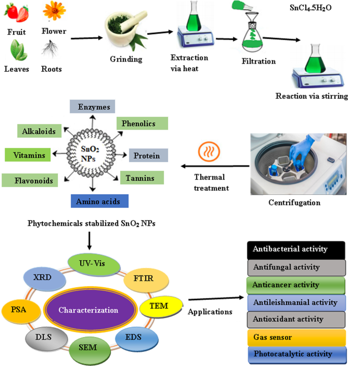
植物提取物介导合成SnO2纳米颗粒的示意图
库拉索芦荟的叶提取物 已被用于从 SnCl2·2H2O 作为前体合成 SnO2 NPs [58]。发现所得 NP 是球形的,平均尺寸从 50 到 100 纳米不等。此外,合成的纳米粒子对硫表现出优异的抗菌活性。金黄色葡萄球菌 和E。大肠杆菌。
在另一项研究中,使用 Plectranthus amboinicus 通过一种高效且廉价的方法完成了 SnO2 NPs 的绿色合成 叶提取物和 SnCl2·2H2O 作为起始材料 [59]。植物提取物充当还原剂和稳定剂。所得纳米颗粒通过 SEM、EDX、XRD 和 PSA 进行表征。 NPs 呈四边形,平均粒径为 63 nm。此外,作者透露,与商业 SnO2 相比,生物合成的 NPs 对罗丹明 B 表现出更大的光催化活性。使用 Nyctanthes arbor-tristis 报道了 SnO2 NPs 的绿色合成 (Parijataka) 花提取物 [60]。从 SEM 和 PSA 分析可以明显看出,合成的纳米粒子呈现出细颗粒的形态,具有微小的团聚阶段,平均粒径为 2-8 nm。该研究还探讨了植物提取物的水解和加帽潜力。
使用Psidium guajava通过低成本和生态友好的工艺进行SnO2 NPs的绿色合成 叶提取物[61]。 UV-Vis 结果显示在 314 nm 处有一个表面等离子体共振峰,证实了 SnO2 NPs 的形成。此外,纳米颗粒是球形的,尺寸范围为 8 到 10 纳米。该研究还表明,在阳光照射下,纳米颗粒在 180 分钟内表现出 90% 的活性黄 186 降解。 Bhosale 等人。 [62] 鉴定了Calotropis gigantea的叶提取物 作为合成 SnO2 NPs 的天然来源。提取物的次级代谢物在氯化锡向 SnO2 NPs 的转化过程中起到稳定剂和封端剂的作用。作者认为 NP 是球形的,平均尺寸在 30 到 40 纳米之间。合成的 NP 在 120 分钟内将甲基橙染料降解高达 80%。辛格等人。 [63] 报道了使用 Piper betle 生物合成 SnO2 NPs 叶提取物。 SEM 和 TEM 分析显示形成了平均尺寸为 8.4 nm 的球形纳米颗粒。 NPs 以伪一级的效率降解活性黄色 186,效率为 92.17%。此外,与活性红 120 和活性绿 119 相比,NPs 在去除活性黄 186 方面表现出优异的选择性。在文献中,不同的植物和植物提取物已用于制备 SnO2(表 1)。从表中可以看出,植物种类的性质和反应条件会影响SnO2 NPs的大小和形状。
细菌介导的氧化锡纳米粒子合成
微生物是重要的纳米工厂,具有作为生态友好和经济高效的工具的巨大潜力,可避免使用有毒化学物质和物理化学合成所需的高能量。各种微生物如细菌、真菌和酵母已被用于在细胞内或细胞外合成金属和金属氧化物纳米颗粒。细胞内合成涉及将金属离子运输到微生物细胞中,并在细胞内存在酶、辅酶和其他生物分子的情况下形成纳米颗粒。在细胞外合成中,金属离子被困在微生物细胞的表面。表面可用的酶和蛋白质会还原金属离子,并负责为 NP 提供稳定性 [73]。然而,与细胞内途径相比,细胞外合成更具有优势,因为它可以用于制造大量的纳米颗粒,并且省去了回收纳米颗粒所需的各个合成步骤[74]。
与化学合成相比,利用细菌进行生物合成是一种绿色且具有成本效益的方法。然而,这种方法有几个缺点:(a) 微生物筛选是一个耗时的过程,(b) 需要仔细监测培养液和整个过程,(c) 难以控制微生物的大小和形态。 NP。由于其固有的代谢过程和酶活性,并非所有细菌都能合成纳米颗粒。因此,需要仔细选择合适的细菌来生产具有明确尺寸和形态的纳米颗粒 [74]。例如,Srivastava 和 Mukhopadhyay [54] 进行的一项研究报告了一种低成本、绿色和最简单的方法,用于使用新鲜干净的 Erwinia herbicola 合成 SnO2 NPs 细菌细胞和氯化锡 (II) 水溶液。合成的 SnO2 NPs 大多是球形,尺寸在 10 到 42 nm 范围内。据报道,细菌蛋白质和其他生物分子在 SnO2 NPs 的合成过程中充当还原剂和稳定剂。这些生物分子还有助于控制 SnO2 NPs 的大小和聚集。
氧化锡纳米粒子的生物分子和其他绿色来源介导的合成
除了植物和细菌介导的 SnO2 NPs 合成外,研究人员还利用氨基酸、维生素、酶和糖等其他生物分子开发了一种无毒、环境友好和绿色的化学方法(表 2)。杨等人。 [75] 使用一种天然可用的生物分子维生素 C(抗坏血酸),使用低成本且环保的方法合成了 SnO2 NP。 TEM 分析表明形成了平均尺寸约为 30 nm 的球形 NP。维生素 C 在合成过程中既充当封端剂又充当还原剂。该研究表明,覆盖在 SnO2 NPs 表面的维生素 C 降低了 SnO2 NPs 对细胞造成的氧化应激,从而减少了新生小鼠的体重减轻。使用碳水化合物(淀粉)制备平均粒径为 13 nm 的球形 SnO2 NP [76]。有人建议碳水化合物作为模板,它可以通过它们的官能团结合几个金属阳离子,因此观察到阳离子的均匀分散。在另一项研究中,从浸泡过的孟加拉豆 (Cicer arietnum L.) 用于合成未掺杂的 SnO2、Ni、Fe 和 Au 掺杂的 SnO2 NPs [77,78,79]。作者认为提取物中的果胶负责合成 SnO2 NP。发现球形未掺杂 SnO2、Ni 掺杂 SnO2 和 Au 掺杂 SnO2 NPs 的平均微晶尺寸分别为 11 nm、6 nm 和 25 nm。
另一项研究显示了一种使用蛋壳膜 (ESM) [80](一种来自鸡蛋壳的天然生物废物)合成 SnO2 NPs 的绿色方法。有人建议 ESM 成分生物分子如糖醛酸和含有醛部分的糖类,它们在合成过程中充当还原剂。形态分析表明,SnO2 NPs 形成棒状、六边形和球形,粒径范围为 13-40 nm。
不同的氨基酸如甘氨酸、精氨酸、天冬氨酸、赖氨酸和酪氨酸 [81,82,83,84,85] 因其良好的封端或络合剂已被用于绿色合成 SnO2 NPs。氨基酸介导的合成消除了合成过程中有毒化学品的使用。球形 SnO2 NPs 是使用精氨酸合成的 [81]。形态学研究表明合成的 SnO2 NPs 是球形的,平均尺寸在 4-5 nm 范围内。巴塔查吉等人。 [83] 使用甘氨酸从氯化亚锡生产 SnO2 NPs。有人提出,在 200°C、400°C 和 600°C 形成的 NPs 是球形、多晶和单分散的,平均尺寸分别为 6、16 和 33 nm。此外,在 400°C 下获得的纳米颗粒是发光的。同样,Begum 等人。 [84] 证明了使用L-赖氨酸合成四方金红石结构的SnO2 NPs,尺寸范围为4-17 nm。
绿色合成氧化锡纳米粒子的形成机制
近年来,不同的植物提取物、微生物和其他生物衍生物已被用于金属和金属氧化物纳米颗粒的绿色合成。研究表明,植物提取物中存在的次生代谢物,如酚类、黄酮类、单宁、皂苷、萜类化合物和碳水化合物,在金属或金属氧化物 NP 的合成中作为还原剂和稳定剂发挥着重要作用 [87]。此外,根据纳米颗粒形成的位置,微生物介导的合成可以在细胞内或细胞外实现。在细胞外合成中,在微生物细胞表面存在可利用的酶的情况下发生生物还原。此外,在细胞内生物合成中,金属离子被转运到微生物细胞中,在细胞内酶存在的情况下,纳米颗粒很容易在那里形成 [73]。塞尔瓦库马里等人。 [53] 利用Camellia sinensis 用于生物合成 SnO2 NPs 的提取物。他们的研究表明,提取物中存在的多酚化合物(表儿茶素、表没食子儿茶素、表儿茶素没食子酸酯和表没食子儿茶素没食子酸酯)既可作为封端剂,也可作为稳定剂。通过该方法形成的 SnO2 NPs 包括一些主要步骤,包括 (i) Sn 2+ 的还原 到 Sn 0 , (ii) 提取物中的酚类化合物 (-OH) 形成 Sn 物质的还原作用,以及 (iii) Sn 物质热转变为 SnO2 NPs。在使用Calotropis gigantean合成的SnO2 NPs中 在叶子提取物中,多酚类化合物负责锡离子的生化转化 [62]。使用花椰菜提取物合成了尺寸在 3.62-6.34 nm 范围内的球形 SnO2 NP [52]。这表明提取物中的多酚和黄酮类化合物与金属离子配位,导致形成 Sn(OH)2 中间体,在煅烧后形成 SnO2 NPs。在另一项研究中,Srivastava 和 Mukhopadhyay [54] 报道了通过细菌 E 分泌的酶在细胞内和细胞外合成球形 SnO2 NP。草本植物 .在这种方法中,Sn 2+ 离子被细菌细胞分泌的胞外酶或细胞表面的膜相关蛋白捕获,还原是由脱氢酶引发的。 Sn 2+ 通过获得两个电子和一个 NAD + 分子减少离子 被氧化形成 NADH,最终导致细胞外 Sn NP 的产生。然后,生物合成的 Sn 纳米粒子被溶液中存在的氧气氧化,从而形成 SnO2 NP。 FTIR 分析显示表面存在类蛋白质分子,这为 SnO2 NPs 提供了天然支持和稳定性。此外,氨基酸起到封端或络合剂的作用,从而最大限度地减少合成过程中有毒化学品的使用。 SnO2 NPs绿色合成的可能机制如图2所示:
<图片>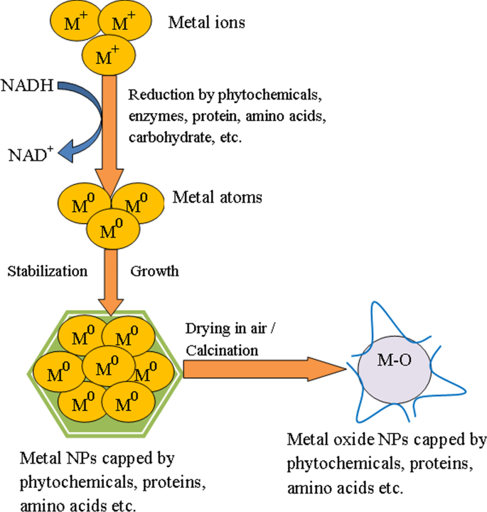
生物介导合成金属氧化物(SnO2)纳米颗粒的可能机制示意图
氧化锡纳米粒子的表征
NPs 的表征对于了解和控制合成过程和应用非常重要。利用各种技术研究了合成 NPs 的表面形态和构象细节,如大小、形状、结晶度和表面积。用于表征绿色合成 SnO2 NP 的一些技术是 UV-Vis、FTIR、XRD、EDS、SEM 和 TEM。 UV-Vis 光谱用于通过研究纳米颗粒的光学特性来监测纳米颗粒的形成。使用 Catunaregam spinosa 合成的 SnO2 NPs 的 UV-Vis 光谱 由于其表面等离子体共振,提取物表现出 223 nm 的最高吸收峰 [57]。 XRD 是一种强大的技术,用于研究材料的晶体结构。巴塔查吉等人。 [81] 表征 SnO2 NPs 在衍射角 (2θ ) 的 26.7, 34.2, 38.07, 51.9, 54.9, 58.1, 62.1, 64.9, 66.08, 71.6, 和 79.07 对应于 (110), (101), (200), (2021), (2021)分别是(310)、(112)、(301)、(202)和(321)平面(图3)。标准衍射峰显示 SnO2 NPs 的四方金红石结构,平均晶粒尺寸为 4.6 nm,使用 Debye-Scherer 方程 [81] 计算。
<图片>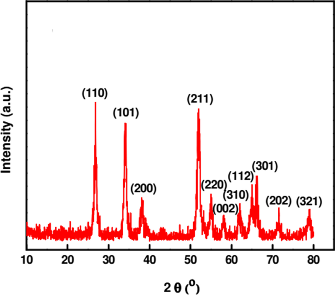
精氨酸在微波辐射下合成的SnO2纳米颗粒的XRD图谱[81]
FTIR 光谱用于研究表面化学并鉴定存在于 NPs 表面的官能团,这些官能团可能负责 NPs 的还原、封端和稳定 [88]。使用 Pruni flos 合成的 SnO2 NPs 的 FTIR 分析 提取物在 3308、2153、1634、423、403 和 383 cm -1 处显示出主要吸收带 .在 3308、2153 和 1634 cm -1 观察到的强波段 由于吸附在 SnO2 表面上的水分子、炔烃的 C-H 伸缩和黄酮类化合物的 C=O 振动,Sn-OH 基团的伸缩振动。 423 到 383 cm −1 之间的波段 归因于反对称 Sn-O-Sn 拉伸 [70]。 SEM 和 TEM 等基于显微镜的技术已被广泛用于确定纳米颗粒的形态特性。然而,与 SEM 相比,TEM 提供了更好的分辨率和内部结构信息,例如晶体结构和形态。使用 FE-SEM 可以获得更准确的表面特性结果。这些技术也可用于估计合成纳米颗粒的平均尺寸 [89]。 FE-SEM 和 TEM 分析显示形成了平均尺寸为 30-40 nm 的轻微团聚的球形 SnO2 NPs(图 4)[62]。
<图片>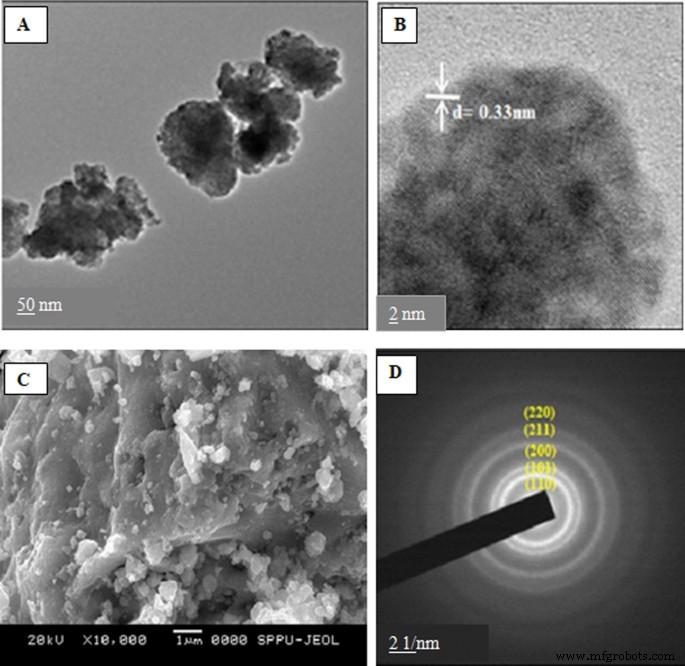
一 生物合成的 SnO2 NPs 的 TEM 图像; b SnO2 NPs 的 HR-TEM 图像; c SnO2 NPs 和 d 的 FE-SEM 图像 SnO2 NPs的SAED图
此外,选择性区域电子衍射 (SAED) 分析表明这些颗粒本质上是纳米晶体(图 4)[62]。使用Piper betle合成的球形SnO2 NPs的形成 水提取物通过 FE-SEM [63] 确定。从 TEM 和 XRD 分析,发现纳米颗粒的平均尺寸为 8.4 nm。
EDS 是一种用于分析样品元素组成的分析技术。使用 Psidium Guajava 合成的 SnO2 NPs 的 EDS 光谱 叶提取物显示存在 Sn 和 O 峰,这证实了纯 SnO2 NPs 的形成 [61]。
绿色合成氧化锡纳米粒子的生物应用
与散装形式相比,绿色合成的 SnO2 NPs 显示出增强的光催化、抗菌、抗氧化和抗癌活性。在本节中,我们讨论了绿色合成 SnO2 NPs 在各个领域的应用,为新研究人员的未来前景提供指导。
抗菌活性
许多研究人员已经观察到 SnO2 NPs 的抗菌活性。例如,使用芦荟合成的SnO2 NPs的抗菌活性 使用E研究植物提取物。大肠杆菌 和S。金黄色葡萄球菌 .有人提出 NPs 对 S 更有效。金黄色葡萄球菌 比E。大肠杆菌 [58]。这可能是因为 E 的细胞壁。大肠杆菌 比 S 更复杂。金黄色葡萄球菌 . S。金黄色葡萄球菌 有一层由厚的肽聚糖组成的膜。然而,E 的细胞壁。大肠杆菌 具有肽聚糖层和由脂多糖组成的外膜。 E的外膜。大肠杆菌 充当屏障,降低 ROS 进入细胞的渗透水平 [90,91,92]。另一个原因可能是它们细胞膜的极性不同。 金黄色葡萄球菌的膜 比 E 具有更多的正电荷。大肠杆菌 ,这会导致带负电荷的自由基的渗透水平更高,从而导致S 中更多的细胞损伤和死亡。金黄色葡萄球菌 比在 E 中。大肠杆菌 [93, 94]. 用石榴合成的SnO2纳米颗粒的抗菌活性 种子提取物已针对E进行了测试。大肠杆菌。 杀菌效果随着纳米颗粒浓度的增加而增加[95]。使用香菇合成的SnO2纳米颗粒 种子提取物对大肠杆菌也表现出类似的抗菌活性。大肠杆菌 [72]。在最近的一项研究中,Clerodendrum inerme 叶提取物用于合成未掺杂和 Co 掺杂的 SnO2 NPs [96]。 The green synthesized un-doped and Co-doped SnO2 NPs were subjected toward antimicrobial activity against five disease-causing pathogens such as E.大肠杆菌 ,B。 subtillis , A. niger , A. flavus , 和 C. Albicans, , and their zone of inhibition diameters (ZOIs), minimum inhibitory concentration (MIC), and minimum bactericidal concentration (MBC) were calculated. The Co-doped SnO2 NPs showed substantial antibacterial activity against E.大肠杆菌 和 B. subtillis in a concentration-dependent manner compared to the un-doped SnO2 NPs, plant extract, and standard drugs in terms of their MIC (22 ± 0.7, 18 ± 0.8 mg/mL) and MBC (31 ± 0.9, 21 ± 0.6 mg/mL) as well as ZOIs (30 ± 0.08, 26 ± 0.06 nm), respectively. The authors suggested that the broad-spectrum antibacterial activity of Co-doped SnO2 NPs is due to cobalt doping, which leads to increased grain size and surface area of the NPs as compared to un-doped SnO2 NPs. As more is the surface area with smaller particle sizes, the greater will be the antimicrobial activity. The associations of biomolecules like flavonoids and phenolic compounds with Co-doped SnO2 NPs are also responsible for the enhancement of their antimicrobial activity. Furthermore, the green synthesized Co-doped SnO2 NPs showed extraordinary antifungal activity with maximum ZOIs of 17 ± 0.04, 23 ± 0.08, and 26 ± 0.06 nm against A. niger , A. flavus, 和C。 Albicans, respectively, in comparison with plant extract, un-doped SnO2 NPs, and standard drugs [96].
The actual mechanism of action of SnO2 NPs against microbial strains is still unknown. However, several mechanisms of action against bacteria have been suggested for metal oxide nanoparticles, such as the decomposition of nanoparticles, electrostatic interaction of nanoparticles with the cell wall of microorganisms, and formation of reactive oxygen species (ROS) by the effect of light radiation [97,98,99]. One possible cause for the antibacterial effect of SnO2 NPs may be the accumulation of the NPs on the surface of the bacterial cell membrane. The ROS generated due to the presence of SnO2 NPs interacts with the cell membrane and disturbs the membrane permeability and respiration system of the bacteria, which leads to cell death [72, 95, 100]. For instance, Khan et al. suggested that the release of Sn 4+ and Co 2+ is responsible for the damage of bacterial DNA and mitochondria, which inactivates the bacterial enzyme and finally leads to cell death [96].
Antioxidant Activity
Many researchers examined the antioxidant activity of NPs by monitoring the ability in quenching of stable DPPH (2,2-diphenyl-1-picrylhydrazyl) radical into non-radical form (DPPH-H). Kamaraj et al. [101] reported the antioxidant activity of SnO2 NPs biosynthesized using Cleistanthus Collinus leaves extract. The antioxidant activity of SnO2 NPs increased with increasing concentration of SnO2 NPs and reaction time. In another study [95], the free radical scavenging activity of green synthesized SnO2 NPs increased in a dose-dependent manner. However, the annealed sample exhibited a lower scavenging activity as compared to the as-prepared sample. The decrease in scavenging activity with increasing annealing temperature may be due to a decrease in surface area-to-volume ratio of the NPs. Moreover, the antioxidant efficacy of SnO2 NPs against DPPH is due to the transfer of electron density between the NPs and the free radical located at nitrogen in DPPH. A similar result was found for SnO2 NPs synthesized using Trigonelle foenum-graecum aqueous extract [72].洪等人。 [67] reported significant antioxidant properties of biosynthesized SnO2 NPs. The antioxidant activity of the biosynthesized SnO2 increased with increasing concentration of the NPs, with IC50 (half-maximal inhibitory concentration) value of 2257.4 µg/ml. Recently, Khan et al. reported significant antioxidant activity of Co-doped SnO2 NPs, as compared to un-doped SnO2 NPs and plant extract [96].
Cytotoxic Activity
SnO2 NPs synthesized using an aqueous extract of agricultural waste of dried peel of Annona squamosa were evaluated for cytotoxicity test against the hepatocellular carcinoma cell line (HepG2) [64]. TEM results revealed the loss of cell volume, considerable swelling of the cells, and nuclear condensation. The nuclear condensation seen in SnO2 NP-treated HepG2 cells might be due to the breakdown of chromatin in the nucleus. SnO2 NPs inhibited cell proliferation in a dose- and time-dependent manner with an IC50 value of 148 µg/mL. SnO2 NPs synthesized using piper nigrum seed extract exhibited higher cytotoxic activity against colorectal (HCT116) and lung (A549) cancer cell lines [69]. The proliferation of both cancer cell lines increased with increasing NPs size. Besides, a gradual decline in cell viability was observed with increasing dosage of SnO2 NPs. The authors concluded that the cytotoxic effect was associated with the generation of oxidative stress from reactive oxygen species (ROS). SnO2 NPs fabricated by using Pruni spinosae flos aqueous extract revealed an excellent cytotoxic effect against non-small cell lung cancer cell A549 and lung fibroblast CCD-39Lu cells, in concentration- and time-dependent manner [70]. Recently, Khan et al. [96] investigated the in vitro cytotoxic effect of green synthesized un-doped SnO2 NPs and Co-doped SnO2 NPs against mammary gland breast cancer (MCF-7), human amnion (WISH), and human lung fibroblast (WI38) cell lines by a colourimetric technique 3-(4,5-dimethylthiazol-2yl)-2,5-diphenyl tetrazdium bromide (MTT) assay. The green synthesized Co-doped SnO2 NPs showed significant and substantial mortality rate compared to un-doped SnO2 NPs, plant extract, and standard drug 5-FU (5-5-5-fluorouracil), while un-doped SnO2 NPs exhibited a similar mortality rate to that of the standard drug, but the lowest cytotoxicity against breast cancer cell line was observed with the plant extract. The cytotoxic effect of the plant extract, green synthesized un-doped SnO2, and Co-doped SnO2 NPs was performed with IC50 values of 31.56 ± 1.4, 26.99 ± 1.9, and 18.15 ± 1.0 µg/mL for MCF-7, and 40.69 ± 0.9, 38.97 ± 0.8, and 36.80 ± 0.6 µg/mL for WI38, and 38.56 ± 0.8, 35.56 ± 0.9, and 31.10 ± 0.7 µg/mL, respectively, and depicted that the cytotoxicity of Co-doped SnO2 NPs was more proficient as compared to plant extract and un-doped SnO2 NPs alone. Additionally, the authors reported excellent observational results showing greater in vitro inhibition of breast cancer MCF-7 cell lines in a concentration- and dose-dependent manner. The green synthesized NPs also showed robust cytotoxicity against MCF-7 as compared to WI38 and WISH normal cell lines with Co-doped SnO2 NPs unveiling higher ROS generation than the un-doped SnO2 NPs. Figure 5 shows the probable cytotoxicity mechanism of green synthesized SnO2 NPs.

Mechanism during cytotoxicity of green synthesized SnO2 NPs
Photocatalytic Activity
The uncontrolled release of toxic chemicals, hazardous textile dyes, and pesticides from various industries into the running water has led to severe environmental problems. These superfluous water contaminants cause long-term adverse effects and pose a real threat to aquatic and human life. Besides, some organic dyes are carcinogenic and toxic. Hence, treating water that contains poisonous chemicals before disposal to the environment is very crucial to reduce environmental pollution. Recent studies have shown that nanostructured semiconductor metal oxides act as an excellent photocatalyst for the removal of various water pollutants [102,103,104,105]. Among the semiconductor metal oxides, SnO2 is extensively used in the removal of common textile dyes and organic compounds owing to its beneficial characteristics, which include physical and chemical stability, high surface reactivity, high photocatalytic efficiency, low cost, and low toxicity [68, 106]. Manjula et al. [107] synthesized SnO2 nanoparticles using glucose. The NPs were effectively used as a catalyst in degrading methyl orange (MO) dye. The effect of calcination temperature (150–500 °C) on the photocatalytic activity of the NPs was investigated, and the results revealed that the as-synthesized SnO2 NPs calcinated at 150 °C is the best photocatalyst for the reaction under study among the studied materials. Moreover, the as-prepared SnO2 nanoparticles degraded methyl orange completely in 30 min, and also, the nanomaterials may be recycled with enhanced efficiency a minimum of five times. In another study [108], the photocatalytic activity of the SnO2 QDs (quantum dots) synthesized by using serine was evaluated by monitoring the optical absorption spectra of eosin Y solution under direct sunlight. It was observed that the rate of degradation of eosin Y using SnO2 QDs (98%) is higher than that using commercial SnO2 (96%) and P25 (88%). Begum et al. [84] reported the synthesis of SnO2 NPs using an amino acid, L-lysine monohydrate. The synthesized nanoparticles were evaluated for their photocatalytic behavior toward toxic organic dyes, namely malachite green oxalate (MGO) and Victoria blue B (VBB) under direct sunlight. The absorption peak of these dyes has begun to reduce, which shows that the chromophore structure has been demolished (Fig. 6). Furthermore, the as-prepared SnO2 NPs exhibited an outstanding photocatalytic degradation of MGO (97.3%) and VBB (98%) dye within 120 min. Literature reports on the photocatalytic activity of green synthesized SnO2 NPs are summarized in Table 3.

Photocatalytic degradation of malachite green oxalate dye under solar irradiation using SnO2 NPs
Gas-Sensing Property
Many metal oxide-based gas sensors are widely used for gas-sensing applications. However, SnO2 NPs has gained tremendous attention in gas sensing under atmospheric conditions because of its beneficial properties, which include high sensitivity, high selectivity, easy reversibility, and cheap manufacturing costs. Green synthesized porous SnO2 nanospheres demonstrated excellent gas sensing capabilities [107]. It was observed that the assimilation of 0.5 wt% Pd into the SnO2 matrix improved the sensitivity and made it highly selective for low-temperature hydrogen detection. Moreover, the fabric was able to respond to even 50 ppm H2 in N2 at room temperature with an interval of 10 s. These sensing properties are due to the synergetic effect of both the porous structure of SnO2 nanospheres and also the catalytic property of Pd nanoparticles. Gattu et al. [77] reported the gas-sensing behavior of biosynthesized and chemically synthesized Ni-doped SnO2 NPs thin films. The biosynthesized Ni-doped SnO2 NPs thin film showed higher NO2 gas-sensing response as compared to the chemically synthesized ones. The sensor response was found to be increased with Ni doping for both biosynthesized and chemically synthesized Ni-doped SnO2 NPs. This may be due to the reduction of particle size with Ni-doping, which results in increased surface area for adsorption of NO2 gas. Furthermore, the Ni-doped SnO2 thin film exhibited excellent selectivity toward NO2 gas when put next to other gases like NH3, LPG and H2S. In another study [78], the gas-sensing properties of un-doped and Fe-doped SnO2 NPs synthesized using Cicer arietnum L. extract were reported. The gas response within the presence of 100 ppm NH3 gas at 200 °C operating temperature was found to be 28% for un-doped SnO2 and 46% for Fe-doped SnO2 thin films. Moreover, the Fe-doped SnO2-based sensor was found to be more selective for NH3 gas as compared to the un-doped SnO2 sensor. The biosynthesized Au-doped SnO2 NPs were found to be highly sensitive to NO2 gas at 200 °C operating temperature [79]. Gas sensor supported Au-doped SnO2 NPs showed the gas response of ~ 30% for 100 ppm of NO2 gas. Additionally, the gas sensor of Au-doped SnO2 NPs showed excellent selectivity toward NO2 gas when put next to other gases like H2S, LPG, and NH3. The improved gas response and selectivity toward NO2 gas are because of the lattice distortion induced by Au-doping and also the oxygen vacancies generation within the SnO2 lattice.
结论
The use of green methods for the production of NPs has been the area of focused research because it is an eco-friendly, inexpensive, nontoxic, and sustainable method. Numerous studies report the possibility of producing SnO2 NPs via a green protocol using a range of plant materials, bacteria, and natural biomolecules. The literature survey shows that the green substrates act as reducing and stabilizing agents or capping agents regardless of their source. Among the various green methods of SnO2 synthesis, plant-mediated synthesis is cost-effective, easy to process, and less hazardous than microorganisms. However, the plant extracts consist of a large number of active compounds in a different composition, which makes it difficult to know the exact amount of the molecules responsible for the reduction of metal ions. Due to this complexity, it is difficult to evaluate the synthesis of nanoparticles. Therefore, further study on the mechanism of formation of SnO2 NPs is required to understand the chemical reactions that occur during the synthesis. With the knowledge of the actual reaction mechanism, it will be possible to monitor and optimize the biosynthesis process, which is essential for the large-scale production of SnO2 NPs. Hence, understanding of the rapidly growing method of synthesis discussed herein will help facilitate future research progress on SnO2 NPs and their enormous potential for industrial-scale production in the near future.
数据和材料的可用性
Not available.
缩写
- NP:
-
纳米粒子
- SnO2 NPs:
-
Tin oxide nanoparticles
- 紫外可见光:
-
紫外可见光谱
- SEM:
-
扫描电镜
- FE-SEM:
-
场发射扫描电镜
- TEM:
-
透射电子显微镜
- HR-TEM:
-
高分辨透射电子显微镜
- FTIR:
-
傅里叶变换红外光谱
- EDS:
-
Energy dispersive X-ray analysis
- PSA:
-
Particle size analyzer
- XPS:
-
X-ray photoemission spectroscopy
- SAED:
-
Selective area electron diffraction
纳米材料
- 特殊氧化物耐火材料及其应用
- 用于改进诊断和治疗应用的多功能金纳米粒子:综述
- 负载 ICA 的 mPEG-ICA 纳米颗粒的制备及其在治疗 LPS 诱导的 H9c2 细胞损伤中的应用
- 氧化铜纳米颗粒对大肠杆菌的生物合成、表征和抗菌潜力评估
- 二氧化钛纳米颗粒对小鼠的潜在肝脏、大脑和胚胎毒性
- 基于苯基三甲氧基硅烷改性氧化铝纳米颗粒的 Al2O3:SiOC 纳米复合材料的形成和发光特性
- Au@TiO2 蛋黄-壳纳米结构的制备及其在亚甲基蓝降解和检测中的应用
- 通过蒸发诱导自组装和增强的气敏特性简便合成虫孔状介孔氧化锡
- 铜纳米粒子合成和稳定方面的环保能力:催化、抗菌、细胞毒性和抗氧化活性
- 桔梗皂苷(桔梗)用于金和银纳米颗粒的绿色合成
- 金属和金属氧化物纳米粒子的绿色合成及其对单细胞藻类莱茵衣藻的影响
- Al-Doped ZnO 薄膜在红外区域的光学特性及其吸收应用