

用于生物医学、药物输送和成像应用的磁性功能化纳米粒子
摘要
医学一直在寻找新的和改进的疾病治疗方法,这些治疗方法需要具有高疗效和成本效益,因此对发现此类新疗法的科学研究产生了大量需求。任何治疗的一个重要方面是能够仅针对疾病而不会对身体的另一个健康部位造成伤害。出于这个原因,金属纳米粒子已经并且目前正在广泛研究其可能的医学用途,包括医学成像、抗菌和抗病毒应用。超顺磁性金属纳米粒子具有的特性允许它们通过磁场引导到身体周围或引导到磁性植入物,这开辟了将各种生物货物与纳米粒子结合的潜力,然后可以引导它们在体内进行治疗。在这里,我们报告了各种金属纳米粒子当前的一些生物医学应用,包括单金属纳米粒子、功能化金属纳米粒子和使用 Fe3O4 核的核壳金属纳米粒子,以及这些核壳纳米粒子的合成方法。
介绍
金属纳米材料代表了未来医学的重要门户。尽管关于金属纳米粒子在医学中的长期安全性仍有很多未知数 [1],但这些粒子已经在各种生物医学应用中找到了自己的位置,例如体内位点特异性成像 [2,3,4]、癌症检测[5, 6],癌症治疗 [7,8,9,10],神经退行性疾病治疗 [11,12,13],HIV/AIDS 治疗 [14,15,16],眼部疾病治疗 [17,18,19] ] 和呼吸系统疾病治疗 [20, 21]。图 1 展示了纳米颗粒在医学中的一系列当前用途。尽管纳米医学最近取得了进展,但纳米治疗的方式仍然存在许多障碍,例如很难实现产生易于重复结果的合成路线,许多纳米颗粒合成方法产生两种尺寸的范围[22 ,23,24] 和纳米粒子的形状 [25,26,27,28] 和/或不生产足够数量的纳米材料以使其具有经济可行性 [29]。另一个关键因素是,由于研究领域相对较新,一些纳米粒子在一段时间内的长期毒性相对未知 [30,31,32]。在金属纳米颗粒的众多可能用途中,包括药物输送领域 [33, 34]。由于纳米颗粒提供的大表面积 [35],它们具有输送大量药物或其他医疗货物的能力 [36]。单一金属纳米颗粒的替代方案是将核加入纳米颗粒,该核对壳材料具有替代性质,其中一个例子是加入磁核。仍然存在的一项挑战是合成核壳纳米粒子;合成纳米粒子的方法有很多[37],但在尝试合成核壳纳米粒子时出现了新的挑战[38]。
<图片>
目前纳米粒子在医学中的一些用途
本综述首先关注目前用于生成核壳纳米粒子的一些方法,重点是使用 Fe3O4 核和金或银涂层。然后,我们研究了单金属纳米粒子当前的生物医学应用、它们的局限性以及如何通过应用磁芯来克服它们。
核壳纳米粒子的合成
金属纳米粒子的合成方法已为人所知多年,例如,Stevenson 等人。 1951 年发表了通过还原 HAuCl4 合成金纳米粒子 [39]。从那时起,出现了许多不同的纳米颗粒合成途径,例如气体沉积[40]、溶胶-凝胶[41]和气溶胶/气相[42]。然而,当试图合成由核壳结构组成的金属纳米颗粒时,一个新的挑战出现了,其中一种金属形成核,另一种金属形成壳,例如 Fe 颗粒在水中降解,而 HAuCl4 是一种强氧化性代理 [38]。将进一步讨论的一个此类示例是使用 Fe3O4(氧化铁)核和金作为涂层壳。在制备这种核壳金属纳米粒子时,最大的两个问题是试图控制涂层的速率和控制涂层的均匀性,以创建形状和尺寸都非常相似的纳米粒子溶液 [43]。将金或银涂覆到氧化铁核上可分为两大类:将金/银直接涂覆到铁上 [44] 或使用中间层作为金和铁层之间的胶水 [45]。这里将讨论前一类。下面介绍一些已设计的合成金和银包覆的 Fe3O4 纳米粒子的方法。
反胶束合成
合成金属纳米粒子的一种流行途径是使用反胶束法,有时也称为微乳液法 [46]。该方法于 1980 年代首次引入,当时首次合成了铑、铂和钯纳米颗粒的胶体溶液 [47]。
当具有疏水和亲水组成部分的分子与水相或疏水相接触时,就会形成胶束 [48]。胶束将以这样的方式组织起来,使亲水部分与水相接触,而疏水成分面对疏水相[49]。本质上,球体由内部屏蔽相形成,其中可以进一步包含货物[43, 50,51,52]。
微乳液途径有多种方法,包括油包水 (w/o) [53] 和超临界 CO2 包水 (w/sc-CO2) [54]。当水分散在基于碳氢化合物的连续相 [53] 中,热力学驱动的表面活性剂自组装,然后产生反胶束时,会产生 w/o 乳液,其中球形胶束是最常见的形状 [43]。添加到该混合物中的任何添加的极性或离子材料都会在胶束内分隔开,然后当胶束膜通过布朗运动相互收缩时形成纳米粒子 [55]。 w/sc-CO2 乳液涉及使用处于超临界状态的流体 (CO2),即高于其临界压力和温度 [56]。这种方法特别令人感兴趣,因为它是一种更“绿色”的纳米颗粒合成方法,因为不需要有毒的有机溶剂。通过简单地降低压力并将流体作为 CO2 气体释放 [57],也更容易回收产品。
反胶束路线已经从合成金属纳米粒子到涂覆先前合成的纳米粒子[58]。第一个在反胶束中合成的镀金氧化铁 (Au-Fe3O4) 纳米粒子是在将近 20 年之前完成的 [59]。 Au-Fe3O4 纳米粒子的这种合成是使用 H2O/CTAB(十六烷基三甲基溴化铵)系统产生的胶束,以硼氢化钠 (NaBH4) 作为还原剂,将氯化金 (HAuCl4) 还原到铁核上。这种合成产生了平均尺寸为 12 nm 的纳米颗粒分散体。由于这是首次使用微乳液生产 Au-Fe3O4 NPs,因此发现了一系列 Au-Fe3O4 NPs 合成路线 [46, 60,61,62,63]。图 2 是如何使用反胶束路线形成纳米颗粒的一般表示。
<图片>
含盐反胶束反应形成金属纳米颗粒的一般表示
林等人。发表了一种稍微改进的方法,使用反胶束方法用金涂覆 Fe3O4 [60]。该合成还采用以CTAB为表面活性剂形成反胶束的体系,但以1-丁醇为辅助表面活性剂,辛烷为油相,加入含有金属离子的水溶液,使用NaBH4将HAuCl4还原到表面氧化铁纳米颗粒。报道的涂覆颗粒的光学结果表明,从金胶体 (526 nm) 到 Au-Fe3O4 (555 nm) 的紫外/可见光谱的吸收峰发生了变化。包覆颗粒的 TEM 结果表明粒径分布为 5-15 nm,平均粒径为 10 nm。 Pana 等人重复了这种方法。 5-35nm 尺寸的 Au-Fe3O4 纳米颗粒的尺寸分布稍大 [63]。此外,Seip 等人使用了一个非常相似的系统。除了使用肼还原 HAuCl4 [64]。
Fe3O4 纳米粒子的涂层不仅限于金;洛佩兹佩雷斯等。报道了使用含有环己烷/Brij-97(辅助表面活性剂)和含有 FeSO4.7H2O 和 FeCl3.6H2O 铁盐的水相的系统合成氧化铁纳米颗粒 [65]。该系统涂有银 [58] 和金 [46],产生 13 纳米粒子。 Tamer 等人报道了另一种方法。用于合成 Au-Fe3O4 纳米粒子 [62]。该方法采用铁盐在 NaOH 中的共沉淀,然后在 HClO4 中洗涤以产生氧化的 Fe3O4 纳米颗粒。通过由 CTAB 胶束输送到系统中的 NaOH 还原 HAuCl4,金在 Fe3O4 NPs 上的涂层发生。 Au-Fe3O4 NPs 的平均尺寸为 23.5 nm。表征后,然后用各种官能团修饰粒子以形成自组装单层(SAM)并进一步用于大肠杆菌的捕获和检测 .
Zhang 等人 完成了反胶束合成的改进版本。涉及使用激光作为用金涂覆铁纳米颗粒的引发剂 [66]。该过程包括制备封装在 CTAB 胶束中的铁纳米粒子、水中的金纳米粉末和辛烷的反应混合物,然后用脉冲激光照射,同时剧烈搅拌反应。激光照射促进金纳米粒子的热分解。在铁纳米粒子周围形成金原子和簇,形成镀金的铁纳米粒子。以这种方式合成的 Au-Fe 纳米粒子的 TEM 结果显示,平均粒径为 18 nm,粒径分布为 ±36 nm。
热合成
在金壳-铁核纳米颗粒合成的各种方法中,有一种热途径,其中反应包括将反应混合物加热到其沸点以上 [67],有时回流 [68, 69]。这种类型的合成有两大类:水热(水基溶剂)[70, 71] 和溶剂热(有机溶剂)[68, 72]。虽然有许多通过热途径合成金属纳米粒子的技术 [73,74,75,76,77,78],但不可能在一锅反应中实现核心和金涂层的合成 [68, 69, 72, 74, 77, 79,80,81],在某些情况下,Fe3O4 核是通过反胶束路线 [70] 或胶体路线 [78] 合成的,然后使用水-或溶剂热技术 [70, 76, 78]。虽然在这些合成方法中使用了多种溶剂系统,但大多数途径都涉及将氧化铁纳米颗粒添加到沸腾的 HAuCl4 中,或者将 HAuCl4 的倒数添加到氧化铁纳米颗粒的沸腾溶液中 [74, 79] .
Rudakovskaya 等人已经完成了一种合成 Au-Fe3O4 纳米颗粒的方法。通过水热技术[76]。该方法的原理遵循将 Fe3O4 纳米粒子添加到沸腾的 HAuCl4 溶液中。这些纳米粒子的 TEM 分析表明平均尺寸为 30 nm,具有一般的球形形状,尺寸分布在 20 和 35 nm 之间;这些图像可以在图 3 中看到。
<图片>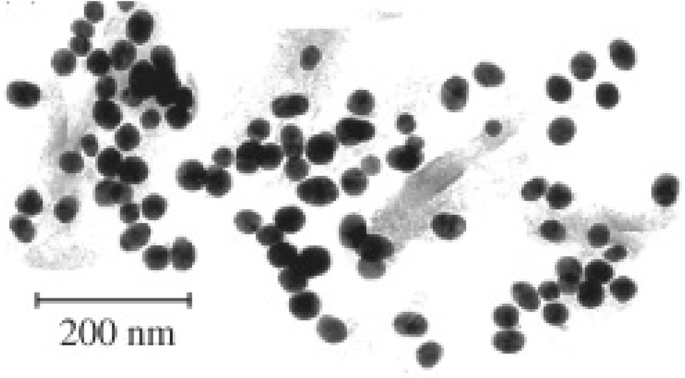
Rudakovskaya 等人合成的纳米粒子的 TEM 图像。可以看出,纳米颗粒大致呈球形,平均尺寸为 30 nm [76]
胶体合成
胶体合成技术提供了一种简单而有效的合成金属纳米粒子的方法 [82]。胶体技术通常比其他纳米颗粒合成技术更简单,不需要不同的溶剂,或者可以在室温下进行 [83, 84]。合成的基本原理包括将不同的金属离子分散在水相中,向混合物中加入还原剂,然后在受控温度下混合以形成不溶性纳米颗粒 [39]。胶体合成路线提供的好处是不必在合成中涉及潜在的有毒溶剂(如果纳米颗粒用于生物用途,则是理想的选择)。然而,胶体途径存在一些限制,例如难以控制最终合成纳米颗粒的尺寸分布[85],并且纳米颗粒的形状会受到试剂浓度的严重影响[85]。然而,从积极的方面来说,生产大量纳米颗粒会更容易 [86]。这种合成金属纳米粒子的方法已经存在多年,被用于合成不同类型的纳米粒子,如银[87]和金[39, 88]。
这种基本方法已经得到改进和发展,以产生不同的合成路线,以形成金包覆的氧化铁纳米粒子 [83, 84, 89,90,91,92,93,94,95,96,97]。大多数合成镀金氧化铁的方法都围绕使用各种还原剂将 HAuCl4 还原到氧化铁表面。纳达古达等人。提供了一种建议的“绿色”合成路线,使用抗坏血酸来减少 HAuCl4 [84]。然而,由于缺乏合成中使用的封端剂(一种结合到纳米粒子外部的试剂,阻止纳米粒子的进一步“生长”),这种方法似乎几乎没有显示出对涂层纳米粒子的大小或形状的控制[98]。 Pal 等人提出了一种对合成包覆颗粒的形状和尺寸进行更多控制的方法。 [95] 这种方法采用醋酸金作为金盐,将其还原到 6 纳米 Fe3O4 纳米粒子的表面,形成 7 纳米大小的 Au-Fe3O4 粒子,这些粒子呈球形。 Rawal 等人提出了一种快速涂覆 Fe3O4 纳米颗粒的方法。这包括将 Fe3O4 纳米颗粒分散在 HAuCl4 溶液中,然后与乙醇混合 [83]。在室温下 15 min 后,停止反应,然后用磁铁分离 Au-Fe3O4 纳米颗粒。纯化溶液的 TEM 分析表明,产生的颗粒大小从 30 到 100 纳米不等,并且在整个样品中具有不同的形状;这些图像可以在图 4 中看到。虽然这种合成技术可以快速生产出包覆的纳米颗粒,但它似乎不是生产形状和尺寸均匀的颗粒的非常有效的合成方法 [83]。
<图片>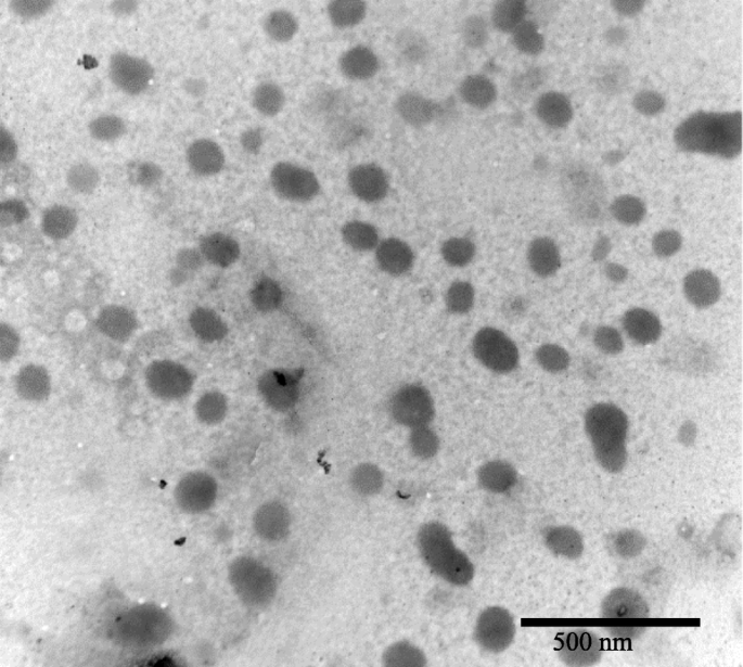
Rawal 等人合成的纳米粒子的 TEM 图像。这些纳米粒子的尺寸分布为 20-100 nm [83]
虽然一些技术仅提供金盐的还原,但其他技术更喜欢将还原剂置于铁表面,例如羟胺 [90, 93]。在许多情况下,当 Fe3O4 纳米粒子涂有金时,金盐的还原也会产生标准的金纳米粒子 [74],因此在铁纳米粒子表面添加还原剂旨在提高涂层的效率和旨在降低作为副产品产生的金纳米粒子的数量[93]。
另一种技术涉及将金播种到磁性纳米粒子的表面上,这提供了一种更直接的途径,使金在纳米粒子的磁核周围成核 [91, 92, 97]。该技术涉及将比溶液中的氧化铁纳米颗粒小的金种子结合到氧化铁的表面。当 HAuCl4 在溶液中被还原时,Au + 离子将种子到氧化铁上并在氧化铁纳米颗粒周围形成壳。这种黄金播种已被多个团体成功采用;古恩等人。使用聚乙烯亚胺控制金在 Fe3O4 表面的播种,产生完全包覆的纳米粒子。 [91] 然而,合成的 Au-Fe3O4 颗粒显示出高多分散性,粒径范围从 40 到 110 nm。莱文等人。设法生产出尺寸范围为 50-70 nm 的金壳磁核纳米粒子,使用有机硅烷分子功能化的核与金种子结合 [92]。金纳米粒子在铁核上的播种可以用各种核形状来证明,例如,Wang 等人。证明了金在水稻形状的“纳米稻”Fe3O4 结构上的播种,然后当金被还原到表面时会导致一个完整的厚金壳[97]。
金属纳米粒子的生物医学应用
抗菌剂
细菌感染非常常见,自 1928 年亚历山大·弗莱明 (Alexander Fleming) 发现青霉素 [99] 以来,抗生素成为主要的治疗方法。纳米医学为我们提供了一种新的、范围广泛的可能的治疗方式,正在探索金属纳米粒子以用于未来的治疗 [100]。表 1 列出了一些已被探索用于抗菌应用的纳米颗粒。一种已被检验其潜在用途的材料是银,它已被证明具有多种生物医学用途 [101],例如,Sreekumar 等人。利用银纳米粒子作为抗菌纤维网络的一部分。纳米颗粒大小从20到120 nm不等,对大肠杆菌具有抗菌功效 与不含银纳米粒子的纤维相比,高达 94.3% [102]。虽然已经证明氨苄青霉素等抗生素能够在E中达到≤ 99.9%的杀灭率。大肠杆菌 [103],同一项研究还报告了某些大肠杆菌菌株对氨苄青霉素的抗性。大肠杆菌 .同样,据报道,E.大肠杆菌 可以对银纳米粒子产生抵抗力;然而,这种阻力不是遗传变化,而是一种试图导致胶体纳米颗粒聚集的物理反应[104]。 Holtz 等人也使用银的抗菌特性。设计了一个 60-nm 钒酸银纳米线系统,用直径为 1-20 nm 的银纳米颗粒“装饰”[105]。该系统显示有希望对抗三种金黄色葡萄球菌 菌株,而且有趣的是对耐甲氧西林金黄色葡萄球菌的生长抑制浓度要低得多 (MRSA) 比抗生素苯唑西林。
Verma 等人报道了银纳米颗粒的合成。他们使用纳米颗粒对抗细菌:荧光假单胞菌 ,E。大肠杆菌 , 和真菌白色念珠菌 [106]。与一些常用抗生素(如氨苄青霉素和新霉素)的最小抑制生长浓度分别为 4.0 μg/ml 和 16.0 μg/ml 相比,银纳米颗粒在三种菌株中的平均最小抑制生长浓度为 5.83 μg/ml。对抗E 菌株。大肠杆菌 [110]。潜在的兴趣是纳米颗粒对 P 显示的特性。荧光剂 C.白化病 ,这两者都与在免疫功能低下的患者中引起疾病有关 [111]。进一步的研究可能会发现,与一些最常用的抗生素(如具有广泛副作用的两性霉素 B)相比,银纳米颗粒是一种更有效的治疗病原体的方法[112]。
Selvaraj 等人报道了硫鸟嘌呤封端的金纳米粒子的合成。其中对几种细菌的抗菌作用增强,包括:E.大肠杆菌 , 烟曲霉 , 和铜绿假单胞菌 [107]。发现硫鸟嘌呤封端的金纳米粒子作为抗癌和抗菌剂比未结合的硫鸟嘌呤更有效,其活性显示出作为抗癌药物载体的潜在用途。以类似的方式,据报道,金纳米粒子对 假结核棒状杆菌 具有抗菌作用 [108],平均尺寸为 25 nm 的纳米颗粒,使用 50 μg/ml 的剂量,在暴露 20 分钟后显示出 95% 的细菌生长抑制。类似地,裸金纳米颗粒显示出对多种革兰氏阴性和革兰氏阳性细菌包括S 具有抗菌作用。金黄色葡萄球菌 , 肺炎克雷伯菌 , 和 枯草芽孢杆菌 [109]。对于S,1.35 μg/ml剂量的AuNP显示出46.4±0.4%、38.3±0.2%和57.8±0.2%的生长抑制。金黄色葡萄球菌 ,K。肺炎 , 和 B.枯草 , 分别。
抗病毒
与抗菌应用一样,金属纳米颗粒已被证明在抗病毒应用中很有前景。表 2 展示了一系列已被证明具有抗病毒特性并可能在治疗病毒时应用的纳米颗粒。裸银纳米颗粒和包覆银纳米颗粒 [113,114,115,116] 在纳米级范围内均已被证明具有一系列抗病毒应用。
根据世界卫生组织的数据,乙型肝炎 (HBV) 是一种病毒感染,目前影响全球 2.57 亿人,并在 2015 年造成 887,000 人死亡 [121]。小的(10-50 nm)裸银纳米粒子已被测试作为一种可能的治疗 HBV 的方法 [113],并显示出与 HBV 有效结合并进一步抑制 HBV RNA 的产生。假设作用模式是由于 AgNPs 与 HBV dsDNA(双链 DNA)结合。罗杰斯等人。已经证明可以使用裸银纳米颗粒和多糖涂层作为抗猴痘病毒 (MPV) 的抗病毒剂 [114]。在 12.5–100 μg/ml 之间的浓度范围内对纳米颗粒进行体外抗 MPV 测试;研究结果表明,所有浓度的多糖包覆银纳米粒子(Ag-PS-NPs)都能在体外减少MPV诱导的斑块形成。
银纳米粒子甚至可能在人类免疫缺陷病毒 (HIV) 的治疗中发挥作用 [115, 116]。艾滋病毒是一个主要的健康问题,据世卫组织估计,截至 2016 年,有 3670 万人感染了艾滋病毒 [122]。快速有效地发现和实施艾滋病毒治疗方法很重要;拉拉等人。已经证明了银纳米粒子(30-50 nm)对 HIV-1 分离株的影响,显示出对所有 HIV-1 分离株的抑制作用 [116]。裸纳米粒子对 HIV-1 的整体 IC50 为 0.44 mg/ml ± 0.3,病毒抑制机制显示为抑制病毒-宿主细胞结合,特别是银纳米粒子抑制 gp120 蛋白之间的相互作用(包膜糖蛋白)和靶细胞膜受体。同一组还证明了涂有聚乙烯吡咯烷酮 (PVP) 的银纳米粒子能够防止 HIV-1 转染到人类宫颈组织外植体模型中 [115]。具体而言,0.15 mg/ml PVP 包覆的银纳米粒子 (PVP-AgNPs) 可抑制 HIV-IIIB 和 HIV-AZT-RV 分离株的感染。与对照样品相比,这种浓度的 PVP-AgNPs 还诱导淋巴细胞(免疫细胞)向感染部位增殖 [115]。
不仅银和涂覆的银纳米颗粒被用于对抗病毒:涂覆有两亲硫酸盐配体的 2 纳米金纳米颗粒也被证明对 HIV-1 有效 [118]。这些颗粒显示出靶向病毒的融合过程,并在体外显示出与 gp120 蛋白结合并直接中和 HIV-1 感染。巯基乙磺酸盐包覆的金纳米粒子 (Au-MES) 显示出对 1 型单纯疱疹病毒 (HSV-1) 感染的抑制作用,这可能是通过抑制病毒与宿主细胞的结合、细胞间病毒的传播或改变细胞对病毒的易感性来实现的。纳米颗粒的存在诱导感染[117]。
碘化铜纳米粒子 (CuI-NPs) 已被证明对几种不同的病毒具有抗病毒特性:猫杯状病毒 (FCV) [119] 和更有趣的是猪源甲型流感病毒 (H1N1) [120]。 100 到 400 纳米的 CuI-NPs 在对抗 FCV 时表现出抗病毒特性,并且假设单价 Cu 离子负责产生活性氧 (ROS),导致随后的衣壳蛋白氧化,导致 FCV 失活。 H1N1 病毒也被证明以非常相似的方式被 CuI-NPs 抑制,即产生羟基自由基,导致蛋白质降解。然而,这些自由基也可能被证明对未感染的组织有毒,这对于在批准使用治疗之前确定这一点很重要[123]。
成像
磁共振成像 (MRI) 扫描是一种非常有用的医学诊断工具,可提供清晰的解剖图像。使用 MRI,人们可以可视化血液流动、生理化学特征以及体内组织和器官的状态 [124]。 MRI 中经常使用造影剂以提高诊断敏感性 [125]。常规使用的造影剂是螯合剂,但当前造影剂的主要缺点是其生物稳定性和在细胞中积累时的毒性水平 [126]。例如,一些造影剂是基于碘的,据报道,碘造影剂暴露与随后发生的甲状腺功能亢进症和明显甲状腺功能减退症的发生有关 [127]。已经开发出替代方案,通过减少造影剂对身体的负面影响来提高扫描效率 [128]。替代品包括金属纳米颗粒,可能与一种试剂结合,该试剂的作用类似于用于 MRI 扫描的造影剂 [129]。图 5 是 AuNPs 处理前后大鼠大脑皮层的 MRI 对比图像 [130]。
<图片>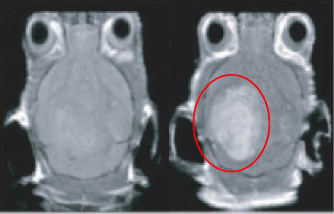
大鼠大脑皮层治疗前(左)和治疗后(右)的 MRI 对比图像。包含 AuNPs 的区域以红色环绕
表 3 显示了一些已被探索用于医学成像的纳米粒子。一些计算机断层扫描 (CT) 造影剂存在的问题包括循环半衰期短 [131] 和潜在的组织损伤 [130]。因此,还研究了金属纳米粒子用于 CT 成像 [132];由于其 X 射线衰减 [133],Au 纳米粒子在成像中显示出有前景的用途。小岛等人。表明金纳米粒子与聚乙二醇化树枝状大分子 (PEG-AuNPs) 缀合,可用于在体外以及 用于 X 射线计算机断层扫描,与市售碘剂碘帕胺相比 [134]。 PEG-AuNPs 显示出比市售碘帕胺更高的对比效率,从体内快速排泄 [135]。作者还指出,PEG-AuNPs 具有光细胞毒性,可用于光热疗法。
李等人。已经证明使用涂层金纳米粒子作为动脉粥样硬化的成像工具; AuNP 被应用于一种称为“单光子发射计算机断层扫描”(SPECT)的医学成像 [136]。这种类型的成像与使用伽马相机非常相似,但它能够提供真正的 3D 图像,可以对其进行切片、旋转和操作,以实现更准确的分析技术 [136]。修饰的纳米颗粒特异性靶向含有凋亡巨噬细胞的动脉粥样硬化斑块,表明是一种有用的工具,可用于侵入性准确检测动脉粥样硬化斑块[136]。
AuNPs 先前已被证明是光声成像 (PA) 的可能试剂,显示出高空间分辨率和灵敏度 [137]。 PA 依赖于对暴露于非电离脉冲激光照射时从组织发出的超声波的检测 [140]。超声波发射的强度/幅度决定了图像的对比度,因此任何能够吸收激光脉冲然后放出热量的物质都会增加超声波发射的幅度,而 AuNPs 具有两种能力这些 [141, 142]。由于有机染料对光漂白和从血液中快速清除的敏感性,AuNP 可能比有机染料更好 [143]。 AuNPs also have use in cell imaging for examining movement of nanoparticles within cells when conjugated with various cargoes. Figure 6 is a darkfield imaging of A431 lung cancer cells treated with AuNPs that target epidermal growth factor receptor, and the bright areas within the cells are the nanoparticles indicating their locations within the cells [144].

Darkfield imaging of A431 lung cancer cells treated with AuNPs; the bright yellow/orange dots are nanoparticles within the cells
Biomedical Cargo Delivery
Nanoparticles make for an ideal molecule for drug delivery due to the huge surface area to the volume ratio they provide when compared to their bulk material [145]. In addition, it is possible to engineer nanoparticles to either avoid or interact with the immune system in specific ways [146, 147]. For example, it has been demonstrated that an increased hydrophobicity of nanoparticles/sub-groups conjugated to the nanoparticles illicit and increased immune response by measuring cytokine mRNA levels in mice [146]. Focusing in the opposite direction, it has been suggested that nanoparticles can be conjugated with various ligands to directly activate the immune system to target the destruction of a tumor [148] or by accumulation in the liver or spleen for the generation of tolerance or immunity, respectively [147].
Gold nanoparticles have been extensively studied for their delivery of medical cargo, for example, Bhumkar et al. have explored the application of AuNPs for trans-mucosal delivery of insulin. Gold nanoparticles were synthesized in the presence of chitosan, which acts as a polymeric stabilizer [149]. These nanoparticles were then loaded with insulin and administered both nasally and orally to diabetic rats. The results showed an overall reduction in the rat’s blood glucose levels, an indication of successful movement of the nanoparticles through the mucosal membranes and into the bloodstream.
More recently “smart” AuNPs have been employed in PA [138]. These nanoparticles are roughly 10 nm in diameter and are functionalized with citraconic amide moieties which are susceptible to hydrolysis. The citraconic amides are converted into positively charged primary amino acids at a mildly acidic pH, while the surface molecules adopt negative charges at physiological pH [138]. Combined, these two properties cause the “smart” nanoparticles to adopt both positive and negative charges allowing them to aggregate rapidly due to electrostatic attraction. These nanoparticles are referred to as “smart” due to the nanoparticles presenting cancer-specific properties and accumulate rapidly and efficiently in cancer tissues and show a much lower accumulation in normal tissues [150].
Gold nanoparticles can also be used as a delivery system for nucleic acids [153], including oligonucleotides [151] and small interfering RNA (siRNA) [154]. Many different methods have been developed to functionalize AuNPs with nucleic acids, for example, Yonezawa et al. have synthesized gold nanoparticles modified with thiocholine, which then bound to DNA and formed a fusion of wire-like structures throughout the DNA [155]. Sandström et al. demonstrated the ability to bind nucleic acids onto gold nanoparticles [151], and a similar modification has been done by Rosi et al. where tetrathiol-modified antisense oligonucleotides were bound to 13-nm gold nanoparticles [152]. Being able to conjugate nucleic acids to nanoparticles opens up the possibility of targeted gene delivery, which could, for example, lead to genes coding for a specific protein to be delivered to a cell that was either deficient in that protein or could not produce the protein themselves [156]. It has also been exhibited that gold nanoparticles modified with DNA can transfect cancer cells [157] (Table 4).
Anticancer Drug Delivery
Cancer is one of the world’s leading killers with large areas of scientific research being dedicated to the fight against cancer, and nanoparticles offer a new doorway into methods to target and treat cancer. Table 5 presents a selection of nanoparticle/drug conjugates that have been tested for anticancer treatments. Paciotti et al. have investigated the application of PEGylated AuNPs as a carrier for tumor necrosis factor (TNF) which is a cell-signaling protein that possess the ability to induce apoptosis in healthy cells [158]. The Au-PEG-TNF nanoparticles were injected intravenously and agglomerated significantly more in MC-38 colon carcinoma cells compared to other healthy cells/tissues. The TNF not only gave therapeutic action on the MC-38 cells, but also seemed to possess a targeting property, indicated by the lack of agglomeration in healthy cells. Another interesting observation reported was the ability for the Au-PEG-TNF nanoparticles to diminish a tumor mass compared to “free” TNF.
Doxorubicin is a widely used cancer therapeutic agent but has dose-limiting associations with cardiotoxicity. A gold nanoparticle-doxorubicin conjugate has been developed that demonstrates little no to cardiotoxicity to mice while being able to treat cancer [160]. Dixit et al. demonstrated the selective delivery of folic acid-coated AuNPs into folate receptor (FR) positive cancer cells, whereas when compared with a cell line that did not have folate receptors, uptake was shown to be minimal [159]. These results demonstrated the use of folate to target metal nanoparticles to FR-positive cancer cells for tumor imaging and ablation.
Limitations of Single Metal Nanoparticles and Overcoming Them
The principal obstacle with nanoparticle drug delivery is the ability to direct the nanoparticle to the target area [162, 163]. There are several methods in use for metal nanoparticle targeting such as antibodies [164,165,166] and homing peptides [167, 168]. There are however limitations to these methods, with the biggest being that before they even reach the desired target cells they have to pass through a variety of other barriers, such as blood vessels and the blood-brain barrier [169]. One way to overcome this targeting limitation is to use magnetic nanoparticles [170]. A magnetic nanoparticle-targeting system works by directing the nanoparticles to a target site using an external magnetic field, it has already been demonstrated that the magnetic anisotropy of the nanoparticle is a very important factor for medical treatments [171], with a change in anisotropy being able to the change the efficacy of hypothermia treatments for example [172]. Superparamagnetic metal nanoparticles have this property (they only present magnetic properties while in the presence of a magnetic field) [173]. However, the benefit of magnetic nanoparticles also presents a potential limitation, due to the toxicity of many magnetic materials [31, 174, 175]. Despite iron being approved for various imaging uses [5, 6, 31], it has been suggested in several studies that naked iron oxide nanoparticles may have some adverse effects when used in cell labeling [176,177,178]. One method that can be used to overcome any potential toxicity limitations is to coat the iron core [179]. A range of materials can be used as the coating material:silica [180,181,182], polymers [183, 184], gold [62, 93, 95, 185], or silver [58, 186]. Gold has low pharmaceutical activity [187] and silver has been used in biomedical applications for many years [188, 189],
The combination of a superparamagnetic core with an inert and safe metal coating produces metal nanoparticles with superior characteristics to non-magnetic metal particles [190]. As well as reducing toxicity, the coating also provides the potential for the conjugation of functionalized molecules onto the surface, such as drugs and biomolecules for application in the medical field [97, 140, 152]. It is of note that a core-shell nanoparticle still possesses the properties and uses of a nanoparticle made from the same material as just the shell, but the superparamagnetic core gives the ability to direct the nanoparticle in the body [191]. For example, a gold nanoparticle with an antibody is classified as a targeting nanoparticle, introducing the core would classify the nanoparticle as a directed targeting nanoparticle [173].
Current Medicinal Uses of Gold-Coated Iron Oxide Nanoparticles
Core-shell superparamagnetic nanoparticles have already been assessed for their biomedical uses, with a wide range of uses already being applied [192] and with a majority of research investigating into the use of gold as a shell for the nanoparticles, in part due to its biocompatibility and ability to easily bind to a variety of materials. As such, this section will deal exclusively with gold shell nanoparticles. One of these uses is as a magnetic carrier for drug targeting [192,193,194,195,196]. Kayal and Ramanujan have tested an in vitro apparatus that simulates the human circulatory system as a test for the magnetic delivery of gold-coated iron oxide nanoparticles (Au-Fe3O4) loaded with doxorubicin [194]. Their system had various magnetic fields of increasing strength next to a capillary through which the doxorubicin-loaded particles were passed. A significant percentage of these nanoparticles were captured within the magnetic fields, strongly indicating the potential for the use of magnetic nanoparticles in drug delivery. Another use for a targeted system is the application of Au-Fe3O4 nanoparticles in photothermal therapy. Bhana et al. demonstrated the use of a core-shell system used in combination therapy deployed against two different cancer cell lines; head and neck (KB-3-1) and breast (SK-BR-3) with a reported decrease in cell viability of 64% when they exposed cell lines to a combined photothermal and photodynamic therapy, compared to each modality used on its own [197]. In photothermal therapy, gold nanoparticles are coated with a ligand, such as PEG [142], and these nanoparticles are irradiated with a laser, with a wavelength that matches the UV-vis λ -max of the gold nanoparticles [194]. The nanoparticles vibrate at the laser’s frequency which causes heat to be released causing the death of the surrounding tissue [198], introducing a core which is superparamagnetic can allow for a more accurate targeting for use in this therapy. Similarly, it has been reported by Kirui et al. that gold hybrid nanoparticles were deployed against SW1222 colorectal cancer in photothermal therapy, showing an increased case of cellular apoptosis after therapy, with their conclusion being that the cells showed an increased uptake, leading to a reduced laser power required to reach threshold therapeutic levels [199]. The use of core-shell nanoparticles for photothermal therapy of cancer has also been reported by other groups [200, 201].
Metal nanoparticles have already shown to have a place in contrast imaging, for example core-shell nanoparticles can also be used in T1- and T2-weighted imaging in MRI [202]. Research by Cho et al. demonstrated that gold-coated iron nanoparticles can be successfully used in MRI imaging, as well as opening the route for conjugating various ligands for use in biosensors [202]. A magnetic carrier capable of imaging and photothermal therapy has been reported by Cheng et al. They demonstrated the magnetic targeting of multi-functional nanoparticles to a tumor in a mouse model, which could be imaged inside the tumor and showed a reduction in the tumor size when combined with photothermal therapy [203]. It is also of note that in this work, both the nanoparticle dosage (1.6 mg/kg) and laser power (1 W/cm 2 ) are among the lowest applied for in vivo photothermal therapy. Moreover, there was no obvious toxicity from the nanoparticles reported. Table 6 presents some of the currently reported uses of core-shell nanoparticles.
Another medical area where such core-shell metal nanoparticles have been suggested to make an impact is in directed enzyme prodrug therapy (DEPT) [170, 191]. DEPT is a promising method of cancer treatment, with several therapies making it through to clinical trials [207, 208]. The main principal of DEPT is the targeted delivery of a prodrug-activating enzyme to a tumor site. Upon arrival at the tumor site, the enzyme enters the target cells where it can later activate an administered prodrug. However, the efficacy of the therapy depends on the ability to direct the enzyme to the tumor site, with current directional techniques relying on passive targeting methods such as viruses [207, 209] or antibodies [210, 211], rather than an active targeting system for enzyme delivery. A novel therapy proposed by Gwenin et al. potentially overcomes the targeting issue [170, 212]. This approach involves conjugating a genetically modified prodrug-activating enzyme onto the surface of a gold-coated iron oxide superparamagnetic nanoparticle (AuMNP), then directing the AuMNP-enzyme conjugate to the target site using a magnetic field to increase the efficacy of the targeted therapy. Figure 7 presents some of the uses of a core-shell nanoparticle.

A pictorial representation of the applications of core/shell nanoparticles
结论
In brief, single metal nanoparticles have been shown to currently possess a wide range of biomedical applications, with more application for these nanoparticles being discovered. One of the limiting factors that these nanoparticles face in medical treatments is to find a way for precise accurate targeting of areas within the body, be it for targeting of a drug delivery or for therapies involving the nanoparticles directly. A way to overcome this is to employ a magnetic core to create core-shell nanoparticles that can then be directed around a body using a magnetic field. There are a variety of methods that can be used to synthesize these core-shell nanoparticles, with each method having its own advantages and disadvantages. There remain many obstacles for core-shell nanoparticles before they can be routinely applied in the medical field and these include
- 1)
Achieving a synthesis route which produces easily repeatable results;
- 2)
Producing particles of a set size [22,23,24] and shape [25,26,27,28]; and
- 3)
Producing large enough quantities to make it economically viable [29].
Another key factor is the relatively unknown toxicity of some nanoparticles over an extended period of time due to how relatively new the field of research is.
数据和材料的可用性
Not applicable
缩写
- (o/w):
-
Oil-in-water
- (w/o):
-
Water-in-oil
- (w/sc-CO2):
-
Water-in-supercritical CO2
- AgNP:
-
Silver nanoparticle
- Ag-PS-NPs:
-
Polysaccharide-coated silver nanoparticles
- Au-Fe3O4 :
-
Gold-coated iron oxide nanoparticle
- Au-MES:
-
Mercaptoethane sulfate-coated gold nanoparticle
- AuNP:
-
Gold nanoparticle
- Au-PEG-TNF:
-
Polyethylene glycol-coated tumor necrosis factor-loaded gold nanoparticles
- CT:
-
Computed tomography
- CTAB:
-
十六烷基三甲基溴化铵
- CuI NPs:
-
Copper-iodine nanoparticles
- DNA:
-
Deoxyribonucleic acid
- FCV:
-
Feline calicivirus
- FR:
-
Folate receptor
- Gp120:
-
Glycoprotein 120
- H1N1:
-
Influenza A of swine origin
- HBV:
-
Hepatitis B virus
- HIV:
-
Human immune-deficiency virus-1
- HSV-1:
-
Herpes simplex virus 1
- KB-3-1:
-
Head and neck cancer
- MC-38:
-
Colon carcinoma
- MPV:
-
Monkeypox virus
- MRI:
-
Magnetic resonance imaging
- PA:
-
Photoacoustic imaging
- PEG:
-
聚乙二醇
- PVP-AgNP:
-
Polyvinylpyrrolidone-coated silver nanoparticle
- RNA:
-
Ribonucleic acid
- ROS:
-
活性氧
- siRNA:
-
Small interfering ribonucleic acid
- Sk-BR-3:
-
Breast cancer
- SPECT:
-
Single-photon emission computed tomography
- TNF:
-
肿瘤坏死因子
纳米材料
- 用于增强药物递送的纳米纤维和细丝
- 用于改进诊断和治疗应用的多功能金纳米粒子:综述
- 用于癌症治疗的纳米粒子:当前的进展和挑战
- 生物相容性 FePO4 纳米颗粒:药物递送、RNA 稳定和功能活性
- 用于合成和生物医学应用的荧光纳米材料的进展和挑战
- 钴掺杂 FeMn2O4 尖晶石纳米粒子的制备和磁性
- 用于癌症应用的基于细胞的药物递送
- 131I 追踪的 PLGA-脂质纳米颗粒作为靶向化疗治疗黑色素瘤的药物递送载体
- 纳米技术:从体内成像系统到受控给药
- 用于体内 CT 成像和肾脏清除特性的新型生物相容性 Au Nanostars@PEG 纳米颗粒
- 用于光热疗法和光声成像的聚吡咯涂层铁铂纳米粒子的合成和体外性能
- 3D 打印微型机器人有望用于药物输送