

碳纳米管及其衍生物对体外肿瘤细胞和生化参数、体内细胞血液成分的影响
摘要
拟议工作的目的是在细胞和生物体水平上分析氧化碳纳米管 (CNTox) 的毒性,由阿霉素 (CNT-Dox) 和荧光素 (CNT-FITC) 功能化。分析了 CNTox、CNT-Dox 和 CNT-FITC 对体外肿瘤细胞(2-D、3-D 培养物)和体内 Balb2/c 小鼠模型的细胞毒性作用。结果,证明了将阿霉素固定在 CNT 表面并从 CNT 表面受控释放阿霉素 (Dox) 的可能性。与游离 Dox 相比,Dox 固定与 CNT-Dox 的细胞毒性作用降低一致。与 CNT 表面的肽键断裂导致阿霉素的释放和 CNT 和 Dox 的细胞毒性作用的剂量依赖性增强。显示了 CNT、Dox 和胰蛋白酶对肿瘤细胞存活的联合细胞毒性作用。在生物体水平上,研究了所获得的纳米结构对实验动物肝酶系统状态、蛋白质代谢和细胞血液组成的影响。 CNTox 体内模型的影响在统计学上与对照相同。与纯阿霉素相比,CNT-Dox 表现出较低的总生物体毒性作用。细胞血液成分的偏差表明 CNT-Dox 的一般毒性作用,但与纯阿霉素相比,毒性作用更温和。根据获得的数据,我们得出结论,将 CNT 与多柔比星结合可以降低多柔比星对血液一般生化指标的毒性和体内血细胞组成的破坏。同时,CNTs和阿霉素在药物释放后的联合作用使我们在体外抑制肿瘤生长方面取得了更大的疗效。
背景
碳纳米管最引人注目和最有前途的应用之一是医学。碳纳米管 (CNT) 具有独特的化学和生物学特性 [1,2,3,4,5]。碳纳米管具有很大的表面积,使它们能够附着多种生物物质 [6]。此外,碳纳米管能够穿透细胞膜、毛细血管,并在细胞和组织中积累[7,8,9]。碳纳米管很容易被细胞吸收,它促进了碳纳米管到达细胞核的能力,暗示了基因治疗的可能性[10]。这就是为什么碳纳米管是将蛋白质、抗原、RNA/DNA 载体 [11]、疫苗和药物输送到细胞 [12] 的有吸引力的载体。特别令人感兴趣的是使用碳纳米管作为个性化载体的前景,控制药物释放用于抗癌 [13]、抗菌 [14] 和免疫治疗 [15]。靶向递送和控制释放是现代药物使用的实际问题,尤其是具有细胞毒性的药物。药物释放的确切机制确保了靶组织中活性物质的有效浓度,而其他组织中的浓度最低。它将减少药物的剂量,同时保持其有效性并减少副作用造成的伤害。目前,有多种有目的的给药方式,例如使用脂质体[16]、聚合物胶束和树枝状聚合物[17]、可生物降解颗粒[18]和其他纳米颗粒[19]。但最近的实验表明,与其他纳米粒子相比,使用 CNT 递送药物有很多好处 [20, 21]。其中之一是碳纳米管具有相当大的活性表面,可以填充所需的化学物质,从小分子到蛋白质、抗体和 RNA/DNA。碳纳米管的开放端使得内部体积和表面可用于功能化。因此,碳纳米管的大表面积为不同类型的功能化提供了许多结合位点。与此同时,随着碳纳米管的发展,碳纳米管的溶解度低、聚集能力强、亲水性好、半衰期长、对整个生物体的影响也存在一些问题[22]。据文献报道,将碳纳米管引入实验动物体内,伴随着碳纳米管及其衍生物在消化道、脾脏、肾脏[23]、肌肉[24]和肺组织[25]等器官中的积累。 .下一步,碳纳米管会影响代谢和炎症过程的活动 [25, 26]、免疫系统状态 [27] 和实验动物的存活 [28]。然而,这些问题是进一步推进使用碳纳米管的积极研究的主题。许多研究已经令人信服地证明了碳纳米管作为用于药物递送的纳米载体的优势。 [29, 30] 的作者报告说,对于药物而言,特定的纳米管可能比纳米载体的危害更小。还表明,简单的官能化(-OH、-COOH、-NH、PEG)[31] 或 CNTs 的封装 [22],增加了水溶性,提高了生物利用度,并降低了 CNTs 的毒性。将 CNTs 引入移植肿瘤的小鼠可能对靶向转化组织有效,并导致肿瘤体积减小并提高动物存活率 [32]。
在之前的研究 [33] 的基础上,我们假设活性抗肿瘤物质(阿霉素)可以固定在碳纳米管肽键的表面。所得化合物,即由阿霉素 (CNT-Dox) 功能化的碳纳米管,可以降低 CNT 作为阿霉素 (Dox) 的细胞毒性。同时,处理过的构建体的分解可能允许实现 CNT 和 Dox 的细胞毒性特性。因此,可以实现提高两种物质的抗肿瘤活性。因此,本研究的目的是确定 CNTs-Dox 构建体在体外蛋白酶(胰蛋白酶)存在下的细胞毒性作用。我们在体外分析了 CNT 及其衍生物对 2D 和 3D 细胞模型中肿瘤细胞的影响。另一个目的是研究获得的化合物(CNTs、CNTox 和 CNT-Dox)对肝酶系统活性、蛋白质周转和体内细胞血液成分的影响。因此,作者实现了对使用碳纳米管及其衍生物治疗胃肠道肿瘤的毒性、生物利用度和有效性的复杂估计。
方法
将白种人结肠腺癌 II 级癌 (HT29) 细胞系用作体外 2D(单层)和 3D(球体)系统中的实验细胞模型。该细胞系购自 Kavetsky 实验病理学、肿瘤学和放射生物学 NAS 乌克兰研究所的细胞系和动物组织银行。细胞在标准细胞培养条件下(95% 湿度,空气中 5% CO2;37°C)在 DMEM 全培养基和 10% FBS 实验室控制级别 2 下处理。
碳纳米管的合成、氧化和功能化
最初的多壁碳纳米管 (MWCNT) 是通过化学气相沉积(丙烯和氢与 Mo/Fe/Al2O3 作为催化剂)获得的。合成的多壁碳纳米管有 10-30 层,内径 5-15 纳米,外径 10-60 纳米,长度 20-500 微米,比表面积 120 米 2 /g 和堆积密度 5–50 g/l [33]。
通过HF处理从金属/催化剂中纯化MWCNT。从碳纳米材料中去除无定形碳是通过在 450-500°C 的空气中氧化来实现的。含有多壁碳纳米管的粗样品与空气中的氧气反应并产生二氧化碳或一氧化碳。催化剂载体的 HF(水)溶解在 450-500°C 的空气中氧化后,已经获得了一定量的碳沉积物。气流石英管式反应器用于此过程 130-150 分钟。我们分析和描述了先前工作中获得的多壁碳纳米管的物理和化学特性[33]。
MWCNTs(JEOL SL6060LA,日本)的扫描电子能谱(SEM)用于确定MWCNTs的结构特征。配体固定后,形态未发生变化。
MWCNTs 的氧化
在下一步中,将纯化的碳纳米管 (CNT) 在 70% HNO3 中在 99°C 下氧化 4 小时,然后用蒸馏水和 10% NH4OH 溶液洗涤 12 小时。结果,合成了碳纳米材料氧化物MWCNTox(CNTox)。之后,将 CNT 用 dH2O 洗涤三次至中性 pH。 CNT 表面的酸性位点用 0.1 M HCl 溶液再生。通过离心分离得到氧化的碳纳米管-ox,充分冲洗,并重新悬浮在 dH2O 水中。
CNT-Dox 粒子的制备
下一步的任务是将 Dox 盐酸盐(Dox,Teva Nederland B.V,Netherlands)固定在 MWCNTs 颗粒的表面。为了通过含氨基的 Dox-乳糖一水合物对氧化的 CNT(200 毫克)的表面进行功能化,将它们与双功能连接剂——N-环己基-N'-(2 吗啉乙基)碳二亚胺甲基对甲苯磺酸盐(C 1011,Sigma Aldrich ,美国)在室温下 15 分钟(图 1a)。之后,将 100 毫克 DOX-乳糖一水合物添加到 10 毫升 0.15 M 磷酸盐缓冲液 pH 6.5 中到每种纳米碳材料。使混合物在 30°C 下反应 24 小时。在这种情况下,形成了 Dox 和 CNT 之间的共价结合(图 1b)。通过以 10,000 rpm 离心 15 分钟收集组合物并用 dH 2 O 洗涤三次。上清液用于分光光度计检测游离DOX的浓度。 FITS 在 CNT 表面的固定由图 2a、b 所示的方案提供。
<图片>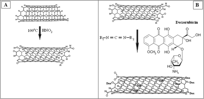
一 碳纳米管与碳二亚胺的反应示意图。 b 阿霉素对碳纳米管进行功能化的示意图
<图片>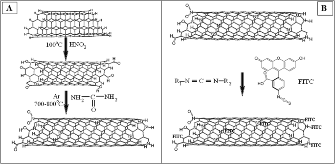
一 碳纳米管与尿素反应的示意图。 b FITC功能化碳纳米管的示意图
MWCNTs 稳定悬浮液的制备
MWCNTs的胶体悬浮分两个阶段进行。在第一阶段,碳纳米材料在磷酸盐缓冲液 (PBS) 中使用超声波分散器 UZDN-2 T 进行超声波处理。处理模式为 I =10 mA, R =22 kHz,持续时间 — 30 分钟。在第二阶段,所得水溶胶在室温下通过离心分散。该过程包括几个离心循环。因此,以这种方式选择了亲水性溶解性 SWCNT 部分。在加入悬浮培养的细胞之前,MWCNT 溶液通过煮沸 30 分钟进行消毒。 CNTs衍生物(CNT-Dox和CNT-FITC)通过100倍浓度的PSGA(青霉素:链霉素:庆大霉素:两性霉素)进行灭菌。
纳米管悬浮液的 Zeta 电位
通过电泳光散射 (M3-PALS) 测量样品的 Zeta 电位。对于测量,使用由 Malvern Instruments (Malvern, UK) 制造的 Zetasizer Nano ZS 分析仪。实验在 25°C 下进行,重复七次。为了测量样品,使用了通用潜水电极(Universal Dip Cell)ZEN1002 和聚苯乙烯比色皿(DTS001),一种光源——H-Ne 633 nm 激光器。
FTIR 光谱
通过傅里叶变换红外(FTIR)光谱研究所得纳米材料的化学成分。红外光谱由 FTS 7000e Varian FTIR 光谱仪测定。用于分析的样品是通过在磨机中研磨 ~ 1 mg CNT 和 150 mg 光谱纯 KBr 的混合物制备的。使用压力为3.0-3.5 × 10 3 的压力机制备样品 公斤/厘米 2 .通过在 600°C 的温度下加热 60 分钟使样品脱水。初步获得KBr的预射光谱,然后从样品的光谱中减去它们。所有光谱均根据有机化合物光谱鉴定目录进行分析 [34]。为了比较,使用氧化的 MWCNTs (CNT-O)、阿霉素 (CNT-D) 功能化的 MWCNTs 和荧光标记 (CNT-F) 功能化的 MWCNTs 样品。三种材料的样品和浓度的所有处理和条件相同。
多柔比星从碳纳米管表面释放的比色分析
为了评估固定在 CNM 上后游离 Dox 的量和 Dox 释放的有效性,使用了游离 Dox 在 495 nm 波长下发出荧光的能力 [35]。 Dox 盐酸盐中 Dox 的活性浓度为 16.7% w /w .为了绘制校准线,Dox Teva 20 mg/ml 稀释十次至 8 × 10 −3 使用毫克/毫升。然后,通过分光光度计读板仪Multiscan(Labsystem,Finland)测量了DOX与CNT复合物上清液中游离Dox的荧光。
CNT-DOX、CNT-FITC 粒子中的官能团浓度
对于 Dox 固定,使用 1 毫升 dH2O 中的 1 克 MWCNT。在 1 ml dH2O 中,Dox-TEVA 的量为 100 mg(16.7 mg 活性 Dox)。反应后上清液中游离 Dox 的量为 0.37 毫克或来自活性物质的 2.2%。因此,我们得出结论,16.23 mg DOX 固定在 1 g CNTox 上。对于 FITC,固定百分比很低。然后,将 8.35 毫克 FITC 固定在 1 毫升 dH2O 中的 1 克 CNTox 上。功能化的有效性为 1.62% w /w 对于 CNT-DOX 和 0.84% w /w 对于 CNT-FITC。 CNT-Dox和CNT-FITC浓度的进一步计算基于这些数据。
CNT 衍生物在 2-D 和 3-D 细胞模型上的细胞毒性分析
使用 MTT 测定法评估了 Dox、CNT、CNT-FITC 和 CNT-Dox 对 HT29 肿瘤细胞的细胞毒性。 MTT 测试基于活细胞中 NAD(P)H 依赖性线粒体氧化还原酶将 3-[4,5-dimetltiazol-2]-2,5-dipheniltetrazolium 盐转化为甲臜晶体。协议由 T. Mosmann [36] 描述。简而言之,1 × 10 4 HT29 接种在 96 孔板中并在全培养基中培养 12 小时。然后,将当前培养基替换为含有 CNTox(样品 #1)、MWCNT(样品 #2)、CNT-Dox(样品 #3)、Dox(样品 #4)和 CNT-FITC(样品 #5)的培养基.样品#1、2、3、5 的浓度为 12.5-25-50-100-200 微克/毫升,样品#4 的储备浓度为 20 微克/毫升,最终浓度为 1 到 10 微克/毫升。细胞在全培养基中培养并用作对照。孵育 24 小时后,通过比色测定用 MTT 分析细胞。我们向 100 μl 细胞悬液中加入 20 μl MTT 溶液(5 mg/ml PBS,Sigma)。之后,在标准条件下将细胞与 MTT 孵育 4 小时。然后,样品在 1500 g 下离心 5 分钟,并提取上清液。总之,孔中加入 10 μl DMSO (Sigma) 用于 MTT 晶体稀释和 20 μl 25 mM 甘氨酸。在分光光度计读板机 Multiscan(Labsystem,Finland)上在 540 nm 处测量反应溶液的吸光度。
多细胞肿瘤球体生成
作为肿瘤微转移模型系统的 HT29 细胞多细胞肿瘤球体 (MTS)(3D 培养)通过前面描述的成熟方法进行培养 [33]。简而言之,使用台盼蓝对细胞悬浮液进行计数并种植相同数量的细胞(5 × 10 4 细胞/毫升)。在标准条件(95% 湿度,空气中 5% CO2,37°C)下,将 3D 细胞培养物维持在含有 10% FBS(Sigma,美国)的 DMEM(Sigma,美国)培养基中。 MTS 的生成是通过我们实验室开发的技术进行的。将肿瘤细胞的培养在用含有 0.24% 羧甲基纤维素的培养基中的 1% 琼脂包被的 24 孔板中保持 24 小时。为了研究 MTS 的大小和数量对 CNT 的浓度和类型的依赖性,MTS 是在各种浓度的 CNT 存在下产生的。在对细胞培养物产生 MTS 之前,将 PBS 中的 CNTs 溶液添加到培养物中至终浓度,如先前针对 MTT 测定所述。进一步培养在 48 小时内在定轨振荡器 (80 rpm) 上的平板不断旋转下进行。在下一阶段,采用“暗场”方法拍摄微照片图像。总共完成了 120 多张图像。然后,使用 Axio Vision Rel 4.7 程序 Zeiss 计算文件上所有 MTS 的体积。我们使用了 Rolf Bjerkvig 的公式:V =0.4 ∙ a ∙ b 2 , 其中球体的几何尺寸 (b <a ) [37]。结果的可视化在Stemy 2000C显微镜Zeiss上进行。
分析碳纳米管对肝酶系统、蛋白质周转和体内细胞血液成分的影响
涉及动物及其护理的程序符合欧洲指令 2010/63 EU,经当地动物实验伦理委员会批准(Protocol №1 21.10.2016)。为了分析所得物质对生物体稳态总体状况的影响,进行了一系列体内实验。体内研究使用了 Balb/2a 系小鼠。 6-8 周龄的雄性和雌性小鼠在各组中相等,每组 10 只。小鼠被关在带有钢丝顶部和玉米芯垫料的笼子里,并保持在受控气氛中,12/12 暗/光循环,温度为 22°C ± 3°C,湿度为 50-70%,可自由进入到食物和淡水。因此,形成了四组小鼠。第 1 组:完整动物用 200 μl PBS 处理,对照。第 2 组:用 1.5 mg/kg 剂量的 CNTox 处理小鼠。第 3 组:用 1.5 mg/kg 剂量的 CNT-DOX 处理小鼠。在第 4 组中,小鼠接受剂量为 20 mg/kg 的阿霉素治疗。小鼠在 4 周内每 3 天接受 200 μl PBS 中的 CNT 肠胃外注射。阿霉素腹膜内给药,每 3 天一次,浓度为 20 毫克/千克体重。
血清生化分析
一个月后,小鼠退出实验。立即从死亡动物身上采集心脏血样。从上午 10 点到 11 点同时从动物身上取血。对于血浆,将血液在 37°C 下孵育 40 分钟,然后离心(20 分钟,2000 转/分钟)。然后用诊断试剂盒(Cormay,Warsaw,Poland)测定血浆中的生化参数,总蛋白、白蛋白、天冬氨酸氨基转移酶(AST)和丙氨酸氨基转移酶(ALT)、碱性磷酸酶(ALP)。实验是在半自动生化分析仪 FP-901M(Labsystems,Finland)上通过统一的实验室协议进行的。血液细胞成分测定采用中国迈瑞BC-3000 Plus血液分析仪。
统计分析
对于 3D 培养的统计分析,所有细胞聚集体根据大小从 1 × 10 -4 分成几组 毫米 3 到 1 × 10 −2 毫米 3 步长为 1 × 10 −3 毫米 3 .然后估计每组的MTS数量和每组MTS体积的中位数。所有测量均重复 3 次。对于微观统计分析正态分布的随机变量,我们使用了小群体的学生系数。指示的 p 值为 *р ≤ 0.05 或 **р ≤ 0.01。
结果与讨论
MWCNTs 的扫描电子显微镜
根据协议 [38],碳纳米管的平均直径为 10-20 nm,氩解吸确定的比表面积为 200-400 m 2 /G。堆积密度在 20–40 g/dm 3 .特别是,在应用 CVD 方法的工业 CNT 生产过程中获得了尺寸为 20-500 μm 的缠结管形式的附聚物(图 3)。
<图片>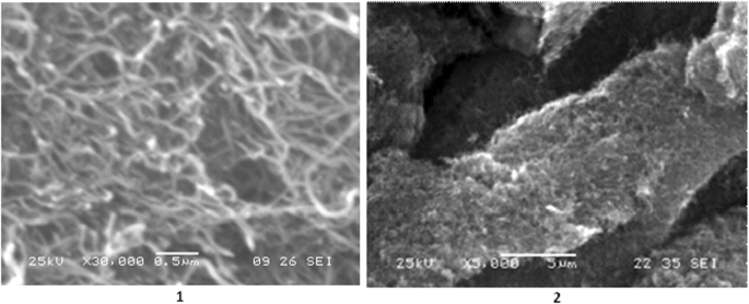
碳纳米管的 SEM 图像,1) 尺度 0.5 μm; 2) 尺度 5 μm
纳米管悬浮液的 Zeta 电位
CNTox 和 CNT 表明聚集的显着稳定性,这直接取决于 zeta 电位的值。 CNT-DOX 具有比 CNTox 和 CNT 更小的 zeta 电位,并且测量的重现性高,这表明颗粒的均匀性(表 1)。 CNT-FITC 的 zeta 电位值较小可以表明颗粒尺寸较大或浓度较低,如测量时的高噪声比所表明的那样。
CNT、CNT-Dox 和 CNT-FITC 的 FTIR 光谱
CNTox 与阿霉素 (DOX) 和荧光素 (FITC) 之间的结合是根据红外光谱数据估算的,如图 4 所示。
<图片>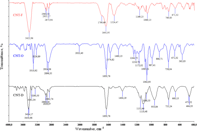
CNTox (CNT-O), CNT-DOX (CNT-D), CNT-FITC (CNT-F)的红外光谱图
在 υ =3311 cm −1 处存在强带 (CNT-O) 和 υ =3451 cm −1 (CNT-D) 归因于 CON-H 耦合波动,以及在 υ =1634 cm -1 处存在强带 (CNT-O) 和 υ =1631 cm −1 (CNT-D)归因于 O-CNH 键合振动,这清楚地表明了 CNT 和 Dox 之间的酰胺键类型。此外,IR 显示了位于 2969-2834 cm -1 的 C-H 键的吸收 以及 1460–1407 cm −1 的宽带 来自 C-O-H 片段。因此,我们可以得出结论,作为化学反应的结果,碳纳米管被抗肿瘤药物(阿霉素)和荧光标记物(FITC)功能化。
单层和球体细胞生长模型不同功能化阶段的碳纳米管的细胞毒性
下一步是确定氧化碳纳米管 (CNTox)、用阿霉素 (CNT-Dox) 功能化的碳纳米管和荧光标记 (CNT-FITC) 的细胞毒性。 HT29单层培养MTT检测结果见图5。
<图片>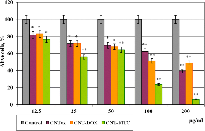
在与 CNT 及其衍生物(CNT-Dox 和 CNT-FITC)孵育 48 小时后,单层培养物中肿瘤细胞 HT29 的存活率。统计显着性:*р ≤ 0.05 或 **р ≤ 0.01
结果表明,CNTox、CNT-Dox 和 CNT-FITC 在 12.5 μg/ml (81%) 的浓度下具有中等的细胞毒性作用。与对照相比,将 CNTox 浓度从 25 增加到 50 和 100 μg/ml 导致肿瘤细胞的生存力呈剂量依赖性降低至 71.8-69.6-62.5%。在高达 200 μg/ml 的 CNTox 浓度下,HT29 的活力降低至 39.2%。当时,与 CNTox 相比,低浓度(12.5-50 μg/ml)的 CNT-Dox 没有显示出统计学上显着的细胞毒性。在高浓度 (200 μg/ml) 下,CTN-Dox 的细胞毒性甚至低于 CNTox (50%)。同时,用荧光素功能化的碳纳米管对肿瘤细胞具有相对较高的细胞毒作用。与浓度为 25 μg/ml 的 CNT-FITC 孵育后,HT29 细胞存活率分别降低至 55% 和 100-200 μg/ml,分别降至 23% 和 7%。因此,在那种情况下,碳纳米管起到了相当惰性的细胞基质的作用。不仅如此,CNT 固定了 Dox 并降低了 Dox 的细胞毒性。在之前的研究中 [33],我们已经证明初级纳米管具有疏水性,中等的细胞毒性作用,并刺激大量细胞聚集体的形成。根据文献[39],碳纳米管的表面电荷在氧化过程中会发生变化。碳纳米管变得更加亲水,并导致形成更小的聚集体并增加碳纳米管的细胞毒性作用。我们在之前的研究中收到的数据证实了这种趋势。与对照、45% 的 CNT-Dox 和 97%(200 μg/ml)的 CNT-FITC 相比,CNTox 将肿瘤细胞的存活率降低了至少 60%。对获得的数据的解释可能在其他作者的报告中,在大多数情况下,功能性配体会改变碳纳米管的表面 zeta 电位,刺激碳纳米管的溶解度,并增加碳纳米管的细胞渗透[40]。碳纳米管在氧化和功能化后形成聚集体的能力降低,而通过细胞膜的渗透性增加,细胞器的反应性增加。不仅如此,高浓度的 CNT-Dox 具有比 CNTox 更低的细胞毒性作用。同时,根据获得的数据,可以假设肽键将阿霉素保持在CNT表面。 CNTs 和 Dox 之间的肽键在细胞培养条件下足够稳定。它不会分解并以非活性形式服用阿霉素。同时,荧光素钠分子 (C20H12O5),一种大小为 332.311 g/mol 的配体,很容易从 CNT 表面解离并进入细胞。结果,DNA结构、细胞核和线粒体发生了不可逆转的破坏[41]。
为了验证碳纳米管及其衍生物的细胞毒性和对肿瘤细胞粘附特性的影响,分析了 HT29 细胞 2D 集落的面积。结果如图6所示,表明CNTs及其衍生物对单层培养中肿瘤细胞的粘附能力和肿瘤细胞集落的形成有影响。与对照相比,用浓度为 12.5、50.0 和 200 μg/ml 的 CNTox(样品 #1)孵育肿瘤细胞导致 2D 细胞集落的剂量依赖性减少 55.2%、57.8% 和 78.3%。同时,相同浓度的 CNT-Dox 样品(样品 #3)不会产生这种影响。 2D 菌落面积分别减少了 34.8%、61.6% 和 82.4%。 CNT-FITC(样品#5)在单层培养中对肿瘤细胞的影响最大。与 CNT-FITC 孵育后的 2D 菌落面积分别减少了 59.8%、85.2% 和 89.8%。应该指出的是,获得的结果与 MTT 分析具有相同的趋势。但是 MTT 分析的结果并没有显示出如此显着的细胞毒性作用。由此产生的差异可能是 CNT 对细胞集落的非细胞毒性作用的结果,部分原因是抗粘附影响。以前,我们已经证明碳纳米管的细胞毒性能力低于抗粘附能力。根据我们的数据,碳纳米管刺激肿瘤细胞迁移到悬浮部分和多细胞肿瘤球体 (MTS) 的形成。在单层培养和球体培养中,细胞对抗肿瘤剂的敏感性是不同的。这就是为什么在下一步中,我们研究了在 CNTox、CNT-Dox 和 CNT-FITC 存在下肿瘤细胞 HT29 形成 MTS 的原因。碳纳米管的浓度与之前的实验相同。在图 7 中,证明 CNTox(样品 #1)能够刺激 MTS 的形成。 CNTox 浓度的增加伴随着 MTS 中值体积的剂量依赖性增加。在 CNTox 浓度从 12.5、50、200 μg/ml 开始时,MTS 的中值体积从 1.79 增加到 2.18 和 10.98 mm 3 ;几乎是十倍。碳纳米管的衍生物不会对肿瘤细胞产生这种影响。 CNT-Dox(样品#3)以 2.83 倍的速度刺激 MTS 的形成。同时,3D 肿瘤细胞培养物与 CNT-FITC(样品 #5)一起孵育导致 MTS 体积减少 2.4 倍。
<图片>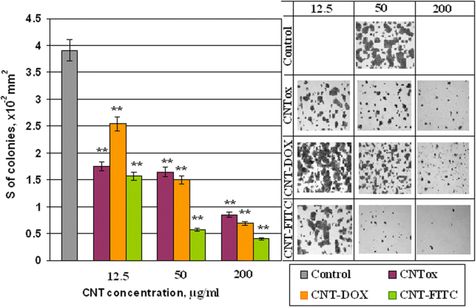
用 CNTox (#1)、CNT-Dox (#3)、CNT-FITC (#5) 孵育的单层培养物中 HT29 肿瘤细胞的集落面积。统计显着性:*р ≤ 0.05 或 **р ≤ 0.01
<图片>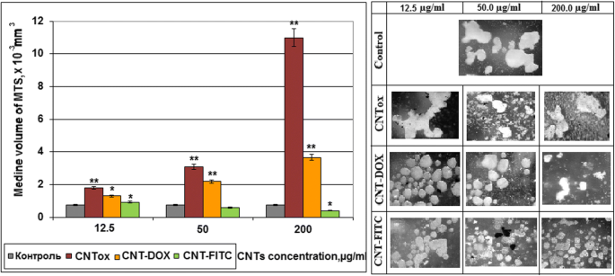
多细胞肿瘤球体体积的中位数。 MTS 是在 CNTox(样品 #1)、CNT-Dox(样品 #3)、CNT-FITC(样品 #5)存在的情况下产生的。统计显着性:*р ≤ 0.05 或 **р ≤ 0.01
值得注意的是,在大多数实验中,无论是在 2D 还是在 3D 培养中,CNT-Dox 对肿瘤细胞都没有这种细胞毒性作用,这可以与单一阿霉素的作用进行比较。阿霉素的细胞毒作用机制基于渗透到细胞核和核苷酸对之间的嵌入、复制和 DNA 修复的破坏、蛋白质合成以及因此导致的细胞死亡。 The cause of the reduction of cytotoxic effect of the CNT-Dox may be the binding of the doxorubicin with CNT surface through peptide bonds. It prevents Dox dissociation from CNT surface and Dox penetration into the cell and cellular organelles. This fact may let to deliver the compound to certain tissues without causing a negative effect on “non-target” objects.
In this case, the next step of the study was controlled release of doxorubicin. For peptide bonds breaking, we used the commonly known peptidase trypsin as the release agent. The effect of trypsin on the Dox release from the surface of the CNT was investigated by spectral analysis. Since the free doxorubicin has a fluorescence peak at 495 nm, bounded doxorubicin has no such ability. It was analyzed how the increasing of trypsin concentration affected on concentration of free doxorubicin. The results are shown in our previous work [42]. Shortly, it was demonstrated that increasing the concentration of 0.05% trypsin from 11 to 20% in culture medium contributed to an increasing the concentration of free doxorubicin from 3.13 to 6.55 μg/ml. A further increasing the concentration of trypsin to 60% did not lead to increasing in free doxorubicin. However, the concentration of trypsin by about 66% stimulated release again and led to increasing the concentration of doxorubicin in two times, to 11.38 μg/ml. Thus, it was concluded that 0.05% trypsin has several effective concentrations. Under these conditions, peptide bonds between doxorubicin and CNT were broken and doxorubicin was released. Therefore, to analyze how CNT-Dox influence the tumor cells after Dox release, we incubated tumor cells in the w/o fetal bovine serum (FBS) nutrient medium with trypsin and in the presence of CNT-Dox. The survival of tumor cells was determined using the MTT test. The ratio of the nutrient medium, trypsin, concentration of CNT-Dox, and doxorubicin are given in Table 2. The results of incubation HT29 during 48 h are demonstrated on Fig. 8. Cell viability in the presence of trypsin—blue columns, alone doxorubicin—orange columns, and CNT-Dox with trypsin—pink columns. In results, it has been found that the simultaneous use of trypsin and CNT-Dox significantly increased the cytotoxic effect of CNT and doxorubicin compared to the separately use of these substances. So doxorubicin alone at concentrations from 0.05 to 1.0 μg/ml causes a dose-dependent decreasing the percentage of living HT29 cells from 7.18 to 45.7% relative to control (Fig. 8, orange columns). Incubation of CNT-Dox at concentrations of 20.0 to 1.25 μg/ml with trypsin at concentration from 0 t0 70% led to a dose-dependent decreasing in the percentage of living cells from 80.9 and 99.8% respectively (Fig. 8, pink columns). At the same time, incubation with trypsin alone leads to decreasing percentage of living cells only by 34.0–42.0% (Fig. 8, blue columns). Thus, we can conclude that we observe the synergistic effect of CNT-DOX and trypsin. Each individual component has a small cytotoxic effect on cells, and together the cytotoxic potential of substances increases several times.

Percentage of alive cells HT29 after 48 h of incubation in the presence of trypsin, doxorubicin, CNT-Dox. Statistical significance:*р ≤ 0.05 or **р ≤ 0.01
As functional groups (Dox) were attached to CNTs surface by peptide bonds as, in vivo Dox release will be realized in organs of the gastrointestinal tract with an increased content of proteases, peptidases. In organism, proteases are used for various metabolic processes. Acid proteases secreted into the stomach (such as pepsin) and serine proteases present in duodenum (trypsin and chymotrypsin) enable us to digest the protein in food [43]. Other proteases are present in leukocytes (elastase, cathepsin G) and play several different roles in metabolic control. This is one of the fastest “switching on” and “switching off” regulatory mechanisms in the physiology of an organism. By complex cooperative action, the proteases may proceed as cascade reactions, which result in rapid and efficient amplification of an organism’s response to a physiological signal. Therefore, the authors suggest that CNT-Dox construction will be the most effective in the case of stomach, pancreas, liver, and small intestine cancer localization in case of parenteral administration of drug.
Influence of CNTs and Their Derivatives on Cell Blood Composition, Hepatic Enzyme System, and Proteins Turnover In Vivo
To determine the impact of the CNTox and CNT-Dox on the protein metabolism and the state of the hepatic enzyme system, the level of albumin (Al), total protein (Tp), alanine aminotransferase (ALT), aspartate aminotransferase (AST), and alkaline phosphatase (ALP) was determined. Data are given in Fig. 9a, b. As a result, it was noted that CNT-Dox and Dox have influence on the hepatic enzymes activity, namely on the AST and ALP. AST level increased in mice serum from 1 group for 21.6%, 2 group for 93.9%, and 3 group for 126.4% compared with control. At the same time, the activity of ALP increased in mice serum from 1 group for 23.5%, 2 group for 119.1%, and 3 group for 147.8%. CNTox administration did not show statistically significant changes in AST, ALT, and ALP levels. In addition, it should be noted that CNTox, Doxorubicin, and CNT-Dox slightly increased the level of total protein in the blood of experimental animals. So, we found that systematic introduction of CNTox have not influence on mice serum enzyme profile. And opposite, administration of CNT-Dox and Dox had toxicity influence and induced a chronic hepatic damage. Notably, symptoms of hepatitis had more manifestation after Dox treatment than CNT-Dox. To determine possible inflammatory processes and systemic effects of CNTox, CNT-Dox on the state of blood cells composition, it was analyzed the blood cell formula of the experimental animals. The results are shown in Table 3. Obtained data are in good agreement with the well-known manifestations of doxorubicin’s hematological toxicity:anemia (decreased red blood cells, hemoglobin, hematocrit), thrombopenia (platelet count), neutropenia (decrease in the number of granulocytes). Interestingly, that it was demonstrated the same direction of the changes of practically all hematological parameters in the 1 (CNTox), 2 (CNT-Dox), and 3 (Dox) groups of animals. However, the change in the 2 and 3 groups of animals was statistically significant accordingly to control and 1 group. In the 2 group (CNT-Dox), it was found pronounced decreasing of the monocyte content related to the system of phagocyte mononuclear cells and cell’s immune response (9.5% and 0.12 × 10 9 /l in control, 4.6% and 0.06 × 10 9 /l in 2 group, and 4.55 and 0.05 × 10 9 /l in 3 group). Animals of 2 and 3 groups demonstrated reducing the content of granulocytes (neutrophils) (12.6 and 0.16 × 10 9 /l in control and 8.5% and 0.2 × 10 9 /l in 2 group and 8.05 and 0.16 × 10 9 /l in 3 group). The number of lymphocytes, both in percentage and absolute, increased in 2 and 3 groups (77.9% and 0.97 × 10 9 /l in control and 86.9% and 2.0 × 10 9 /l in 2 group and 87.8% and 2.3 × 10 9 /l in 3 group). The main function of the lymphocytes is recognition of the antigen and participation in the adequate immunological response of the body. T lymphocytes perform regulatory and effectors functions. B lymphocytes take part in humoral immunity, providing immunoglobulins in response to stimulation of other people’s antigens. So, it can be assumed that an increase in the index of lymphocyte content in experimental 1, 2, and 3 groups can be associated with the introduction of foreign antibodies (CNTs) and/or tissue distraction [44]. Some decrease in the absolute number of leukocytes in 2 (1.2 × 10 9 /l) and 3 (0.85 × 10 9 /l) groups compared with the 1 group of animals (2.0 × 10 9 /l) can be regarded as the result of a gradual accumulation of the reversal of toxic effects of Dox. It is described the reaction of WBC in experimental animal groups as well-known toxic hematologic impact from doxorubicin in the development of leucopenia. The analysis of indicators reflecting the number and state of RBC (erythrocytes and hemoglobin) also showed the same trend of misbalance in the 2 and 3 groups of animals. Statistically significant thrombocytopenia and anemia are more indicative in animals of 2 and 3 groups:the number of erythrocytes (5.54 × 10 12 /l in control, 3.97 × 10 12 /l in 2 group, and 3.12 × 10 12 /l in 3 group), the amount of hemoglobin (78 g/dl for intact animals, 55.5 g/dl for 2 group, and 46.2 g/dl for 3 group), hemoglobin (25.25% for intact animals and 16.75% for 2 group and 15.12% for 3 group). The amount of erythrocytes decreased but the average content of hemoglobin in one erythrocyte did not decrease. And, as a result, concentration of hemoglobin per one erythrocyte increased. Analysis of the RBW counts let us suggest that in 2 and 3 group of animals, there are several processes:decreasing the formation of erythrocytes in the bone marrow, the acceleration of erythrocyte destruction, the violation of the structure of membranes of red blood cells, and molecular defects (oxidation) of hemoglobin. Thrombocytopenia (27 × 10 9 /l in intact animals, 14.0 × 10 9 /l in 2 group, and 12.05 × 10 9 /l in 3 group), a dose-dependent reversible myelosuppression, leucopenia and granulocytopenia (neutropenia) are the predominant manifestations of doxorubicin hematologic toxicity and is the most common acute dose-limiting toxicity of this drug. The direction of changes in the platelet content in the blood of experimental animals treated with CNTox and CNT-Dox is the same; however, significantly more pronounced in the group of animals receiving free Dox.

Monitoring of clinical parameters following CNTox (1 group), CNT-DOX (2 group), free DOX (3 group) administration in Balb/2a mice. Mice were treated by CNTox, CNT-DOX, DOX every 3 days during 4 weeks, in concentrations:CNT—1.5 g/kg and Dox—20 mg/kg of weight. Each experiment was done in triplicate. Statistical significance:*р ≤ 0.05 or **р ≤ 0.01. ALT alanine transferase, AST aspartate transferase, AP alkaline phosphatase
Thus, according to our results from in vivo experiments, it was demonstrated that systematic introduction of CNTox have not influence on mice serum enzyme profile and protein turnover. And opposite, administration of CNT-Dox and Dox had toxicity influence and induced a chronic hepatic damage. More than that, dose-dependent reversible myelosuppression, leucopenia, and granulocytopenia (neutropenia) was shown for the group of animals receiving free Dox. This predominant manifestation of hematologic toxicity was less pronounced in the group of animals receiving free CNT-Dox and CNTs. So, we can assume that, in the case of parenteral administration of CNT-Dox under action of gastric juice peptidases, CNT-Dox particles breaks up into free CNT and Doxorubicin. After that, Doxorubicin enters to the stomach cells and partly in the bloodstream. That also causes the specified effect on blood cells composition. For modeling Dox release in vivo, trypsin were used in vitro.
One of the goals of this investigation was to minimize the side effects of carbon vehicle on the body. Previously, the authors demonstrated that CNTs may be a potential threat to tumor development due to its ability to stimulate cell migration and support cells in suspension fraction [33]. In this case, CNTs themselves play a role of artificial extra cellular matrix. Another way CNTs can stimulate suspension cells to aggregation. If this process will be coincident with antitumor drug accumulation on CNTs surface, CNTs will attract substrate-independent cells and kill them. Simultaneously, it was found that pure CNTs did not have statistically significant cytotoxicity to tumor cells. So, CNTs alone are not dangerous as cytotoxic agents and can act as carrier for antitumor drugs to target cells. Then, studies were conducted to the functionalization of the CNTs by antitumor antibodies. It was demonstrated that CNTs are able to carry on the surface not only the Dox but also specific tumor antibodies—anti EGFr [42]. Under the action of trypsin, Dox was released and realized strong cytotoxic effect on tumor cells. Recent studies have shown the synergy cytotoxic effect of CNTs and Doxorubicin after release from CNT-Dox construct in the presence of trypsin. Such effect was not demonstrated by CNTs and Dox separately. Therefore, the benefits from the use of CNTs seems in the usage CNTs as vehicle for antitumor antibody and drug. On the other hand, the negative effects of Doxorubicin, like many other early antitumor drugs, are totally cytotoxic effects. Binding of the Dox to the CNTs partially blocks it. This effect allows ensuring local accumulation of the Dox in the focus of tumor activity with subsequent release and action. Thus, greater efficacy can be achieved with a smaller dose of administration. This effect can be achieved only if there are tumor-specific antibodies on the surface of the CNTs. The possibility of creating such construct was demonstrated by the authors in previous work. Undoubtedly, that CNTs dissemination, accumulation, and Dox effective should be tested on mixed culture in vitro and on a tumor model in vivo. For this purpose, synthesized CNT-FITC was created and conducted. The ability of the CNTs to act as an extra-cellular matrix and the significance of this process for the “tumor-body” system should be tested on the animal model. The effectiveness of the drug will be investigated on the model of Ehrlich carcinoma and colon rectal carcinoma [44]. The distribution of CNTs in the tissues of the body will be investigated using the construct created by the CST-FITC, as was mentioned in this work. The accumulation of CNTs in the tissues of the organs of the gastrointestinal system, namely the stomach, liver, and intestines, will be analyzed by histological assay. These studies are already conducted by the authors.
According to Shang-Lin Wang [4] and our data, free doxorubicin causes similar side effects, but more pronounced than CNT-Dox. Other authors reported that toxicity effect of CNTs depends on way of administration, dose, and time of exposure and varies according to the size and type of the cells [45,46,47,48,49]. Settling of CNTs in the tissues of the liver, kidneys, and stomach with prolonged exposure can cause oxidative stress, DNA damages, compromise cell proliferation, necrosis of tissues, and chronic inflammation [50]. According to Manna [51], CNTs can cause oxidative stress and compromise cell proliferation. In the case of in vitro studies, the cytotoxicity of CNT highly depends on the degree of CNTs purification, functionalization, size, and surface charge [52,53,54,55]. According to our results and previous data, the obtained CNTs did not demonstrate a significant cytotoxic effect [33]. More than that, our data let us suggest that there is a combined cytotoxic effect of CNTs and DOX that occurs as in vitro. That is why we assumed that obtained CNTs can be used against tumor cells, in case of the target accumulation of CNTs/CNT-Dox in tumor tissue. However, mechanisms of the cytotoxic effect of CNTs are not completely clear. Some authors suggested that CNT particles activate NF-kappaB pathway depending on the dose and that the mechanism of activation was due to activation of stress-related kinases [51]. Other authors reported that CNTs have a direct pro-apoptotic effect in vitro in different cancer cell lines and tumor cells obtained from surgical specimens [56], CNTs able to modify fatty acids in cell membranes [57] or erythrocyte membrane damage [58]. For further investigation of accumulation CNTs in cells and tissue, we plan to use CNT-FITC composite.
Other unclear questions are as follows:what is the prolonged impact of CNTs/CNT-Dox on organism level and how does it stimulate accumulation CNTs/CNT-Dox in tumor tissue? To investigate the influence of chronical introduction of CNTs/CNT-Dox, long-time studies on model of transplanted or initiated tumors of various localizations will be realized. Other authors analyzed the distribution of nanotubes throughout the body of mice after pulmonary exposure [59]. In results, accumulation of MWCNTs was documented in several organs, including notably the white pulp of the spleen and the bone marrow. The CNTs usually deposit in the liver, spleen, or lungs after they have served their purpose from where they are expelled gradually out of the body through the renal excretion route [60]. Zhao and Liu reported that accumulation of CNTs in the body can lead to granulomatous inflammation or alveolar septal thickening. Zhuang Liu et al. reported that SWNT-PTX affords higher efficacy in suppressing tumor growth than clinical Taxol® in a murine 4T1 breast-cancer model, owing to prolonged blood circulation and tenfold higher tumor paclitaxel (PTX) uptake by SWNT delivery likely through enhanced permeability and retention [61]. Accumulation of CNTs in the target tissue can be enhanced by placing on the CNTs surface specific antibodies which over expressed by tumor cells. The possibility of using antibodies for the targeted delivery of nanostructured preparations has been described by many authors [62, 63].
Conclusion
In 2D culture CNTox concentration from 25 to 50 and 100 μg/ml led to dose-dependent decreasing the viability of tumor cells to 71.8–69.6–62.5% accordingly compared with control. At concentration of CNTox up to 200 μg/ml, the viability of HT29 decreased to 39.2%. At that time, CNT-Dox at concentrations 12.5–25–50 μg/ml did not show statistically significant cytotoxicity, compared with CNTox. At high concentrations (200 μg/ml), CTN-Dox had even less cytotoxicity than CNTox (50%). After incubation with CNT-FITC in concentration of 25 μg/ml, HT29 cell survival reduced to 55% and at 100–200 μg/ml to 23 and 7% respectively. In 3D culture, increasing of CNTox concentration is accompanied with dose-dependent increasing of median volume of MTS. At concentration CNTox from 12.5, 50, 200 μg/ml median volume of MTS increases from 1.79 to 2.18 and 10.98 mm 3 . CNTs-Dox and CNT-FITC did not cause such effect on tumor cells. Doxorubicin alone at concentrations from 0.05 to 1.0 μg/ml causes a dose-dependent decrease in the percentage of living HT29 cells from 7.18 to 45.7% relative to control. Incubation CNT-Dox at concentrations of 20.0 to 1.25 μg/ml with trypsin at concentration from 0 to 70% led to a dose-dependent decreasing the percentage of living cells from 80.9 and 99.8% respectively. At the same time, incubation with trypsin alone leads to decreasing percentage of living cells only by 34.0–42.0%. So, the synergistic effect of CNT-DOX and trypsin was observed. Each individual component has a small cytotoxic effect on cells, and together the cytotoxic potential of substances increases several times. In vivo, systematic introduction of CNTox have not influence on mice serum enzyme profile. And opposite, administration of CNT-Dox and Dox had toxicity influence and induced a chronic hepatic damage, thrombocytopenia, a dose-dependent reversible myelosuppression, leucopenia, and granulocytopenia (neutropenia). Notably, symptoms of hepatitis had more manifestation after Dox treatment than CNT-Dox.
So, the possibility of targeted delivery and controlled release of any highly toxic drug with the use of CNT depends on strategies of reducing toxicity of CNTs and realizing potential of CNTs and anti-tumor drugs “in right place in right time.” According to the data obtained by authors and literature, it is possible. Our data support the assumption that this approach allows to reduce toxicity of the doxorubicin on the general biochemical indicators of blood and violations in the blood cells composition. At the same time, doxorubicin releasing is realized under certain conditions. And combined effect of CNTs and doxorubicin let us achieve greater efficacy in suppressing tumor cell growth in vitro.
缩写
- Al:
-
Albumin
- ALP:
-
Alkaline phosphatase
- ALT:
-
丙氨酸氨基转移酶
- AST:
-
Aspartate aminotransferase
- CNT-Dox:
-
Carbon nanotubes which were functionalized by doxorubicin
- CNT-FITC:
-
Carbon nanotubes which were functionalized by fluorescein
- CNTox:
-
Oxidized carbon nanotubes
- CNTs:
-
Carbon nanotubes
- Dox:
-
阿霉素
- FTIR:
-
Fourier-transform infrared spectroscopy
- MTS:
-
Multi-cellular tumor spheroid
- MWCNTs:
-
Multi-walled carbon nanotubes
- PTX:
-
Paclitaxel
- Tp:
-
Total protein
纳米材料
- 制造和成像环碳
- 在 FTO 上电沉积 SnO2 及其在平面异质结钙钛矿太阳能电池中作为电子传输层的应用
- Au 纳米颗粒对 HT29 和 SPEV 细胞系影响的体外研究
- 石墨烯和氧化石墨烯的体外和体内生物安全和抗菌能力
- 5-氨基乙酰丙酸-角鲨烯纳米组件用于肿瘤光检测和治疗:体外研究
- 磁性金纳米粒子标记乙酰肝素酶单克隆抗体及其在肿瘤磁共振成像中的后续应用
- 嵌入TiO2致密层的不同尺寸和浓度的Ag纳米颗粒对钙钛矿太阳能电池转换效率的影响
- 吸入多壁碳纳米管对血压和心脏功能的影响
- 碳纳米管在水净化中的毒性真相:透视图
- 基于紫杉醇的靶向脂质纳米颗粒的抗增殖和细胞凋亡触发潜力,通过转铁蛋白受体增强细胞内化——白血病细胞研究
- 一种新型 Ho3+-Yb3+-Mg2+ 三掺杂 TiO2 上转换材料及其在钙钛矿太阳能电池中的应用
- 用于增强维拉帕米细胞摄取的冻干混合纳米结构脂质载体:统计优化和体外评估