

上调 MicroRNA-410 或下调 Wnt-11 可增加成骨细胞并减少破骨细胞以缓解股骨头坏死
摘要
背景
关于 microRNA-410 (miR-410) 在股骨头坏死 (ONFH) 中的功能作用知之甚少;因此,本研究旨在探讨靶向Wnt-11的miR-410调控成骨和破骨细胞机制预防ONFH。
方法
收集了 15 个 ONFH 样本和 15 个正常样本。观察临床标本中股骨头、成骨细胞、破骨细胞的病理变化。 ONFH大鼠模型注射agomir-miR-410、Wnt-11-siRNA或oe-Wnt-11。和平号-410; Wnt-11;成骨细胞相关因子碱性磷酸酶 (ALP)、骨γ-羧基谷氨酸蛋白 (BGLAP) 和 Collα1 表达;通过 RT-qPCR 和蛋白质印迹分析测试破骨细胞相关因子酸性磷酸酶 5 (ACP5)、组织蛋白酶 K (CTSK) 和 MMP9,以及 Bcl-2 和 Bax 的表达。 ELISA检测血清中成骨功能指标ALP、OCN以及破骨细胞功能指标NTX-1、CTX-1。
结果
在临床样本的 ONFH 组织中,MiR-410、ALP、BGLAP 和 Collα1 降解以及 Wnt-11、ACP5、CTSK 和 MMP9 增强。上调miR-410和下调Wnt-11可增强大鼠的骨密度(BMD)和BV/TV,提高股骨干、股骨头和脊柱的BMD水平,并提高大鼠的血清钙和磷水平,同时抑制骨细胞凋亡,升高OCN、ALP、BGLAP和Collα1的表达,降低大鼠ACP5、CTSK、NTX-1、CTX-1和MMP9的表达。
结论
本研究表明,上调 miR-410 或下调 Wnt-11 可增加成骨细胞并减少破骨细胞,从而减轻 ONFH 的发生。因此,miR-410可能成为治疗ONFH的潜在靶点。
介绍
股骨头坏死(ONFH)是影响髋关节的最常见疾病之一,会导致严重的疼痛或关节功能障碍,主要发生在中年人[1]。在中国有812万患者的成人ONFH的治疗仍然是外科医生面临的挑战[2]。许多危险因素与ONFH的发生有关,如高脂血症、自身免疫性疾病、凝血障碍、酒精中毒和皮质醇增多症[3]。手术治疗包括有或没有辅助的核心减压,例如自体骨髓,而全髋关节置换术 (THR) 则保留用于未通过关节保留治疗的老年患者或晚期骨坏死 [4]。然而,对于那些经常需要 THR 的患者,ONFH 的预后并不令人满意 [5]。因此,迫切需要探索治疗ONFH的准确治疗靶点。
MicroRNAs(miRNAs)是细胞功能和基因表达的主要调节因子,对内皮稳态发挥着巨大的作用,可能是一种新的治疗方法[6]。一项研究报告称,已发现 miR-410 在几种人类恶性癌症类型中异常表达,可用作子宫内膜癌、肺癌、骨髓瘤和乳腺癌的肿瘤抑制剂 [7,8,9,10]。另一项研究表明,miR-410通过抑制PTEN/AKT/mTOR信号通路对6-羟基多巴胺诱导的帕金森病细胞模型具有神经保护作用[11]。此外,已发现 miR-410 通过靶向 FKBP5 和 AKT 信号通路调节急性淋巴细胞白血病儿童的恶性生物学行为 [12]。一篇文章还证明了 miR-410 靶向血管紧张素 II 1 型在胰腺癌中的抑制作用 [13]。 Wnt 蛋白是一种富含半胱氨酸的分泌性脂糖蛋白,对疾病的发生发展发挥着巨大的作用[14]。 Wnt-11 属于 Wnt 信号通路,是一种在致癌过程中起关键作用的正调节因子 [15]。有研究报道了鳞状细胞癌抗原和Wnt11在宫颈癌中表达的临床意义[16]。此外,有人提出 TGF-β1 和 Wnt11 协同信号通过 Rho 激酶-肌动蛋白-MRTF-A 信号传导驱动平滑肌中 sm-α-肌动蛋白的表达 [17]。基于上述文献,本研究旨在探讨 miR-410 调节 Wnt-11 对预防 ONFH 的作用,并提出靶向 Wnt-11 的 miR-410 可调节成骨和破骨细胞的假说。 ONFH的预防机制。
材料和方法
道德声明
该研究是在首都医科大学北京世纪坛医院机构审查委员会的批准下进行的,并遵循了赫尔辛基宣言的原则。参与者提供了参与本研究的书面知情同意书。所有动物实验均符合美国国立卫生研究院实验动物护理和使用指南。该方案经首都医科大学北京世纪坛医院动物实验伦理委员会批准。
学习科目
选取2017年1月至2018年9月在首都医科大学附属北京世纪坛医院骨科收治的患者共30例。其中ONFH患者15例行THR手术治疗,患者中位年龄50.6±4.3 岁,体重57.0±5.6 kg,男7例,女8例(ONFH组)。另15例股骨颈骨折行THR手术治疗,中位年龄59.6±3.3 岁,体重50.0±5.6 kg,男9例,女6例(对照组)。两组性别、年龄、体重无显着差异(P> 0.05) 具有可比性。沿股骨头纵线凿取软骨下坏死区的组织,保存在-80 °C。每组分别进行病理学和分子生物学检查。
苏木精-伊红 (HE) 染色
样品用4%多聚甲醛固定,10%乙二胺四乙酸脱钙,每周更换一次脱钙液。观察骨样品的颜色并测量脱钙程度。完全脱钙后,将样品包埋在石蜡中并切成4 μm的厚度。烘焙切片依次浸入二甲苯I和二甲苯II中10 min,脱蜡切片分别浸入无水酒精I、无水酒精II、95%酒精、80%酒精和70%酒精中2 min。切片用苏木精染色3 min,1%盐酸乙醇染色2 min。接下来,切片依次在50%、70%和80%酒精中浸泡2 分钟,然后在伊红中浸泡5 s。接着将切片分别浸入95%酒精、无水酒精I和无水酒精II中3 min,然后依次浸入二甲苯I和二甲苯II中5 min。最后用中性胶封片,显微镜下观察。
碱性磷酸酶 (ALP) 染色
在每个样品中选择一个切片并在 60 °C 下烘烤 60 分钟。石蜡切片分别用二甲苯Ⅰ和二甲苯Ⅱ脱蜡15 min,依次浸入无水酒精Ⅰ、无水酒精Ⅱ、95%乙醇、90%乙醇、80%乙醇和75%乙醇中5 min,用三蒸水2 min清洗3次。切片滴加ALP染色试剂盒(南京建诚生物工程研究所,南京)制备的底液,使底液完全覆盖样品,37 ℃避光孵化15 min。弃去多余染料溶液,切片立即滴加显色剂A 5 min,三蒸水清洗30 s。然后切片用显色剂B染色30 s,再用试剂复染30 s。照片在200×光学显微镜下拍摄,并通过显微镜图像分析系统(Image-Proplus 6.0)计算成骨细胞数量。
抗酒石酸酸性磷酸酶 (TRAP) 染色
每个样品选择一个切片,并在 60 °C 下烘烤 60 分钟。石蜡切片经二甲苯Ⅰ、二甲苯Ⅱ脱蜡15 min后,依次浸入无水酒精Ⅰ、无水酒精Ⅱ、95%乙醇、90%乙醇、80%乙醇、75%乙醇中5 min。切片用一些准备好的固定液固定30 s。将切片插入染色架,放入装有新鲜配制的TRAP染色液(Sigma,St. Louis,MO,USA)的暗盒中,染色液应完全覆盖切片,然后在37 °C水浴锅1 小时。接下来,切片用苏木精复染 2 分钟并干燥。由于 TRAP 染色会随时间衰减,因此切片将直接通过显微镜检查而无需密封。照片在200×光学显微镜下拍摄,用显微镜图像分析系统(Image-Proplus 6.0)计算破骨细胞数。
免疫组化染色
脱钙后,切片用石蜡包埋,切片,厚度为4 μm。切片用常规二甲苯脱蜡,梯度酒精脱水,3% H2O2(Sigma-Aldrich Chemical Company, St Louis, MO, USA)在7 °C孵化30 分钟,然后用0.01 M柠檬酸缓冲液在30℃下煮沸95 °C 20 分钟。切片用正常羊血清工作液封闭10 min,加入一抗Wnt-11(1:100,Invitrogen,Carlsbad,California,USA)4 °C过夜,二抗(即用型二抗)试剂盒(PV-6000),ZSGB-Bio,北京,中国)20 分钟,辣根过氧化物酶标记的链霉菌卵清蛋白工作液(SA/HRP,北京康威生物科技有限公司,北京,中国)2 分钟。切片由二氨基联苯胺(DAB)(Sigma-Aldrich Chemical Company,St Louis,MO,USA)显色,苏木精复染(上海博高生物科技有限公司,中国上海),然后密封。磷酸盐缓冲盐水 (PBS) 代替一抗作为阴性对照 (NC)。光镜下,阳性细胞胞质内呈褐色反应物,强阳性表达呈深棕黄色,弱阳性表达呈淡棕黄色,阴性表达无着色。光镜下观察细胞染色情况,每切片观察5个高倍视野(400×),每视野计数100个细胞。平均值为各断面的观察结果。根据阳性细胞的比例和分布情况确定为:阴性(-),单细胞染色,阳性细胞小于5%;弱阳性(+),散在或小细胞团染色,阳性细胞数为5-24%;阳性(++),片状或簇状细胞染色,阳性细胞数为25-50%;强阳性(+++),细胞染色呈弥漫性,阳性细胞数在50%以上。免疫组化结果由两人独立进行双盲评分。
逆转录定量聚合酶链反应 (RT-qPCR)
通过 Trizol 提取试剂盒(Invitrogen,Carlsbad,California,USA)从组织样本中提取总 RNA。测定RNA的浓度和纯度。引物由 Takara Bio Inc. (Otsu, Shiga, Japan) 设计和合成(附加文件 1:表 S1)。然后使用 PrimeScript RT 试剂盒(Takara Biotechnology Ltd.,大连,中国)将 RNA 逆转录成 cDNA。反应液用于荧光定量PCR,参照SYBR® Premix Ex Taq TM 说明书 II 试剂盒(Takara Biotechnology Ltd.,大连,中国)。荧光定量 PCR 在 ABI PRISM® 7300 系统(Applied Biosystems,Massachusetts,USA)中实施。 U6是miR-410的上样对照,磷酸甘油醛脱氢酶(GAPDH)是Wnt-11、ALP、骨γ-羧基谷氨酸蛋白(BGLAP)、Collα1、抗酒石酸酸性磷酸酶5(ACP5)、组织蛋白酶的内参K (CTSK)、基质金属肽酶 (MMP)9、Bcl-2 和 Bax。目的基因的相对转录水平按2 −△△Ct 计算 方法[18]。
蛋白质印迹分析
从股骨头组织中提取总蛋白质。测量每个样品的蛋白质浓度,并通过去离子水调整上样量以确保大小一致。制备十二烷基硫酸钠分离凝胶和间隔凝胶(10%)。冰浴和离心后,用等量的微量进样器电泳分离样品,然后将凝胶上的蛋白质转移到硝酸纤维素膜上。硝酸纤维素膜用 5% 脱脂奶粉在 4 °C 下密封过夜,并与一抗 Wnt-11(1:150,Santa Cruz Biotechnology,Santa Cruz,CA,USA)、ALP(1:100,Sigma,St . Louis, MO, USA)、BGLAP、Collα1 和 MMP9 (1:1000)、ACP5 (1:100)、CTSK(1:500,Abcam,Cambridge,MA,USA),以及 Bcl-2 和 Bax(1: 500,Proteintech,芝加哥,美国)并在一夜之间孵化。滴加HRP标记的二抗IgG(1:1000,湖北武汉博斯特生物科技有限公司),孵育1小时。将膜浸入增强型化学发光反应溶液(Pierce, Rockford, IL, USA)中 1 分钟。除去液体后,样品被食品保鲜膜覆盖。显影固定后观察效果。 GAPDH为内参,采用ImageJ2x软件分析蛋白质印迹图像。
ONFH大鼠模型的建立
选择 90 只雄性 Sprague-Dawley (SD) 大鼠(上海 SLAC 实验动物有限公司,上海,中国),体重在 300 g 和 350 g 之间。环境温度设置为 18-25 °C,湿度为 45-70%。大鼠在单独的笼子里饲养,避免噪音。适应性喂养1周后进行后续实验。
该模型是由外伤性ONFH建立的。造模方法如下:SD大鼠腹腔麻醉,脱毛、备皮。切开大鼠皮肤,分离组织,暴露股骨头后切断圆韧带。刮除大鼠股骨颈骨膜,破坏股骨头周围血供。关节囊、肌肉、皮肤逐层缝合,再次消毒。
动物分组
70只成功造模的SD大鼠分为7组,每组10只:ONFH组; agomir-NC组(0.5 mL miR-410激动剂NC(200 nM,广州瑞博生物有限公司,广东,中国)在成功建立ONFH后1周在髋关节周围注射),agomir-miR-410组(0.5 mL miR-410激动剂(200 nM,广州瑞博生物有限公司,广东,中国)在成功建立ONFH后1周在髋关节周围注射),siRNA-NC组(NC为0.4 mL沉默Wnt -11 向量(包含 1 × 10 9 成功建立ONFH后1周,在髋关节周围注射斑块形成单位(PFU)(上海基因制药有限公司,中国上海),Wnt-11-siRNA组(0.4 mL沉默的Wnt-11载体(含1 × 10 9 成功建立ONFH后1周在髋关节周围注射PFU)(中国上海基因制药有限公司),agomir-miR-410+过表达(OE)-NC组(0.5 mL miR-410激动剂0.4 mL 上调 Wnt-11 载体(含 1 × 10 9 成功建立ONFH后1周在髋关节周围注射PFU)(上海基因制药有限公司,中国上海)和agomir-miR-410 + OE-Wnt-11组(0.5 mL miR-410激动剂和 0.4 mL 上调的 Wnt-11 载体(包含 1 × 10 9 成功建立ONFH后1周,在髋关节周围注射PFU)(上海基因制药有限公司,上海,中国)。同时以正常组(仅腹腔注射生理盐水,10只)为对照组。 ONFH 4 周后进行显微CT、骨计量学分析、X线观察、骨密度测定、血清钙磷水平测定。
Micro-CT 测试和骨测量分析
将大鼠右侧股骨头置于微型CT机(美国马萨诸塞州通用电气(GE)公司)上,同时对随机配备的标准体模进行扫描。扫描参数如下:分辨率27 μm×27 μm×27 μm,扫描电流450 mA,扫描电压80 kV,单次扫描时间88 min。详细观察了大鼠股骨头的微观结构。根据规范标定后,从不同组大鼠中随机选取三个长方体感兴趣区域(ROI)进行重建(0.5 × 0.5 × 0.5 cm 3 )。使用机器软件GE Microview进行图像处理,通过骨计量重建和分析ROI。选择骨密度(BMD)和骨体积分数(BV/TV)参数。
X射线观察、BMD测定、血清钙磷水平测定
术后4周,各组大鼠经腹注射10%水合氯醛。麻醉后,将大鼠取仰卧位,固定四肢,然后进行X线摄影检查。观察股骨头形状和密度的变化。用双能X线骨密度测量仪检测左侧股骨骨干、股骨头和脊柱的骨密度。术后4周,各组大鼠在采血前禁食12 h。早上通过心脏收集血液样本(5 mL)并放入真空清洁的凝固管中。通过3000 r/min离心15 min分离血清。全自动生化分析仪测定血清钙、磷水平。
电子显微观察
大鼠骨组织块用3.5%戊二醛固定,用5%盐酸溶液和1%锇酸脱钙,然后用梯度丙酮脱水,用醋酸铀和柠檬酸双染色,最后用Epon-61包埋。制备半薄片后,用透射电子显微镜观察超薄片。
TdT 介导的 dUTP-生物素尼克末端标记 (TUNEL) 检测
石蜡包埋切片经常规脱蜡脱水预处理。切片用胃蛋白酶(0.25~0.5%盐酸溶液)孵育25 min,滴加50 μL TUNEL反应混合液,湿箱孵育60 min,滴加50 μL试剂-过氧化物酶,孵育湿箱 30 分钟。切片用DAB试剂显色,切片是否着色,显微镜下观察。切片加水停止显影,苏木精染色2 分钟。然后将切片分别浸入95%乙醇I-II、无水乙醇I-II 3-5 min,二甲苯I-II 3-5 min,中性胶密封。结果在光学显微镜下进行分析。
酶联免疫吸附试验 (ELISA)
大鼠麻醉后,取大腿动脉血,静息1h后离心取血清。根据ELISA试剂盒(上海酶联生物技术有限公司,中国上海)的说明书,在7个标准孔中依次加入不同浓度的标准品(100 μL),空白孔中依次加入100 μL标准稀释液,其余孔加待测样品100 μL。酶板涂膜,放置1 h,弃液。向所有孔中加入溶液(100 μL),酶板用薄膜覆盖1 h。洗涤后,在所有孔中加入100 μL B溶液,酶板盖上薄膜30 min。然后加入四甲基联苯胺底物溶液(90 μL),酶板盖上薄膜10-20 min,所有孔加入50 μL终止液。酶标仪测定各孔的光密度(OD)值,并计算出样品的实际浓度。
双荧光素酶报告基因检测
使用生物信息学软件 http://www.targetscan.org 预测 miR-410 和 Wnt-11 3' 非翻译区 (UTR) 的结合位点和靶标关系。包含 miR-410 结合位点的 Wnt-11 3'UTR 启动子区序列被构建并插入 pMIR-REPORT TM 用于配制 Wnt-11 3'UTR 野生型质粒 (Wnt-11-WT) 的荧光素酶载体质粒 (Ambion, Company, Austin, TX, USA)。并在该质粒和突变结合位点的基础上构建了Wnt-11 3'UTR突变质粒(Wnt-11-MUT)。按照购买的质粒提取试剂盒(Promega,Madison,Wisconsin,USA)的步骤,将对数细胞接种到 96 孔板中。当细胞融合度约为70%时,采用Lipofectamine 2000进行转染。 Wnt-11-WT 和 Wnt-11-MUT 分别与模拟物 NC 和 miR-410 模拟物(上海基因制药有限公司,上海,中国)混合,然后共转染到 293 T 细胞。转染48 h后收集裂解细胞,用荧光素酶检测试剂盒(BioVision,San Francisco,CA,USA)和Glomax 20/20光度计(Promega,Madison,Wisconsin,USA)检测荧光素酶活性。
统计分析
所有数据均由 SPSS 21.0 软件(IBM Corp. Armonk, NY, USA)处理。测量数据通过平均值±标准偏差表示。对多组比较和 t 进行了单向方差分析 (ANOVA) 检验两组比较和 Fisher 最小显着差异t 方差分析后使用测试(LSD-t)。 P 值 <0.05 表示存在统计学显着差异。
结果
临床样本 ONFH 组织的病理变化
采用HE染色观察股骨颈骨折组(对照组)和ONFH组股骨头的病理变化;结果显示,对照组骨小梁密度分布均匀,结构完整。而ONFH组骨小脑内有许多空骨腔隙,骨细胞减少,骨小梁不断变化。此外,骨小脑周围还有许多其他组织增生(图1a)。
<图片>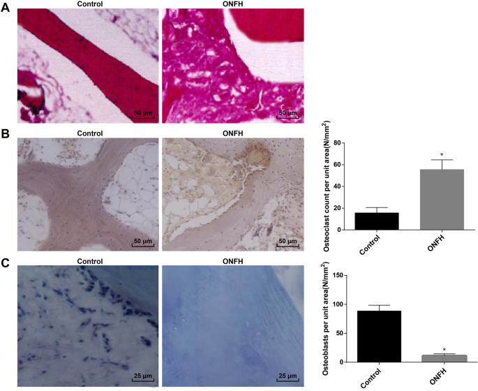
临床样本ONFH组织的病理变化。 一 各组HE染色结果(200×)。 b TRAP染色结果及各组TRAP阳性细胞数(200×)。 c 各组ALP染色及ALP阳性细胞计数结果(400×)。 *P <0.05 vs. 对照组
TRAP染色显示,TRAP主要存在于破骨细胞中,常用于鉴定苋菜中的成熟破骨细胞。与对照组相比,ONFH组TRAP阳性破骨细胞数量增多,破骨细胞形态多样,细胞大,多核外观。与破骨细胞相邻的骨小脑可见明显的骨缺损,表现为骨吸收(图1b)。
ALP染色结果表明,ALP是成骨细胞分化成熟的标志酶,参与骨基质成熟钙化的调控。因此,ALP 表达常用于鉴定成骨细胞。成熟成骨细胞的细胞质中可见褐色或咖啡色颗粒。与对照组相比,ONFH组ALP阳性成骨细胞数量减少(图1c)。
临床样本 ONFH 组织中 MiR-410、ALP、BGLAP 和 Collα1 降解,Wnt-11、ACP5、CTSK 和 MMP9 增强
Western印迹分析和RT-qPCR结果表明ONFH组miR-410的表达较对照组有所减弱,而Wnt-11的表达有所升高;成骨细胞相关因子ALP、BGLAP、Collα1的表达降低;破骨细胞相关因子ACP5、CTSK、MMP9表达升高(均P <0.05)(图 2a-c)。
<图片>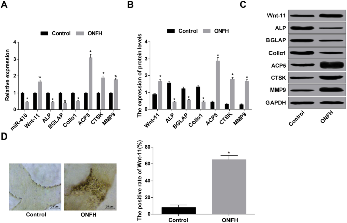
在临床样本的 ONFH 组织中,MiR-410、ALP、BGLAP 和 Collα1 减少,Wnt-11、ACP5、CTSK 和 MMP9 增加。 一 通过 RT-qPCR 检测 miR-410、Wnt-11 以及成骨细胞和破骨细胞相关基因。 b 通过蛋白质印迹分析检测到 Wnt-11 和成骨细胞和破骨细胞相关蛋白。 c Wnt-11 和成骨细胞和破骨细胞相关蛋白的蛋白条带。 d Wnt11表达(400×)与免疫组化染色检测Wnt11蛋白阳性率比较。 *P <0.05 vs. 对照组
免疫组化检测Wnt-11的表达,结果显示Wnt-11蛋白主要在细胞质中表达,阳性表达基本呈棕黄色或褐色。与对照组相比,ONFH组Wnt-11蛋白表达阳性率升高(P <0.05) (图 2d).
上调 miR-410 和下调 Wnt-11 提高大鼠的 BMD 和 BV/TV
Micro-CT提示正常组股骨头外观呈圆形,颈粗细均匀,骨小梁结构连续均匀分布。 ONFH组、agomir-NC组、siRNA-NC组和agomir-miR-410+OE-Wnt-11组大鼠股骨头逐渐塌陷,骨吸收区逐渐出现,股骨颈变细,骨小梁断裂,连续性破坏。 agomir-miR-410组、Wnt-11-siRNA组、agomir-miR-410+OE-NC组大鼠外观完好,无塌陷,无明显骨吸收区出现,骨小梁排列正常,结构完整(图3a)。
<图片>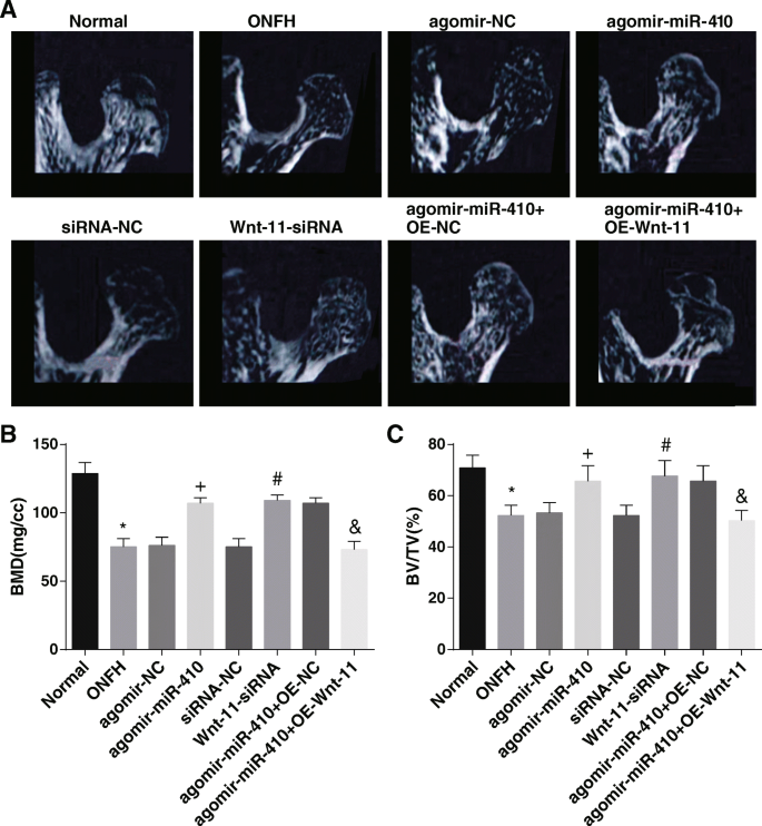
高表达的 miR-410 和低表达的 Wnt-11 会增加大鼠的 BMD 和 BV/TV。 一 各组大鼠显微CT结果。 b BMD 分析结果。 c BV/TV 分析结果。 *P <0.05 与正常组相比。 + P <0.05 与 agomir-NC 组相比。 # P <0.05 与 siRNA-NC 组相比。 & P <0.05 vs. agomir-miR-410 + OE-NC组
骨计量结果显示,与正常组相比,ONFH组的BMD和BV/TV均降低(均P <0.05)。与agomir-NC组和siRNA-NC组相比,agomir-miR-410组和Wnt-11-siRNA组的BMD和BV/TV均升高(均P <0.05)。与agomir-miR-410 + OE-NC组相比,agomir-miR-410 + OE-Wnt-11组的BMD和BV/TV均下降(均P <0.05) (图 3b, c).
miR-410 的过度表达和 Wnt-11 的低表达会提高股骨干、股骨头和脊柱的 BMD 水平,并提高血清钙和大鼠的磷水平
X线片观察正常组股骨头外观呈圆形,股骨头密度均匀,结构完整。 ONFH组、agomir-NC组、siRNA-NC组、agomir-miR-410+OE-Wnt11组大鼠股骨头外观变薄,出现骨吸收区。 agomir-miR-410组、Wnt11-siRNA组、agomir-miR-410组+OE-NC组大鼠股骨头外观均匀、厚度完整(图4a)。 <图片>
上调的 miR-410 和下调的 Wnt-11 增加了股骨干、股骨头和脊柱的骨矿物质密度水平,也提高了大鼠的血清钙和磷水平。 一 动物X线观察结果。 b Comparison of the bone mineral density of the femoral shaft, femoral head, and spine in each group. c Comparison of the serum calcium and phosphorus levels in each group. *P <0.05 vs. the normal group.
+
P <0.05 vs. the agomir-NC group.
#
P <0.05 vs. the siRNA-NC group.
&
P <0.05 vs. the agomir-miR-410 + OE-NC group
BMD and serum calcium and phosphorus level determination reported that compared to the normal group, the BMD of the femoral shaft, femoral head, and spine, as well as the serum calcium and phosphorus levels, fell in the ONFH group (all P <0.05)。 In contrast with the agomir-NC group and siRNA-NC group, the BMD of the femoral shaft, femoral head, and spine, as well as serum calcium and phosphorus levels, ascended in the agomir-miR-410 group and Wnt-11-siRNA group (all P <0.05)。 In relation to the agomir-miR-410 + OE-NC group, the BMD of the femoral shaft, femoral head, and spine, as well as the serum calcium and phosphorus levels in the agomir-miR-410 + OE-Wnt-11 group, was abated (all P <0.05) (Fig. 4b, c).
The results of HE staining showed that the bone trabeculae were neat and clear and arranged regularly and tightly. Bone cells filled the bone lacunae, and the calcified zone was well connected to the subcartilage bone trabeculae in the normal group. In the ONFH group, agomir-NC group, siRNA-NC group, and agomir-miR-410 + OE-Wnt-11 group, the bone trabecula was sparse; thinned, and even broken; the structure was disordered; and some fragments appeared. Some osteocytes in the bone lacuna were necrotic and a large number of bone lacuna was in emptiness that no bone cells filled in, and obvious proliferation of granulation tissue was observed in the necrosis bone trabecular space, which was wrapped around the necrotic bone trabeculae. In the agomir-miR-410 group, the Wnt-11-siRNA group, and the agomir-miR-410 + OE-NC group, the appearance of the rats remained intact, no obvious bone resorption area appeared and the bone trabeculae were arranged normally and the structure was complete (Fig. 5a).
Poor expression of Wnt-11 and overexpression of miR-410 alleviate pathological changes of rats tissues and restrain apoptosis of osteocytes. 一 Morphological observation of the femoral head of rats in each group (200 ×). b Ultrastructural observation of bone cells in rats of each group (8000 ×). c The results of TUNEL staining (200 ×). d Detection of apoptosis rate by TUNEL assay. e Detection of the Bax and Bcl-2 mRNA expression by RT-qPCR. f , g Detection of the Bax and Bcl-2 protein expression by western blot analysis. *P <0.05 vs. the normal group.
+
P <0.05 vs. the agomir-NC group.
#
P <0.05 vs. the siRNA-NC group.
&
P <0.05 vs. the agomir-miR-410 + OE-NC group
Electron microscopic observation observed that the shape of the bone cells in the normal group was consistent with the lacunae, and there was a small gap between the cell and the lacunae wall. The organelles were abundant, the cells in the lacunae were normal, the nucleus was egg-shaped, the nuclear membrane was intact, the chromatin did not agglutinate, and the cytoplasmic pseudo-foot stretched to the peripheral bone small tube and was connected with the adjacent bone cells. Osteoblasts were located on the surface of the bone trabecula, the sample was long, and there were many organelles. In the ONFH group, agomir-NC group, siRNA-NC group, and agomir-miR-410 + OE-Wnt-11 group, a large number of lipid deposits were found in the cytoplasm of osteoblasts, the gap between capsule and lacunae well was further widened, edema bright bands appeared between the nuclear membrane and cytoplasm, nuclear margination showed with compression, nuclear membrane was intact, mitochondria in cytoplasm was swollen, and the endoplasmic reticulum and Gore apparatus disappeared. In the agomir-miR-410 group, Wnt-11-siRNA group, and agomir-miR-410 + OE-NC group, the morphology of rat osteoblasts was normal, chromatin aggregation was found in the nucleus of a small number of cells, no obvious lipid droplets were found in the cytoplasm, and the nuclear membrane was intact (Fig. 5b).
The results of TUNEL staining demonstrated that compared to the normal group, the apoptosis rate of osteocytes in the ONFH group was raised (P <0.05)。 In contrast with the agomir-NC group and the siRNA-NC group, the apoptosis rate of osteocytes abated in the agomir-miR-410 group and the Wnt-11-siRNA group (both P <0.05)。 In relation to the agomir-miR-410 + OE-NC group, the apoptosis rate of osteocytes enhanced in the agomir-miR-410 + OE-Wnt-11 group (P <0.05) (Fig. 5c, d).
Western blot analysis and RT-qPCR reported that the Bcl-2 expression reduced and Bax expression raised in the ONFH group compared to the normal group (both P <0.05)。 By comparison with the agomir-NC group and the siRNA-NC group, Bcl-2 expression ascended and Bax expression descended in the agomir-miR-410 group and the Wnt-11-siRNA group (all P <0.05)。 In contrast with the agomir-miR-410 + OE-NC group, the Bcl-2 expression declined and Bax expression appended in the agomir-miR-410 + OE-Wnt-11 group (both P <0.05) (Fig. 5e–g).
TRAP staining revealed that in the normal group, the morphology of osteoclasts with positive TRAP staining was different, most of them were shuttle or fusiform, few large osteoclasts were polygons, and most of them were distributed around the bone cerebellum. In the ONFH group, agomir-NC group, siRNA-NC group, and agomir-miR-410 + OE-Wnt-11 group, TRAP-positive cells were ascended, the morphology of cells was diverse in large polygons and polykaryotes, bone resorption was found in the bone trabeculae adjacent to osteoclasts, and typical resorption lacunae were formed. In the agomir-miR-410 group, Wnt-11-siRNA group, and agomir-miR-410 + OE-NC group, the number of cells positive for TRAP staining was reduced, the cells were in a long strip shape and the morphology was more regular, the polygonous polynuclear osteoclasts were rare, and the bone structure of adjacent bone trabeculae was relatively complete (Fig. 6a).
Overexpression of miR-410 and low expression of Wnt-11 increase the number of osteoblasts and decrease the number of osteoclasts. 一 Results of TRAP staining in osteoclasts of rats (200 ×). b Count of positive cells in TRAP staining. c Results of ALP staining in osteoblasts of rats in each group (200 ×). d Count of positive cells determined by ALP staining. *P <0.05 vs. the normal group.
+
P <0.05 vs. the agomir-NC group.
#
P <0.05 vs. the siRNA-NC group.
&
P <0.05 vs. the agomir-miR-410 + OE-NC group
ALP staining presented that in the normal group, in osteoblasts, which were positive for ALP staining, the morphology was small and round. The cells were distributed in aggregation, most of which were located in the bone trabecular space of bone marrow cavity and on the surface of some bone trabeculae. In the ONFH group, agomir-NC group, siRNA-NC group, and agomir-miR-410 + OE-Wnt-11 group, the number of osteoblasts positive for ALP staining was dispersedly distributed in the trabecular space of the bone marrow cavity. In the agomir-miR-410 group, Wnt-11-siRNA group, and agomir-miR-410 + OE-NC group, the number of osteoblast-positive cells enhanced, and they were distributed in the trabecular space of the bone marrow cavity and the surface of the part of the bone trabecular (Fig. 6c).
By comparison with the normal group, the number of osteoblasts per unit area was suppressed and the number of osteoclasts was heightened in the ONFH group (both P <0.05)。 In relation to the agomir-NC group and the siRNA-NC group, the number of osteoblasts per unit area was heightened and the number of osteoclasts was declined in the agomir-miR-410 group and Wnt-11-siRNA group (all P <0.05)。 In contrast to the agomir-miR-410 + OE-NC group, the number of osteoblasts per unit area was declined and the number of osteoclasts was raised in the agomir-miR-410 + OE-Wnt-11 group (both P <0.05) (Fig. 6b, d).
The results of ELISA revealed that in relation to the normal group, the level of osteogenic function index ALP and OCN degraded while the levels of osteoclast function index NTX-1 and CTX-1 heightened in the ONFH group (all P <0.05)。 In contrast with the agomir-NC group and the siRNA-NC group, ALP and OCN ascended and NTX-1 and CTX-1 descended in the agomir-miR-410 group and the Wnt-11-siRNA group (all P <0.05)。 By comparison with the agomir-miR-410 + OE-NC group, ALP and OCN dropped and NTX-1 and CTX-1 appended in the agomir-miR-410 + OE-Wnt-11 group (all P <0.05) (Fig. 7a).
Highly expressed miR-410 and lowly expressed Wnt-11 elevate the expression of OCN, ALP, BGLAP, and Collα1 as well as abate NTX-1, CTX-1, ACP5, CTSK, and MMP9 expression in ONFH rats. 一 Detection of osteogenesis and osteoclast function indices in the serum of rats in each group by ELISA. b Detection of osteoblast- and osteoclast-related genes by RT-qPCR. c , d Detection of osteoblast- and osteoclast-related genes protein expression by western blot analysis. *P <0.05 vs. the normal group.
+
P <0.05 vs. the agomir-NC group.
#
P <0.05 vs. the siRNA-NC group.
&
P <0.05 vs. the agomir-miR-410 + OE-NC group
The results of western blot analysis and RT-qPCR revealed that in relation to the normal group, the expression of osteoblast-related factors ALP, BGLAP, and Collα1 degraded, and osteoclast-related factors ACP5, CTSK, and MMP9 expression ascended (all P <0.05)。 In contrast with the agomir-NC group and the siRNA-NC group, the expression of ALP, BGLAP, and Collα1 elevated, and ACP5, CTSK, and MMP9 expression abated in the agomir-miR-410 group and the Wnt-11-siRNA group (all P <0.05)。 By comparison with the agomir-miR-410 + OE-NC group, the expression of ALP, BGLAP, and Collα1 depressed, and ACP5, CTSK, and MMP9 expression heightened in the agomir-miR-410 + OE-Wnt-11 group (all P <0.05) (Fig. 7b–d).
The targeting relationship between miR-410 and Wnt-11 gene was analyzed by an online analysis software. It was displayed that a specific binding region existed between Wnt-11 gene sequence and miR-410 sequence, implying that Wnt-11 was the target gene of miR-410 (Fig. 8a). Luciferase activity assay was utilized to verify this relationship (Fig. 8b). The results presented that compared to the NC group, the luciferase activity depressed in the miR-410 mimics group (P <0.05), but there was no significant difference in the luciferase activity of MUT 3′UTR (P> 0.05), indicating that miR-410 could specifically bind to Wnt-11.
Increased Wnt-11 and decreased miR-410 are found in ONFH tissues as well as Wnt-11 is the target gene of miR-410. 一 Prediction of binding sites of miR-410 on Wnt-11 3′UTR. b Luciferase activity detection for verifying the relationship between miR-410 and Wnt-11. c Detection of miR-410 and Wnt-11 expression by RT-qPCR. d , e Wnt-11 protein expression determined by western blot analysis. f Expression of Wnt-11 in femoral head of rats in each group detected by immunohistochemistry (200 ×). g Comparison of the positive rate of Wnt-11 protein expression in each group. *P <0.05 vs. the normal group.
+
P <0.05 vs. the agomir-NC group.
#
P <0.05 vs. the siRNA-NC group.
&
P <0.05 vs. the agomir-miR-410 + OE-NC group
The results of western blot analysis and RT-qPCR revealed that in relation to the normal group, miR-410 expression declined and Wnt-11 expression raised in the ONFH group (both P <0.05)。 By comparison with the agomir-NC group, miR-410 raised and Wnt-11 reduced in the agomir-miR-410 group (both P <0.05)。 In contrast with the siRNA-NC group, Wnt-11 declined in the Wnt-11-siRNA group (P <0.05)。 Wnt-11 enhanced in the agomir-miR-410 + OE-Wnt-11 group relative to that in the agomir-miR-410 + OE-NC group (P <0.05) (Fig. 8c–e).
>Wnt-11 expression was verified by immunohistochemistry. Wnt-11 protein was mainly expressed in the cytoplasm, and the positive expression was mainly brownish yellow or brown. Compared to the normal group, the positive rate of Wnt-11 protein expression in the ONFH group was raised (P <0.05)。 By comparison with the agomir-NC group and the siRNA-NC group, the positive rate of the Wnt-11 protein expression descended in the agomir-miR-410 group and Wnt-11-siRNA group (both P <0.05)。 In relation to the agomir-miR-410 + OE-NC group, the positive rate of Wnt-11 protein expression enhanced in the agomir-miR-410 + OE-Wnt-11 group (P <0.05) (Fig. 8f, g).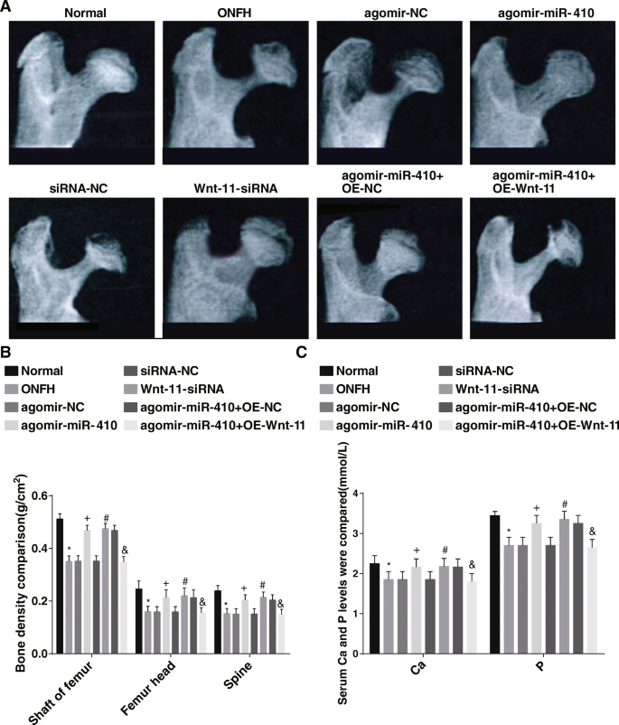
Silencing Wnt-11 and Upregulating miR-410 Alleviate the Pathological Changes of Rat Tissues and Restrain Apoptosis of Osteocytes

Overexpression of miR-410 and Low Expression of Wnt-11 Increase the Number of Osteoblasts and Decrease the Number of Osteoclasts

Highly Expressed miR-410 and Lowly Expressed Wnt-11 Elevate the Expression of Osteocalcin (OCN), ALP, BGLAP, and Collα1, as well as Abate C- and N-terminal Telopeptides of Type I collagen (NTX-1, CTX-1), ACP5, CTSK, and MMP9 expression in ONFH Rats

Elevated Wnt-11 and Decreased miR-410 are Found in ONFH Tissues of Rats as well as Wnt-11 is the Target Gene of miR-410

Discussion
ONFH, a kind of bone destruction disease, is caused by blood supply failure and coagulation and fibrinolysis system disorder and finally causes the femoral head to collapse [19]. A previous study has discussed miRNA expression in bone marrow mesenchymal stem cells induced by hormone in mice with ONFH [20]. Also, a recent study has provided a proof that the expression profile of miRNA of bone marrow-derived mesenchymal stem cells in osteogenesis related to steroid-induced ONFH [21]. Furthermore, it was revealed the Li-nHA/GM/rhEPO stents can elevate both Wnt and HIF-1/VEGF pathways and promote osteogenesis and angiogenesis, which is beneficial to the repair of ONFH induced by glucocorticoids [22]. Based on these facts, the study aimed to explore the effects of miR-410 in the prevention of ONFH by targeting Wnt-11.
Our study has provided substantial evidence in relation to the notion that miR-410, ALP, BGLAP, and Collα1 degraded and Wnt-11, ACP5, CTSK, and MMP9 enhanced in ONFH tissues. A recent study has promoted that the expression of miR-410 in osteosarcoma is declined, and the anti-tumor effect is shown [23]. Another study has presented miR-410 expression was reduced in human estrogen receptor-positive tissues of breast cancer [24]. It is reported that secreted factor Wnt-11 expression is ascended in several types of cancer, containing colorectal cancer, where it advances the migration and invasion of cancer cells [25]. Similarly, a previous study has proved that Wnt-11 expression is heightened in hormone-independent prostate cancer [26]. ALP is a useful index for the state diagnosis and clinical prognosis of the disease [27]. OCN (bone gamma-carboxyglutamate protein; BGLAP) is a widely conserved molecule related to mineralization of bone matrix [28]. ACP5 is necessary for osteoclast differentiation and bone resorption, and it promotes cell movement by regulating adhesion kinase phosphorylation [29]. CTSK is a critical protease in charge of osteopontin, degrading type I collagen and other bone matrix proteins [30]. MMPs can degrade and modify most of the components of extracellular matrix and basement membrane and push forward an immense influence on cancer invasion and metastasis [31]. Also, our study revealed that Wnt-11 is the target gene of miR-410. Similarly, Zhang et al. found that miR-410 targeted the inferred binding site in the Wnt3a 3′-UTR to modulate the Wnt signaling pathway [32].
In addition, it was revealed that upregulated miR-410 and downregulated Wnt-11 increased BMD and BV/TV of ONFH rats. It has been suggested previously that a decrease of spinal BMD and an increase of urinary DPD/Cr ratio in non-traumatic ONFH patients [33]. Another study has verified that BMD of the femoral head and lumbar vertebrae in the ONFH group were degraded relative to that in the control group [34]. Additionally, imaging analysis revealed muscone can restore BMD and BV/TV ratio of the necrotic femoral head, while histologic examination further confirmed the protective effect of muscone on alcohol-induced ONFH [35]. A result emerged from our data that highly expressed miR-410 and lowly expressed Wnt-11 restrained apoptosis of osteocytes. A study has demonstrated that silencing miR-410 can induce cell proliferation and reduce the apoptosis of human umbilical vein endothelial cells induced by oxidized low-density lipoprotein [36]. It is reported that the descended miR-410 expression and ascended SOCS3 expression could reduce the expression of anti-apoptosis factor Bcl-2 and promoted the apoptosis of cells [37]. In addition, in androgen deficient LNCaP cells, the downregulation of Wnt-11 can prevent neuroendocrine-like differentiation and lead to apoptosis of prostate cancer cells [26]. The study also showed that the downregulation of Wnt-11 and upregulation of miR-410 enhanced ALP, BGLAP, and Collα1 expression and reduced ACP5, CTSK, and MMP9 expression. A study has indicated that Wnt-11 expression heightened in cervical cancer cells may result in activation and phosphorylation of JNK-1 by activating Wnt/Jnk pathway and boosts the proliferation and migration/invasion of tumor cells [15]. It has been suggested that Wnt-11 gene silencing in colorectal cancer cell lines reduced the invasive ability of cells [25]. Furthermore, the overexpression of miR-410 can restrain the invasion, migration, proliferation, and epithelial mesenchymal transformation of osteosarcoma cells via directly targeting TRIM44 [23]. Furthermore, a previous study stated that upregulated miR-410 inhibited the growth of cholangiocarcinoma in a xenograft mouse model by inducing apoptosis [38].
Conclusion
In summary, our investigation revealed that miR-410 was lowly expressed and Wnt-11 was highly expressed in ONFH, and upregulating miR-410 or downregulating Wnt-11 increased osteoblasts and reduced osteoclasts to alleviate ONFH. However, clinical researches might be further carried out to detect the efficacy of miR-410 and Wnt-11 for the treatment of ONFH.
数据和材料的可用性
Not applicable
更改历史
缩写
- ALP:
-
碱性磷酸酶
- ANOVA:
-
Analysis of variance
- BMD:
-
Bone mineral density
- EDTA:
-
Ethylene diamine tetraacetic acid
- ELISA:
-
酶联免疫吸附试验
- GAPDH:
-
Glyceraldehyde phosphate dehydrogenase
- 通用:
-
General Electric
- HE:
-
Hematoxylin-eosin
- LSD-t:
-
Least significant difference t 测试
- miR-410:
-
MicroRNA-410
- miRNAs:
-
MicroRNAs
- NC:
-
Negative control
- OD:
-
Optical density
- ONFH:
-
Osteonecrosis of femoral head
- PBS:
-
磷酸盐缓冲盐水
- ROI:
-
感兴趣的区域
- RT-qPCR:
-
Reverse transcription quantitative polymerase chain reaction
- 标准差:
-
斯普拉格-道利
- SDS:
-
十二烷基硫酸钠
- THR:
-
Total hip replacement
- TRAP:
-
Tartrate resistant acid phosphatase
- TUNEL:
-
TdT-mediated dUTP-biotin nick end-labeling
纳米材料