

设计载有适体金纳米颗粒的 pH 敏感脂质体包裹 Morin 以治疗癌症
摘要
本研究提出合成一种抗癌纳米颗粒、适体和 Au 纳米颗粒 (Apt-Au) 修饰的 Morin pH 敏感脂质体 (MSL),其具有靶向特性。肿瘤难以治愈,因为它们的微环境与正常组织的微环境不同;它的pH值低于正常组织的pH值,这通常会阻碍药物的有效性。因此,pH响应性药物引起了广泛的关注。金纳米粒子(AuNPs)由于其体积小、生物相容性好、易于表面修饰和细胞渗透性强而具有作为药物载体的潜力。 Apt-Au@MSL 表现出优异的单分散性和肿瘤靶向特性,可以通过透析在部分酸性环境中释放。我们通过 MTT 法筛选了我们的模型癌细胞,发现 SGC-7901 细胞可以有效抑制增殖。体内结果表明,Apt-Au@MSL 的给药可以抑制异种移植小鼠模型中的肿瘤生长。 H&E染色和TUNEL实验进一步证实Apt-Au@MSL可以促进肿瘤细胞凋亡。 Apt-Au@MSL 可能通过触发活性氧 (ROS) 的过度产生和调节多信号串扰来诱导细胞凋亡。血液生化测试和 H&E 染色均表明这些材料在体内表现出可忽略不计的急性毒性和良好的生物相容性。凭借其强大的功能,Apt-Au@MSL可作为靶向抗癌材料用于未来临床癌症治疗。
介绍
癌症是最常见的死因之一 [1],可导致重大经济损失和对人类的伤害。已经为开发用于癌症治疗的智能药物做出了相当大的努力[2]。由于它们能够保护药物直到它们到达目标部位,聚合物纳米粒子可以潜在地改善治疗药物的递送 [3]。为确保将治疗剂递送至活性部位,使用高反应性纳米颗粒。这种纳米粒子可以设计成在不同的生物刺激下改变材料特性。反应性纳米粒子使用不同的刺激进行设计,包括外部刺激(例如温度和光)和生物刺激(例如 pH 或氧化还原条件)。由于纳米颗粒内吞作用后 pH 值发生变化,因此对 pH 值敏感的 NPs 引起了研究兴趣。响应 pH 值的材料也引起了人们的注意,因为 pH 响应功能可以很容易地集成到一系列聚合物结构中,以设计一组响应 pH 值的纳米粒子。纳米颗粒可以通过改变表面化学、改变颗粒大小或形状以及分解或释放物质来响应 pH 值。纳米粒子特性的这种变化可用于调节细胞摄取和控制释放。因此,pH响应性纳米粒子为治疗给药系统的设计提供了强有力的策略。
Morin hydrate (3,5,7,2',4'-pentahydroxyflavone)(图 1)是一种从中草药或植物中分离出来的天然活性物质 [4]。它是一种孕酮化合物,是植物中苯酚的次级代谢产物。 Morin在自然界中分布广泛,具有良好的抗氧化、抗癌[5]和显着的抗炎作用。 Morin 的各种应用潜力引起了广泛关注 [6],但这种潜力受到该物质低水溶性和生物利用度的限制 [7]。
<图片>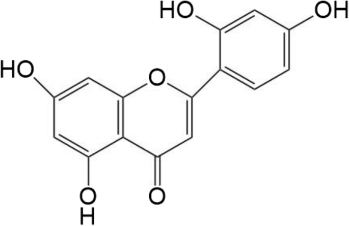
Morin水合物的化学结构
由卵磷脂和神经酰胺等组成的脂质体(中空)具有由两亲脂质分子组成的双层结构 [8]。脂质体提供了理想的特性,例如生物相容性、功能性、减少的副作用以及封装大量药物的能力 [9, 10]。它们可以有效地携带亲水性和疏水性试剂并保护它们免受外部条件的影响,成功地将它们运输到目标组织区域 [11]。设计中加入了改进的功效,以促进通过 pH 敏感脂质体在肿瘤间质内由 pH 触发的抗癌物质的释放 [12,13,14,15]。这些pH敏感性脂质体是一种特殊的脂质体,在组织环境中释放药物,在酸性环境中迅速失稳,如癌组织内体[16,17,18]。
为了增强肿瘤治疗的选择性和疗效,应修改 pH 敏感脂质体以形成靶向肿瘤的纳米颗粒 [19]。 DNA适体最近被开发为高度选择性和敏感的生物传感和成像传感器,以及癌症靶向治疗的潜在药物[20]。单链 DNA 适体 AS1411 已被证明可用作化疗剂,因为它对癌细胞核具有高结合亲和力 [21]。这种适体 (Apt) 与 Au NPs 相结合,制造出一种双组分纳米结构,该纳米结构由负载 Apt 的 Au NPs 组成,可以与癌细胞核相互作用。一些研究还表明,脂质体-纳米颗粒混合物的设计可以为制造这种多功能模式提供丰富的工具箱 [22]。一种由阳离子脂质体和Au NPs组成的混合脂质体-Au NP囊泡系统旨在提高药物在肿瘤间质内的渗透并增强抗癌活性[23, 24]。
金纳米粒子 (Au NPs) 被认为是良好的药物载体,可以用生物相关分子进行修饰,以增强癌症靶向特异性 [25]。通过Au NPs的表面修饰,可以利用巯基的适配体修饰Au NPs的表面,可以通过Au-S共价键靶向,将纳米金表面适当的巯基连接到纳米金表面 [26]。从工程和应用的角度来看,配体结合的金纳米材料为促进靶向识别、检测和治疗提供了一个强大的平台。
在目前的研究中,我们使用了包裹 Morin 的 pH 敏感脂质体并组装了在脂质体表面修饰的 Au-Apt(Apt-Au@MSL,图 3a)。以低水溶性和生物利用度为特征的 Morin 被转化。表征了制备的 Apt-Au@MSL 的形态、尺寸和其他特性。新的纳米颗粒显示出靶向肿瘤的特性和高抗癌活性。体外和体内探索了Apt-Au@MSL的抗癌活性(图2)。
<图片>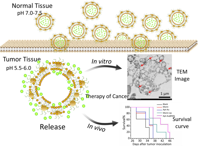
我们设计的含有 Morin 的 Apt-Au@MSL 用于将药物递送至具有肿瘤靶向特性的癌细胞的示意图。
材料和方法
材料
L-α-磷脂酰胆碱 (PC)、胆固醇 (Chol)、胆固醇半琥珀酸酯 (CHEMS)、莫林 (≥ 99.99%)、柠檬酸钠 (99.9%)、HAuCl4·3H2O (≥ 99.9%)、聚乙烯吡咯烷酮 (PVP0, wt 00) 、NaOH (≥ 98.0%) 和 HCl (37%) 购自 Sigma-Aldrich Chemical Co. (USA)。具有二硫键修饰的 AS1411 适配体由 TaKaRa(中国大连)合成,序列如下:5'-HS-T-(C6-S-S-C6)-TTG GTG GTG GTG GTT GTG GTG GTG GTG G-3'。噻唑蓝溴化四唑 (MTT)、碘化丙啶 (PI)、钙黄绿素和 Alexa Fluor 488 Annexin V 购自 Sigma-Aldrich Chemical。所有水溶液均在双蒸水中制备。所有其他试剂均为市售最好的。
金纳米粒子的合成
将高达 1 mL 的 HAuCl4·4H2O(1%)溶解在 25 mL Milli-Q 水(pH =5.5)中,然后将 50 mg PVP 加入溶液中。将所得溶液倒入三颈烧瓶(150 mL)中,并在机械搅拌下通过微波(MW)照射处理5 分钟。随后,将 1.5 mL 柠檬酸钠溶液 (1%) 快速加入该溶液中,并进行 MW 辐照 3 分钟。 10000 rpm离心5 分钟收集溶液。
Apt-Au NPs 的合成
使用三(2-羧乙基)膦裂解适体的二硫键。 30 分钟后,将适体(100 μL,100 μM)溶液添加到10 mL 5 nM Au NPs溶液中,然后孵育24 小时以形成Au-Apt NPs。 1 天后,我们用 2.5 mL 的 500 mM NaCl 溶液将混合物溶液加盐两次,相隔 4 小时 [27]。
pH 敏感脂质体的制备
通过膜水合制备由摩尔比为 33:13:5 的鸡蛋 PC 与 CHEMS 和 Chol 和 5% Morin 组成的脂质体。 Egg PC、CHEMS、Chol 和 2 mg Morin 溶解在 8 mL CHCl3 中,并在 40 °C 下用旋转蒸发器除去有机溶剂。对于 Morin-脂质体,所得脂质薄膜使用 2% (w/v) PBS 水合,然后使用冰浴探针超声 15 分钟。最终的脂质体在胶体系统中合成并稳定。对于 Apt-Au@Morin pH 敏感脂质体 (Apt-Au@MSL),脂质膜在 5% 葡萄糖中与 475 μg/mL Au-Apt NPs 水合,然后超声处理 [23]。
特征化
进行了紫外-可见光谱(UV-Vis,S-3100 光电二极管阵列,Scinco Co., Ltd.,Korea)和傅里叶变换红外光谱(FT-IR)(Nicolet iS50,美国,Thermo Fisher Scientific)。通过透射电子显微镜(TEM,HT7700,Tokyo Japan,Hitachi)检查 Apt-Au@MSL 的形态。 Morin 的释放百分比通过紫外-可见光谱法检测。还通过扫描电子显微镜(SEM,S-4800,Hitachi,Japan)观察了 Morin-脂质体和 Apt-Au@MSL 的形态。动态光散射 (DLS) 和 zeta 电位测量用于在 Brookhaven ZetaPALS 电位分析仪上表征 Morin-脂质体和 Apt-Au@MSL 的光学性质和大小。
细胞培养
人癌细胞系 SGC-7901、BGC-823、A549、HeLa、MCF-7 和 Hs68 购自美国典型培养物保藏中心 (ATCC),并在含有 10% 胎牛血清的 RPMI 中维持在 37 °C 和5% 二氧化碳。
体外抗癌活性研究
SGC-7901、BGC-823、A549、HeLa、MCF-7和Hs68接种于96孔板(2.0 × 10 3 细胞/孔),然后与不同浓度(0、5、10、15、20 和 30 μg/mL)的 Apt-Au@MSL 一起培养。 Au NPs和Morin被指定为对照组。空白组由未经处理的细胞组成。 96 孔板在加湿培养箱中培养。孵育后,每孔加入9.6 mL MTT溶液(5 mg/mL),继续孵育2 h。每组一式三份进行测试,IC50 值源自三份一式三份测试的平均 OD 值与药物浓度曲线的关系。通过共聚焦荧光显微镜获得各组的亮度[28]。
使用实时细胞电子传感系统(RT-CES;ACEA Biosciences, Inc.)每 10 分钟监测一次细胞粘附,持续 75 小时。为了确定细胞粘附,在板中的每个孔中接种细胞(1.0 × 10 4 细胞/孔)用新鲜培养基至终体积为 200 μL,然后在不同浓度(10、20 和 30 μg/mL)的 Apt-Au@MSL 溶液中孵育 10 小时,孵育 12 小时 [29] .空白组由未经处理的细胞组成,对照组由Morin组和Morin-脂质体组组成。
荧光显微镜
SGC-7901 细胞在 48 孔板中在 37 °C、5% CO2 下生长过夜。然后将细胞用不同浓度的 Apt-Au@MSL 溶液(10 和 30 μg/mL)处理 12 小时。对照组由 Morin 和 Morin-脂质体组组成。随后,除去培养基。空白组由未经处理的细胞组成。用PBS洗涤细胞3次。细胞被染色,然后通过活细胞和死细胞的共染色(LIVE/DEAD 测定)进行测量。与Calcein-AM/PI共染色30 min后,用PBS洗涤细胞两次以去除多余的染料,通过共聚焦激光扫描显微镜(CLSM)获得荧光图像(对于calcein-AM,Ex =488 nm和 Em =515 nm;PI Ex =535 nm 和 Em =615 nm [30]).
凋亡细胞死亡分析
为了测量 Apt-Au@MSL 的抗癌作用,在六孔板的每个孔中接种 SGC-7901 细胞(1.0 × 10 5 细胞/孔),然后在不同浓度(5、10、20 和 30 μg/mL)的 Apt-Au@MSL 溶液中孵育 12 小时。空白组由未经处理的细胞组成,对照组由 Morin 和 Morin-脂质体组组成。处理后,收集细胞并用PBS洗涤两次。细胞用 PI 和 Annexin V-FITC 染色,然后使用 CytoFLEX 分析仪(Beckman Coulter)进行分析(对于 PI,Ex =535 nm 和 Em =615 nm;Annexin V-FITC,Ex =488 =nm 和 Em 52 ) [31]。
关于墙壁破坏的研究
为了研究抗癌试验中的细胞壁破坏,SGC-7901 细胞在 6 孔板中在 37 °C 和 5% CO2 条件下生长。无材料培养的细胞作为空白组。将细胞与不同浓度(5、10、20 和 30 μg/mL)的 Apt-Au@MSL 培养 12 h 后,用 0.25% 的胰蛋白酶-EDTA 溶液处理细胞。以2000 r/min 5 min收集所有细胞(包括培养基中的漂浮细胞)。然后将收集的细胞在 2.5% 戊二醛溶液中后固定 4 小时 [32]。固定的细胞在丙酮梯度系列中脱水 20 分钟。细胞最终经过一系列程序,安装在铜网上,并通过TEM(HT-7700,Hitachi,Japan)进行观察。
蛋白质印迹分析
通过Western印迹分析检测SGC-7901细胞的蛋白表达。 SGC-7901 细胞(4.0 × 10⁵ 个细胞/孔)接种在 9 厘米培养板和 20 μg/mL Apt-Au@MSL 上 1、6、12 和 24 小时。处理后收获细胞并悬浮在冰上的细胞裂解缓冲液中1 小时。将混合物以 11,000g 置于离心机中 4 ℃保温10 min,收集含有总细胞蛋白的上清液。使用BCA方法测定上清液中的总蛋白质浓度。然后将蛋白质重新悬浮在上样缓冲液中,并在 100°C 下煮沸 10 分钟。对含有等量蛋白质(40 μg/泳道)的样品进行 SDS-PAGE(10% tricine 凝胶)。然后在 100 V 下将它们转移到硝化纤维 (NC) 膜上 1.5 小时,并使用含 0.1% Tween-20 (TBST) 的 Tris 缓冲盐水中的 5% 脱脂牛奶封闭 1 小时。然后,NC 膜分别与一抗和二抗一起孵育。使用 ECL 蛋白质印迹检测试剂在膜上可视化目标蛋白条带。 β-肌动蛋白用作等量蛋白质加载和转移的内部对照。用Quantity-One软件对蛋白表达量进行定量,并在条带下标记表达率[33]。
动物模型
BALB/c雄性裸鼠(~ 17 g)购自南京大学模型动物研究中心,在无菌环境中饲养(无特定病原体动物)。通过皮下注射细胞悬液(SGC-7901细胞,100 μL,1 × 10 6 )建立肿瘤模型 /mL) 注入裸鼠肩部。皮下注射肿瘤细胞后 15 天生成明亮的肿瘤图像。所有动物实验均按照安徽农业大学实验动物中心批准的方案进行(许可证编号:SYXK 2016-007)。使用游标卡尺确定每个肿瘤的最大纵向直径(长度)和最大横向直径(宽度)。然后使用公式length × width 2 计算肿瘤体积 × 0.5 [34].
体内抗癌研究
当肿瘤体积达到50 mm 3 时进行随访测试 .将小鼠随机分为五组,接受以下静脉治疗:PBS(100 μL)、Morin、Morin-脂质体、Au-Apt 和 Apt-Au@MSL。以2 mg/kg的治疗材料的剂量将小鼠静脉注射到尾部。 24 天后测量小鼠的肿瘤大小。还测定了小鼠的体重和存活率。对小鼠实施安乐死,取小鼠肿瘤和器官组织进行H&E染色[35, 36]。
封闭和透化后,用 PBS 洗涤肿瘤载玻片,用 TUNEL 测定染色,并进行 DAPI 复染。通过 CLSM 获得荧光图像。对于 DAPI 成像,Ex =358 nm 和 Em =461 nm,对于 TUNEL 检测,Ex =450–500 nm 和 Em =515–565 nm [37]。
毒性评估分析
为了评估 Apt-Au@MSL 治疗对重要器官的毒性,4 个治疗组的小鼠在 30 天后被处死。采集各组小鼠重要器官(肝、脾、肾、心、肺、肿瘤)并进行H&E染色。还收集了血液样本,并测量了血糖。此外,还测定了小鼠的体重[38]。
结果与讨论
特征化
本研究旨在开发一种新型脂质体,该脂质体具有纳米颗粒(载 Apt 的 Au NPs)的优点,可有效用作抗癌剂。新脂质体的基本特性对于进行进一步的研究很重要。 Apt-Au@MSL 按照之前的研究 [23] 中的描述合成,稍作修改,然后使用各种方法进行表征(图 3)。图 3b 显示了 UV-vis 光谱的结果,它显示了特征峰并且可以监测 Au NPs 的形成。在约 530 nm 处的 Au NPs 的 UV-vis 光谱中观察到特征峰。具体而言,紫外-可见光谱分析表现出强烈的紫外吸收,这受表面 Apt 修饰的影响。观察到相同的结果。在 pH 敏感脂质体用 Morin 包被后,特征峰被取代。制备的 Apt-Au@MSL 的特征峰位于 362 和 550 nm。这些结果表明合成成功完成。为了进一步表征,进行了 FT-IR 光谱来验证合成结果。 Morin-脂质体和 Apt-Au@MSL 的特征峰红移进一步表明 Morin 的成功修饰(图 3c)。我们在不同 pH 水平的 PBS 中进行了 Morin 释放测定,以确定 Apt-Au@MSL 的 pH 敏感性。这三个pH值模拟了血液循环的中性环境、肿瘤的弱酸性环境和细胞内的酸性环境。 Morin 的释放百分比通过紫外-可见光谱法检测。在pH 5.0时,约54%的Morin在24 h内释放,在随后的96 h内观察到持续释放;同时,在 pH 7.4 时,24 h 内仅释放约 10% 的 Morin。这些结果表明Apt-Au@MSL在正常生理条件下表现出良好的稳定性,防止药物泄漏;然而,药物在核体内迅速释放。这种药物释放行为可以有效提高治疗效果(图 3d)。进行 TEM 和 SEM 以阐明脂质体和 Apt-Au@MSL 的结构(图 3f)。进行TEM以阐明AuNP的结构。如图 3g 所示,AuNP 的粒径约为 10 nm,这与脂质体修饰的大小一致(图 3g)。图 3e 中的 SEM 结果表明原始脂质体仅形成单层囊泡,其直径不均匀约为 120 nm,这与 DLS 确定的大小一致。相比之下,Apt-Au@MSL 表明混合囊泡的形态归因于 Au-Apt 修饰。 150 nm 的直径也与 DLS 确定的尺寸一致(图 3i)。值得注意的是,对 pH 敏感脂质体和 Au-Apt 之间相互作用的 TEM 研究表明,Au NPs 几乎完全与囊泡脂质双层相关并位于囊泡的外围(图 3h)。 ζ 电位测量值如图 3j 所示,Au NPs 和 Au-Apt 的 zeta 电位为负。结果表明,Au NPs(- 57.1 ± 0.3 mV)和Au-Apt都带负电(- 31.7 ± 0.2 mV)。 Apt-Au@MSL 用带正电荷的 Morin-脂质体修饰,电位变为 36.4 ± 0.3 mV。正负电荷吸附是 Au-Apt 和 Morin-脂质体结合的机制。此外,图 3k 表示粒子包封率随时间的变化,表明粒子在一段时间内的稳定性。如图 3k 所示,颗粒的包封率在 24 h 内几乎没有变化,表明颗粒在一段时间内表现出良好的稳定性。制备 Morin 浓度与 Morin 紫外-可见光吸收的标准曲线以研究 Morin-脂质体和 Apt-Au@MSL 的包封率。结果表明,Morin-脂质体和Apt-Au@MSL的包封率分别达到90.2%和89.6%,如表S1和图S1
<图片>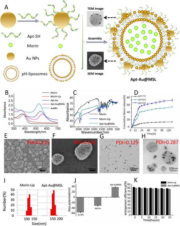
表征图像。 一 Apt-Au@MSL 合成示意图。 b Morin、Au NPs、Au-Apt、Morin-脂质体和 Apt-Au@MSL 的紫外吸收光谱。 c Morin、Morin-脂质体和 Apt-Au@MSL 的 FT-IR 光谱仪。 d 不同 pH 条件下 Morin 从 Apt-Au@MSL 中的释放行为。 e Morin-脂质体的 SEM 图像。 f Apt-Au@MSL 的 SEM 图像。 g Au NPs的TEM图像。 h Apt-Au@MSL 的 TEM 图像。 我 Morin-脂质体和 Apt-Au@MSL 的直径至少通过 DLS 确定三次。 j Au NPs、Apt-Au 和 Apt-Au@MSL 的 ζ-电位。 k Morin-脂质体和Apt-Au@MSL的包封率(EE%)
Apt-Au@MSL 抗癌活性试验
体外
Morin 具有优异的抗肿瘤活性,但由于其水溶性低,因此生物利用度低。 Morin被pH敏感性脂质体包裹并转化为水溶性物质后,利用Au-Apt修饰脂质体表面,获得靶向抗肿瘤作用。图 4a 显示了 Apt-Au@MSL 通过体外 MTT 测定的抗癌活性。药物浓度由 Morin 浓度定义。与空白组相比,Morin 治疗组没有表现出明显的抗癌活性。然而,用pH敏感脂质体包裹的Morin处理的组表现出增强的抗癌活性。结果表明,桑椹脂质体的抗肿瘤活性优于生桑椹。同时,我们发现Apt修饰的脂质体(Apt-Au@MSL)的抗肿瘤活性得到提高。表 1 列出了 Morin、Morin-脂质体、Apt-Au@MSL 和顺铂的 IC50 值。 Morin、Morin-脂质体和 Apt-Au@MSL 对肿瘤细胞表现出广泛的抗癌活性。他们对正常人体细胞具有高效能和低毒性的 SGC-7901 细胞表现出明显的偏好。对于 SGC-7901 细胞,Apt-Au@MSL 的 IC50 值为 15.6 ± 1.5 μg/mL。 RTCA 测试进一步证实了这些结果。 SGC-7901细胞用不同浓度的Apt-Au@MSL培养;空白组用PBS处理,对照组由Morin和Morin-脂质体组成。使用 iCELLigence 监测粘附和扩散(图 4b)。结果与通过MTT测试获得的结果基本相似。 Apt-Au@MSL 的抗肿瘤活性显着高于 Morin 和 Morin-脂质体。 Apt-Au@MSL 可以随着浓度的增加而抑制 SGC-7901 细胞的增殖。荧光显微镜还观察到 Apt-Au@MSL 处理后细胞形态的变化。如图 4c 所示,没有 Apt-Au@MSL 的 SGC-7901 细胞保持了细胞的结构完整性。相比之下,细胞用不同浓度的Apt-Au@MSL处理后细胞完整性受到损害。
<图片>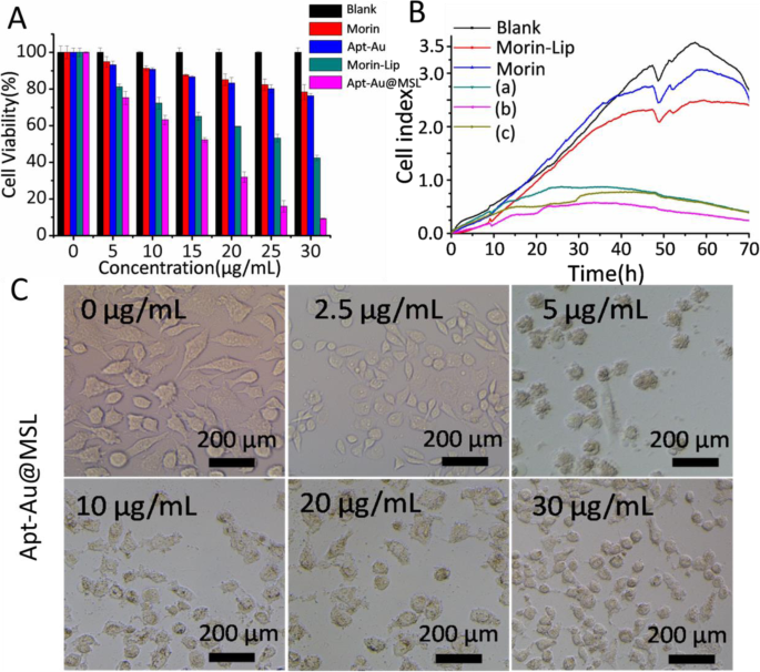
一 SGC-7901 细胞与不同浓度(0、5、10、15、20 和 30 μg/mL)的不同材料(Morin、Au-Apt、Morin-脂质体和 Apt-Au@MSL)一起孵育的细胞活力。 PBS处理过的细胞作为空白组。 b RT-CES系统检测SGC-7901细胞增殖曲线。在 10 h 时加入不同的化合物。 a 的浓度 , b , 和 c 分别是不同的 Apt-Au@MSL(10、20 和 30 μg/mL)。 c 不同浓度(0、2.5、5、10、20和30 μg/mL)下的亮细胞图像
凋亡细胞死亡分析
使用膜联蛋白-FITC 染色通过流式细胞术对细胞凋亡进行定量分析。细胞用 PI 和膜联蛋白 V-FITC 染色,然后使用 CytoFLEX 流式细胞仪(Beckman Coulter)进行分析。 Annexin V 用于检测与暴露的磷脂酰丝氨酸结合的早期凋亡细胞,PI 标记用于染色晚期凋亡细胞。空白组的细胞凋亡率为1.57%。用 Morin 处理的细胞显示出 3.51% 的细胞凋亡率。不同浓度的 Apt-Au@MSL 表现出更高的诱导能力,细胞凋亡率分别为 7.44%、10.75%、15.53% 和 40.77%(图 5)。增强的细胞凋亡也证实了 Apt-Au@MSL 诱导的出色抗癌活性。 SGC-7901细胞凋亡率随着Apt-Au@MSL浓度的增加而增加。
<图片>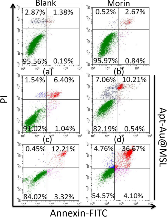
基于Annexin V-FITC/PI染色的SGC-7901不同方法处理后细胞凋亡的流式细胞术分析。 a的浓度 , b , c , 和 d 分别为 5、10、20 和 30 μg/mL
荧光检测抗癌活性研究
通过 LIVE/DEAD 荧光测定直观地评估了 Apt-Au@MSL 对 SGC-7901 细胞的抗肿瘤作用。细胞与不同浓度的 Morin、Morin-脂质体和 Apt-Au@MSL 孵育后,在黑暗条件下用 LIVE/DEAD 试剂盒共染色 30 分钟。在图 6 中,空白组中的细胞显示所有绿色荧光细胞都代表活细胞。对照组,包括 Morin 和 Morin-脂质体,显示红色荧光细胞的数量。然而,用不同浓度的 Apt-Au@MSL 处理的细胞明显表现出大量的凋亡细胞。随着浓度的增加,活细胞逐渐减少。死亡细胞的百分比显着增加,细胞密度降低。结果证实Apt-Au@MSL能有效促进肿瘤细胞凋亡。
<图片>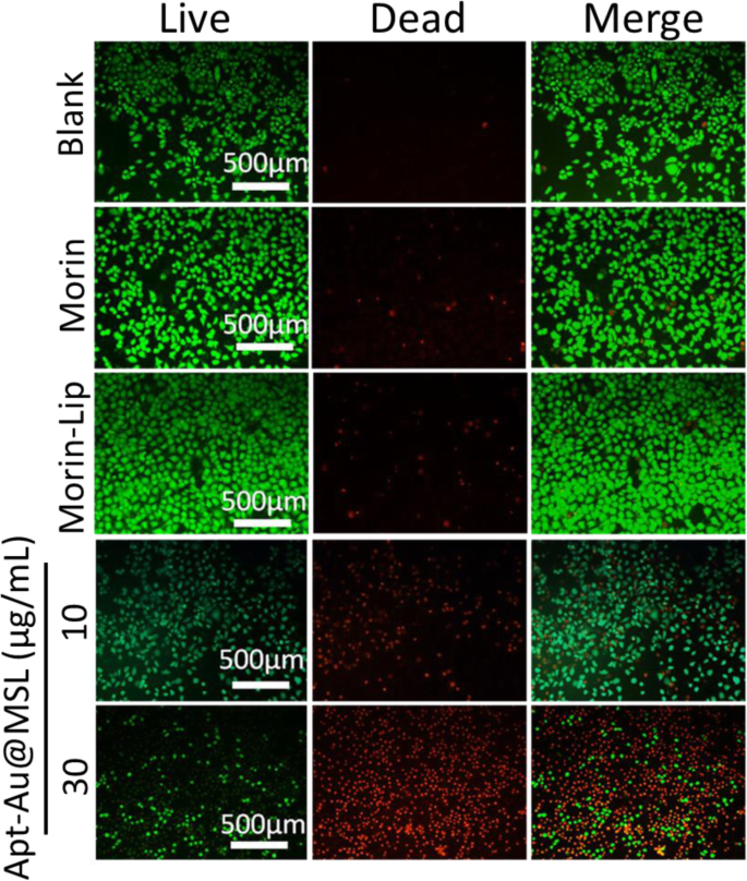
SGC-7901 的荧光显微图像与不同浓度的 Apt-Au@MSL 孵育并随后进行短暂染色。空白组为PBS。 Morin和Morin-脂质体为对照组
细胞完整性研究
为了确认 Apt-Au@MSL 对 SGC-7901 细胞的摄取和运输的影响,我们进行了 TEM 测定。通过TEM观察细胞形态的变化。进行TEM图像以观察细胞的内部结构并为抗癌机制提供参考。如图 7 所示,未经处理的 SGC-7901 细胞的空白图像显示出透明细胞壁形态和外观的显着变化。在对照组(Morin 和 Morin-脂质体)中,细胞形态显示部分损伤,核区收缩。然而,暴露于 Apt-Au@MSL 后也观察到细胞壁和内部结构的显着变化。细胞质泄漏,核结构变得不清楚。许多细胞碎片在空心细胞周围形成。此外,细胞壁解体或被破坏。整个轮廓变得不清楚,细胞受损,细胞质泄漏。红色箭头代表 Au NP 区域。随着 Apt-Au@MSL 浓度的增加,更多的 Au NP 区域出现在细胞核中。 This occurrence suggested the release of Morin after Apt-Au@MSL entered the cell interior, hence the appearance of a large amount of Au NPs. These aforementioned results suggest that antitumor activity was associated with compromised cell integrity and nuclear structure.

TEM images of SGC-7901 cells treated with different concentration of Apt-Au-MSL (10, 20, and 30 μg/mL). The blank group was PBS. The Morin and Morin–liposome were set as control group. The red square is the enlarge area. The red arrow point to Au NPs
Molecular Mechanism Induced by Apt-Au@MSL
To explore the molecular mechanism of Apt-Au@MSL-induced apoptosis, we detected the expression levels of caspases and PARP. Activation of caspase-3, -8, and -9 was first detected with specific substrates. As shown in Fig. 8a, Apt-Au@MSL treatment induces dose-dependent activation of caspase-3, -8, and -9. Caspase-9 promotes mitochondria-mediated apoptosis more than does caspase-8, indicating that the mitochondria-mediated apoptosis signaling pathway is dominant. Western blot analysis further confirmed the existence of Apt-Au@MSL-induced apoptosis at the protein level. Figures 8b and c show that after cells are treated with Apt-Au@MSL, activation of caspases and cleavage of PARP shows time and dose dependence (Figs. 8b, c). In Fig. 8d, e, the Western blot analysis confirmed our results. In conclusion, Apt-Au@MSL inhibits the growth of SGC-7901 cells mainly by inducing apoptosis.

Apt-Au@MSL induced apoptosis in SGC-7901 cells. 一 SGC-7901 cells were treated with indicated concentrations of Apt-Au@MSL for 24 h. Then, the total protein was extracted and incubated with synthetic Apt-Au@MSL substrates for measuring caspase activities. Dose-dependent (b ) and time-dependent (c ) effects of Apt-Au@MSL on PARP and caspases expression. d , e Quantitative analysis of PARP, caspase-7, caspase-9, and caspase-3 expressions. Data are means ± SD, *P <0.05, **P < 0.01
Apt-Au@MSL Inhibits Tumor Growth
In Vivo
The in vivo antitumor activities of Apt-Au@MSL were evaluated using an SGC-7901 tumor xenograft model. A comparison of the images of the tumors with those of the control group (Morin and Morin–liposomes) showed that mice treated with Apt-Au@MSL markedly reduced the weight and size of the tumor (Fig. 9a). The relative tumor volume curves and the mice weight curves indicate that the Apt-Au@MSL in vivo exhibits a higher anticancer efficiency (Fig. 9b) than those of the other treatment groups. No significant difference in average tumor volume was indicated between the control group (Morin and Morin–liposomes) and the blank group. The tumor volume of the Apt-Au@MSL group was only nearly a tenth of the blank group and nearly a sixth of the control group. The result indicated that raw Morin and Morin–liposomes at 40 mg/kg exerted no effect on the growth rate of tumors. However, Apt-Au@MSL could inhibit tumor growth. The body weight of mice in the different groups (PBS, Morin, Morin–liposomes, and Au-Apt groups) showed no marked fluctuation (Fig. 9c) during the treatment period. This result suggested that the treatment was tolerated and caused no acute side effects. Notably, we found that the mice with Apt-Au@MSL treatment showed markedly prolonged survival (Fig. 9d). The surviving mice in this group behaved normally, showing no apparent sign of unhealthy condition. These results demonstrated that the administration of Apt-Au@MSL could inhibit tumor growth in xenograft mouse models.

一 In vivo applications of Apt-Au@MSL and photographs of the mice tumor taken 24 days. A dosage of 2 mg/kg was administrated intravenously for all mice (n = 6–8). b Tumor weight of mice in different groups after 24 days. c Tumor volume index for the different treatment groups. The tumor sizes were measured at the indicated time points. d Survival rate of the mice in different group after tumor inoculation. Data are means ± SD (n = 6-8). The intravenous injection of PBS was set as blank group (100 μL); the treated by Morin and Morin–liposome were set as control group. In vivo therapeutic effects of Apt-Au@MSL in SGC-7901-bearing mice. Data are means ± SD, *P <0.05, **P < 0.01
Histological Analysis of Anticancer Activity
H&E staining of tumor tissue and organ samples was conducted after fixation and treatment. Treatment efficacy with respect to tumor cell death was also evaluated by H&E staining of tumor tissue from different groups. In Fig. 10a, the mice treated with Au-Apt, Morin, and Morin–liposomes show the same extent of thermal damage as that of the mice in the blank group. No apparent apoptosis was observed in the blank group. The tumor tissue sections consisted of tightly packed tumor cells. However, Apt-Au@MSL treatment exhibited the most significant damage to the tumor tissue, with moderate cell apoptosis in the tumor. The result suggested anticancer activity in the mouse models treated with Apt-Au@MSL. To further investigate the ratio of apoptotic cells in tumors tissue in vivo, TUNEL assay was performed for the detection of apoptotic cells. As shown in Fig. 10b, the apoptotic cells in tumors can be stained with green fluorescence to indicate apoptosis. The merged images show fewer green fluorescent regions in the blank group and the control group (Morin and Au-Apt), indicating the presence of fewer apoptotic cells. The cells of the mice treated with Morin–liposomes appear partly apoptotic. Moreover, a large number of green fluorescent regions were observed in the group treated with Apt-Au@MSL, indicating a large amount of apoptotic cells. The results were consistent with that of H&E staining, confirming that Apt-Au@MSL can promote tumor apoptosis in vivo.

一 H&E staining analysis of the tumors in mice. Histological analysis of the tumors in mice following different treatments as PBS, Morin, Morin–liposome, Au-Apt, and Apt-Au@MSL group. b Apoptotic cells were detected by a TUNEL assay (green) and co-stained by nuclear staining DAPI (blue)
In Vivo Toxicity Evaluation
The potential in vivo toxicity is often a significant concern for the clinical application of anticancer medicine. To verify the applicability of Apt-Au@MSL in vivo, the mice were evaluated under different treatments (Morin, Morin–liposomes, Au-Apt, and Apt-Au@MSL). Blood biochemical assays were also conducted to examine possible changes in the biochemistry of mice after treatment. As shown in Fig. 11a, the blood glucose index for blood function of the Apt-Au@MSL groups was similar to those of the blank and control groups. No difference in body weight was found in each group. A steady increase was observed, indicating that the drug exhibited no toxicity (Fig. 11b). H&E staining of organ sections (Fig. 11c) showed no sign of damage or inflammation in the group treated with Apt-Au@MSL, compared with the blank and control group. This finding indicated that PBS, Morin, Morin–liposomes, Au-Apt, and Apt-Au@MSL were negligible side effects in vivo. These results, as well those of H&E staining, further indicate safety in the use of Apt-Au@MSL for tumor treatment.

In vivo toxicity evaluation. 一 Blood glucose data detected in the mouse toxicity model. b Weight of mice in different groups after 24 days. c Images of H&E-stained major organs. Each value represents the mean ± SD (n =3)
Conclusions
In conclusion, this study presents the synthesis of an antitumor nanomaterial, Apt-Au@MSL. Apt-Au@MSL exhibited excellent monodispersity and tumor-targeting properties. The polarity of Morin was modified, and the antitumor activity was enhanced. The pH of the solution was 5.0, and the release rate of Morin from Apt-Au@MSL was the maximum in the characterization experiments. Apt-Au@MSL showed that the morphology of hybrid vesicles was attributable to Au-Apt modification. The diameter of 150 nm was consistent with the size determined by DLS. We screened our model cancer cell by MTT assay and found that SGC-7901 cells could effectively suppress proliferation. The IC50 of Apt-Au@MSL was 15.6 ± 1.5 μg/mL for the SGC-7901 cells. Fluorescent flow cytometric assays confirmed that Apt-Au@MSL could be used as an effective anticancer material and induced apoptosis in vitro. The Apt-Au@MSL found in the internal cell, as shown in the TEM images, suggested that Apt-Au@MSL could target the cancer cell. The administration of Apt-Au@MSL could inhibit tumor growth in xenograft mouse models, as determined from tumor weight testing. H&E staining and TUNEL assay further confirmed that Apt-Au@MSL could promote tumor apoptosis in vivo. Both blood biochemistry testing and H&E staining suggested that these materials exhibit negligible acute toxicity and good biocompatibility in vivo.
数据和材料的可用性
The authors declare that the materials, data, and associated protocols are promptly available to the readers without undue qualifications in material transfer agreements. All data generated and analyzed during this study are included in this article.
缩写
- Apt:
-
Aptamers
- Apt-Au:
-
Aptamers and Au nanoparticle
- MSL:
-
Morin pH-sensitive liposome
- AuNPs:
-
金纳米粒子
- ROS:
-
活性氧
- Apt-Au@MSL:
-
Aptamers and Au nanoparticle-modified Morin pH-sensitive liposome
- PC:
-
L-α-Phosphatidylcholine
- Chol:
-
Cholesterol
- CHEMS:
-
Cholesteryl hemisuccinate
- PVP:
-
聚乙烯吡咯烷酮
- PI:
-
碘化丙啶
- FT–IR:
-
傅里叶变换红外光谱
- UV–Vis:
-
Ultraviolet–visible spectroscopy
- TEM:
-
透射电子显微镜
- SEM:
-
扫描电镜
- DLS:
-
动态光散射
- PDI:
-
多分散指数
- ATCC:
-
American Type Culture Collection
- CLSM:
-
共聚焦激光扫描显微镜
- TUNEL:
-
TdT-mediated dUTP Nick-End Labeling
- H&E staining:
-
Hematoxylin-eosin staining
- RTCA:
-
Real-time unlabeled cell analysis
纳米材料
- IBM 科学家率先演示用于纳米粒子的摇摆布朗马达
- 用于化学传感器的金纳米粒子
- 用于癌症治疗的纳米粒子:当前的进展和挑战
- 用于癌症应用的基于细胞的药物递送
- 用于神经保护的脑靶向聚山梨醇酯 80 乳化多奈哌齐载药纳米颗粒
- 用贵金属纳米粒子装饰的电纺聚合物纳米纤维用于化学传感
- 以叶酸受体为靶点的生物类黄酮染料木素负载壳聚糖纳米颗粒可增强对宫颈癌的抗癌作用
- 合成单分散二元 FePt-Fe3O4 纳米粒子的后处理方法
- 适体修饰的磁性纳米增敏剂,用于表达 HER2 的癌症的体内 MR 成像
- 用于高效结肠癌基因治疗的基于阳离子胶束的 siRNA 递送
- 环境兼容的生物共轭金纳米粒子作为炎症诱导的癌症成像的有效造影剂
- 用于人类癌症检测的双靶向磁性纳米粒子