

源自黄柏皮质碳点的碳点对锐齿象毒液诱发的急性肾损伤的保护作用
摘要
背景
作为一种新兴的纳米材料,碳点 (CD) 已成为生物医学应用的极大关注焦点。然而,关于其抑制蛇毒所致急性肾损伤(AKI)的生物活性知之甚少。
方法
本研究报告了使用黄柏皮层 (PCC) 作为唯一前体合成 CDs 的绿色一步热解工艺的开发,以及它们作为保护剂的潜在应用,以防止Deinagkistrodon acutus (D. acutus) 首次研究了毒液诱发的 AKI。 AKI模型是通过注入D建立的。尖锐的 毒液进入小鼠腹腔以及 PCC Carbonisata-CDs (PCCC-CDs) 在六个时间点(1、3 和 12 小时,和1、2、5 天)进行了调查。
结果
这些结果表明 PCCC-CDs 显着抑制肾功能障碍(降低的血清肌酐 (SCR)、血尿素氮 (BUN)、尿总蛋白 (UTP) 和微量白蛋白尿 (MALB) 浓度)和趋化剂(单核细胞趋化蛋白)的产生1 (MCP-1))、促炎细胞因子(白介素 (IL)-1β)和抗炎细胞因子 (IL-10) 以响应腹膜内注射 D。尖锐的 毒液。 PCCC-CDs对毒液小鼠的有益作用与肾脏组织学改变和血小板减少的作用相似。
结论
这些结果证明了 PCCC-CD 对 D 诱导的 AKI 具有显着的保护作用。尖锐的 毒液,这不仅将拓宽 CD 的生物医学应用,而且还为开发治疗由 D 诱导的 AKI 的新药物提供了潜在的靶点。尖锐的 蛇咬伤。
介绍
Deinagkistrodon acutus (D. acutus) 属于蝰蛇科,被认为是中国最危险的陆生蛇之一 [1]。 D 的毒液。尖锐的 与一系列症状有关,如出血、血小板减少和可能对肾脏的直接损害[2, 3]。急性肾损伤 (AKI) 是 D 毒液中毒最严重的全身反应和常见并发症。尖锐的 这直接导致持续的肾功能障碍和高发病率 [4, 5]。目前的局部治疗涉及使用马来源的超免疫抗蛇毒血清作为解毒剂。然而,其在中和局部组织损伤方面的效果有限,尤其是在 AKI 的发生中,并且具有过敏和热原反应等多种不理想的效果[6]。此外,抗蛇毒血清相对较高的成本和较差的稳定性也导致被D咬伤的人的治疗效果不佳。尖锐的 ,尤其是在野外或农村地区 [7,8,9]。因此,迫切需要有效、安全且负担得起的补充药物来治疗 D。尖锐的 毒液诱发的AKI。
碳点 (CDs) 是一类尺寸 <10 nm 的新型碳纳米材料,于 2004 年通过单壁碳纳米管的分离和纯化偶然发现 [10] 并在过去十年中引起了广泛关注,因为它们具有显着的新的特性,如可观的生物相容性、低毒性、高水溶性和丰富的原材料 [11,12,13]。 CDs的出现促进了各种“智能”纳米系统的发展,主要包括生物成像[14]、生物医学[15]、药物递送[16]和光催化[17]。
值得注意的是,由于上述显着优势,具有固有生物活性潜力的 CD 的开发为发现用于有效控制或治疗某些疾病的新一代药物提供了许多策略。在治疗细菌性角膜炎的抗菌[18]、止血[19]、类过氧化物酶[20]、抗癌、抗病毒和抗炎活性[21]等多种生物活性方面已有报道。这些影响引起了科学家的注意,以研究 CD 的其他药物和生物医学应用。特别是,来自 Schizonepetae Spica Carbonisata [22] 的 CD 对 D. 的缓解活性。尖锐的 毒液引起的出血为研究 CD 对 D 引起的 AKI 的有益影响提供了新的视角。尖锐的 蛇咬伤,直到现在还不太了解。
黄柏 (PCC) Carbonisata (PCCC)-CDs 是通过使用一步热解处理直接热解 PCC(一种中药,已使用> 1000 年)合成的。 PCCC-CDs 是低毒 CDs,直径范围从 1.2 到 4.8 nm。据报道,PCCC-CDs具有显着的止血作用,这不仅拓宽了CDs的生物医学应用,而且开创了阐明PCCC止血物质基础的先河[23]。值得注意的是,PCCC是一种通过木炭加工制成的中药,最早见于太平救灾圣方 (公元978-992 AD,在中国)。 PCCC 的安全性和令人满意的治疗效果,如止血和抗炎,促使其在临床上持续应用了 1000 多年 年,获得了中华人民共和国药典的认可。 (PPRC, 2015)。然而,对 PCCC-CD 的其他潜在生物活性的研究一直是一个挑战。特别是,关于D 诱导的 AKI 抑制的信息很少。尖锐的 毒液。
此外,据报道,蛇咬伤可能通过肾毒性成分或通过激活或调节免疫和炎症介质直接危害肾脏生理 [24]。这些影响主要是生物医学参数[25](血清肌酐(SCR)、血尿素氮(BUN)、尿总蛋白(UTP)和微量白蛋白尿(MALB)浓度)、炎症细胞因子(白细胞介素(IL)-1β)、抗炎细胞因子 (IL-10) 和单核细胞趋化蛋白 1 (MCP-1)、肾脏组织学的变化和血小板 (PLT) 计数。在本研究中,我们使用绿色的一步热解方法合成了 PCCC-CD,并首次研究了它们对小鼠腹腔注射 D 诱导的 AKI 异常发展的保护作用。尖锐的 毒液。
材料和方法
化学品
PCC 材料购自北京千草中药饮片有限公司(中国北京),PCCC 在我们实验室制备。分子量为1000 Da的透析膜购自北京瑞达恒辉科技发展有限公司(中国北京)。细胞计数试剂盒 (CCK)-8 购自 Dojindo Molecular Technologies, Inc. (Kumamoto, Japan)。 D 的冻干蛇毒。尖锐的 由安仁蛇场(中国江西省鹰潭市余江县)提供,用于实验。其他分析纯化学试剂购自北京国药化学试剂有限公司(中国北京)。小鼠 MCP-1、IL-10 和 IL-1β 酶联免疫吸附测定 (ELISA) 试剂盒购自 Cloud-Clone Crop。 (中国武汉)。所有实验均使用去离子水(DW)进行。
动物
动物管理和研究方案得到了北京中医药大学动物实验伦理委员会的支持和批准。昆明雄性小鼠(体重30.0 ± 2.0 g)购自北京维塔尔河实验动物技术有限公司,具有实验动物合格证,饲养于恒温恒湿笼内,光照12小时/ 随意的黑暗循环 获得食物和水。
毒液的制备
冻干毒液溶于生理盐水,温和混合20 分钟(终浓度:5 mg/mL),-20 ℃保存备用。
PCCC-CD 的准备
根据先前的方法,PCCC 是使用一步热解法制备的,PCC 作为唯一的前体 [23]。简而言之,将 PCC 样品置于单独的瓷坩埚中,并在预热的炉中在 350 °C 下加热 1 小时。冷却至30 °C后,将所得均质黑色残留物研磨成细粉,在100 °C的水中煮沸两次,每次1 小时。然后,通过0.22 μm醋酸纤维素膜预过滤悬浮液来收集溶液。通过对 DW 透析 72 小时(保留分子量:1000 Da)除去非碳质杂质。将 PCCC-CDs 溶液浓缩并储存在 4 °C 下直至使用。图1中的流程图说明了上述过程。
<图片>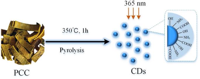
黄柏(PCC)热解处理碳点(CDs)形成过程示意图
PCCC-CD 的特征
使用电子能量为 200 kV 的透射电子显微镜(TEM,JEM-2100 电子,JEOL,日本)研究 CD 的形态。使用 Tecnai G2 20 电子显微镜(FEI,美国)使用高分辨率 TEM(HRTEM)检查结构细节。分别使用 UV-Vis(CECIL,Cambridge,UK)和荧光(F-4500,Tokyo,Japan)光谱记录和测量紫外-可见(UV-Vis)光谱和荧光特性。傅里叶变换红外(FTIR)光谱用于在400和4000 cm − 1 之间的光谱窗口分析表面官能团信息 .
细胞毒性分析
人 L02 肝细胞和人胚胎肾 293 T 细胞系用于使用 CCK-8 测定评估 PCCC-CD 的潜在细胞毒性。 L02 细胞在含有 10% 胎牛血清 (FBS) 的 Roswell Park Memorial Institute 1640 (RPMI 1640) 培养基中培养,而 293 T 细胞在含有 10% FBS 的高葡萄糖 Dulbecco 改良 Eagle 培养基 (DMEM) 中生长。两种细胞系均在 37 °C 下在 5% CO2 加湿中培养。
L02和293 T细胞以2.0 × 10 4 的密度接种 细胞/孔在 96 孔板中,并在 37 °C 和 5% CO2 的潮湿气氛中培养 24 小时。然后用 100 μL 不同浓度(5000、2500、1250、625、156、78、39 和 19.5 μg/mL)的 PCCC-CD 溶液处理两种细胞类型,并在无血清培养基中再孵育 24 H。随后,去除含有 PCCC-CD 的培养基,并用磷酸盐缓冲盐水 (PBS) 洗涤细胞两次。细胞毒性通过加入10 μL CCK-8试剂后立即在450 nm处读数并在37°C下孵育4小时来确定。
D.尖锐的 毒液诱发的 AKI 和治疗
D.尖锐的 毒液诱导的 AKI 模型
AKI小鼠模型是通过给小鼠腹腔注射D建立的。尖锐的 蛇毒。将小鼠随机分成以下五组,每组36只:对照;模型;和高、中、低剂量 PCCC-CDs 治疗组。模型组获得D。尖锐的 1 mg/kg (0.15 mg/mL, 0.2 mL) 体重的毒液和 0.5 mL 生理盐水腹腔注射,而高、中、低剂量 PCCC-CD 治疗组接受等量的蛇毒和 PCCC -CD 提取物的剂量分别为 8.0、4.0 和 2.0 mg/kg。仅用生理盐水 (NS) 腹腔注射的小鼠作为对照。在施用 NS、D 后 1、3 和 12 小时处死每组的 6 只小鼠。尖锐的 毒液和 PCCC-CDs 按上述时间表进行,而在第 1、2 和 5 天处死的小鼠则连续施用 NS,D。尖锐的 蛇毒,每天两次 PCCC-CD。
UTP 和 MALB 浓度分析
给予相应治疗后,对照组、模型和 PCCC-CDs-(4.0 mg/kg) 治疗组的动物立即置于适当的代谢笼中收集尿液,直至潜伏期结束(24 小时)。使用自动生化分析仪分析UTP和MALB的浓度。
肾功能生物标志物分析
通过测量 SCR 和 BUN 的水平来评估肾功能。在对小鼠进行安乐死之前,从眶后神经丛中抽取血样,然后将其放入塑料管中,在 4°C 下至少放置 4 小时。 750×g离心获得血清 15 min,采用全自动生化分析仪分析BUN和SCR水平。
炎症细胞因子水平的检测
取出小鼠的右肾,在干冰中快速冷冻并储存在-80°C直至用于以下程序。将来自不同组的组织样品(100 mg)在冰上用 PBS 匀浆,然后以 750×g 离心 15 分钟。收集上清液,根据制造商的说明使用相应的ELISA试剂盒测定MCP-1、IL-10和IL-1β的水平。
肾脏组织学
小鼠左肾组织样本在10%中性缓冲福尔马林中4 °C固定48 h以上,脱水,石蜡包埋,切片,苏木精伊红染色(H&E)。比较对照组、模型和PCCC-CDs治疗组的形态学变化。
血小板减少活动
对从小鼠眼眶后丛抽取的血液进行 PLT,并使用自动血液分析仪(XS-800i,Sysmex Corporation Co., Ltd., Kobe, Japan)检测。
统计分析
使用社会科学统计软件包(SPSS,版本 13.0)进行统计分析。正态分布数据和齐次方差表示为平均值 ± 标准偏差 (SD)。使用单向方差分析 (ANOVA) 和最小显着差异 (LSD) 检验进行多重比较。 P <0.05 被认为具有统计学意义。
结果
PCCC-CD 的特征
TEM 用于直接观察 PCCC-CD 的形态和尺寸分布(图 2a、c 和 d)。制备的 CD 呈球形且大小均匀,大多数在 2.84 ± 0.89 nm 处,没有明显的聚集。 HRTEM图像显示CD的晶格条纹(0.24 nm),对应于石墨碳,如图2b所示。
<图片>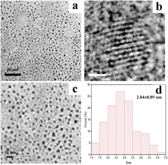
一 和 c 黄柏碳点 (PCCC-CD) 的透射电子显微镜 (TEM) 图像,b PCCC-CD 的高分辨率 TEM 图像,d PCCC-CDs的尺寸分布
如图 3a 所示,CD 显示出明显的蓝色荧光,在 370 nm 激发后在 445 nm 处具有最大峰值。相应地,PCCC-CDs在UV-Vis光谱中的光学信息在265 nm处显示出强吸收峰,对应于CDs表面共轭有机分子的π-π*跃迁(图3b)。
<图片>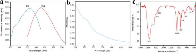
Phellodendri Cortex Carbonisatus 碳点 (PCCC-CD) 的光学特性。 一 荧光光谱,b 紫外-可见光谱 (UV-Vis) 和 c 傅里叶变换红外光谱(FTIR)
此外,根据 FTIR 光谱详细确定了配制的 CD 的官能团,如图 3c 所示。 3433 cm − 1 处的峰 被分配到O-H和N-H伸缩振动的吸收带,而C-H伸缩振动出现在2923 cm − 1 和 2853 cm − 1 .此外,在1624 cm − 1 处的吸收峰 表明存在 C=O。在 1109 cm − 1 处观察到 C-O-C 键 , 以及 1400 cm − 1 处的峰值 归因于 C-N 拉伸。这些发现与之前的报道一致。
体外细胞毒性
为了评估细胞毒性,将 L02 和 293 T 细胞暴露于不同浓度的 PCCC-CD 24 小时,并使用 CCK-8 测定检查活力。如图 4a 所示,即使浓度高达 5 mg/mL,用 PCCC-CD 处理的 L02 细胞的生存力与对照细胞相比也> 85%。类似地,PCCC-CD 在高达约 2500 μg/mL 的浓度下不影响 293 T 细胞的生长(图 4b)。这些结果表明PCCC-CDs表现出低细胞毒性。
<图片>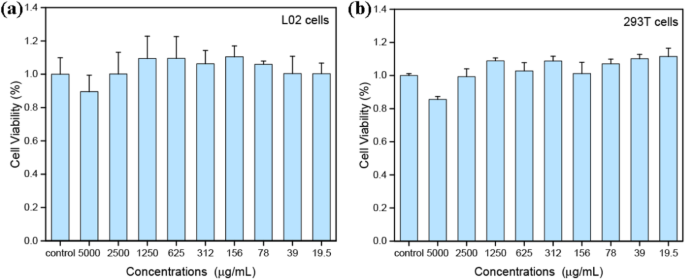
(a 的 CCK-8 测定的细胞活力 ) L02 细胞和 (b ) 293 T细胞与PCCC-CDs孵育4 h
PCCC-CD 对 UTP 和 MALB 浓度的影响
与对照组的UTP水平(0.98 ± 0.38 mg/L)相比,D中的UTP水平显着升高。尖锐的 毒液处理 (3.08 ± 0.92 mg/L, P <0.01) 和 PCCC-CDs (2.36 ± 0.83 mg/L, P <0.05) 组(图 5a)。此外,与毒液给药后的水平相比,PCCC-CD 治疗后 UTP 水平降低(P <0.05)。此外,腹腔注射D后24 h MALB水平明显升高。尖锐的 毒液 (17.78 ± 5.96 mg/L, P <0.01) 与对照组 (2.02 ± 0.91 mg/L) 相比。相比之下,PCCC-CD处理的小鼠MALB水平呈下降趋势(14.25 ± 4.16 mg/L,图5b)。
<图片>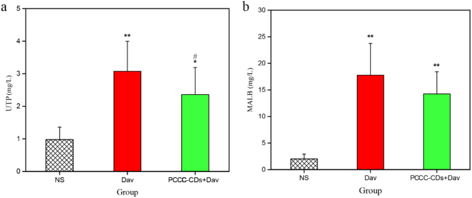
黄柏皮质碳点(PCCC-CDs)对尿液中尿总蛋白(UTP)和微量白蛋白尿(MALB)的影响。 一 UTP 和 b 马尔堡。用生理盐水 (NS)、D.acutus 处理小鼠 毒液(Dav,1 mg/kg)和 PCCC-CDs(4 mg/kg) + Dav(1 mg/kg)。数据表示为每组六只动物的平均值 ± SD。 *p < 0.05 和 ** p < 0.01 与用 NS 治疗的对照组相比。 # p < 0.05 与 Dav 组相比
PCCC-CD 缓解了 D 中的肾功能障碍。尖锐的 毒液诱导的 AKI 小鼠
通过测量 BUN 和 SCR 的水平来评估肾功能,如图 1 和图 2 所示。 6和7。用D处理的小鼠。尖锐的 毒液具有显着更高水平的 BUN (12.07 ± 1.00 mmol/L [1 天], P <0.01; 11.43 ± 1.37 mmol/L [2天],P <0.01; 14.83 ± 2.53 mmol/L [5 天],P <0.01) 和 SCR (12.83 ± 1.43 μmol/L [1 天], P <0.01; 13.48 ± 2.26 μmol/L [2天]; P <0.01。 13.80 ± 1.90 μmol/L [5天],P <0.01) 比 NS 处理的小鼠 (BUN:8.95 ± 1.04 [1 天]、9.53 ± 1.40 [2 天]和 9.07 ± 1.57 [5 天] mmol/L;SCR:9.9± 1.04 [1 天] 、10.27 ± 2.04 [2 天] 和 9.85 ± 1.99 [5 天] μmol/L) 从第 1 天到第 5 天(图 6a 和 7a)。值得注意的是,与模型组相比,低剂量PCCC-CDs治疗显着抑制了SCR水平的升高(9.77 ± 0.79 μmol/mL,P <0.01) 和 BUN (10.50 ± 1.38 mmol/L, P <0.05), 而高 (9.62 ± 1.87 μmol/mL, P <0.01) 和培养基 (10.75 ± 1.48 μmol/mL, P <0.05) 剂量抑制了由 D 诱导的 SCR(图 7b)但不抑制 BUN(图 6b)的增加。尖锐的 毒液在第 1 天。高、中、低剂量 PCCC-CD 治疗组的 SCR(图 7c)水平降低(10.97 ± 0.88、10.42 ± 1.75 和 10.68 ± 1.41μmol/mL,分别;P <0.05)比模型组在第2天。此外,与模型组相比,BUN水平在高-(9.57 ± 0.52 mmol/L,P <0.01) 和低剂量 (10.72 ± 2.04 mmol/L, P <0.05)第2天PCCC-CDs组而不是中剂量组(图6c)。 6d和7d,在高(12.28 ± 1.65 mmol/L,P <0.01 (BUN); 11.38 ± 1.80 μmol/mL,P <0.05 (SCR), 分别) 和中等 (12.40 ± 1.33 mmol/L, P <0.05 (BUN); 10.83 ± 2.57 μmol/mL,P <0.05 (SCR), PCCC-CDs。此外,对照组和PCCC-CDs治疗组的SCR无显着差异。
<图片>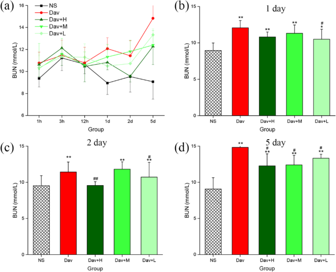
黄柏Cortex Carbonisata - 碳点(PCCC-CDs)对血尿素氮(BUN)的影响。 一 BUN浓度在1 h、3 h、12 h、1 天、2 天和5 天发生变化。 (b 中 BUN 的水平 ) 1 天 (c ) 2 天 (d ) 5 天。用生理盐水 (NS)、D.acutus 处理小鼠 毒液 (Dav, 1 mg/kg) 和高 (H)、中 (M) 和低 (L) 剂量的 PCCC-CDs + Dav (1 mg/kg)。数据表示为每组六只动物的平均值 ± SD。 *p < 0.05 和 ** p < 0.01 与用 NS 治疗的对照组相比。 # p < 0.05 与 Dav 组相比
<图片>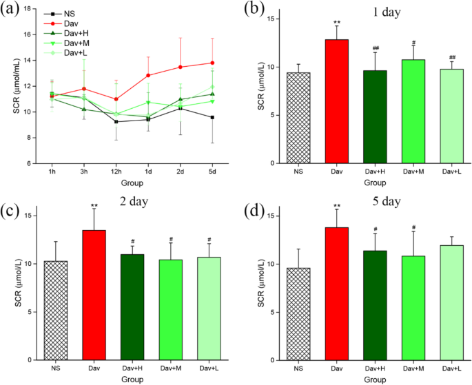
黄柏皮质碳点(PCCC-CDs)对血清肌酐(SCR)的影响。 一 SCR浓度在1 h、3 h、12 h、1 天、2 天和5 天发生变化。 (b 中 SCR 的水平 ) 1 天 (c ) 2 天 (d ) 5天。用生理盐水 (NS)、D.acutus 处理小鼠 毒液(Dav,1 mg/kg)和高(H)、中(M)和低(L)剂量的 PCCC-CDs + Dav(1 mg/kg)。数据表示为每组六只动物的平均值 ± SD。 *p < 0.05 和 ** p < 0.01 与用生理盐水 (NS) 处理的对照组相比。 # p < 0.05 与 Dav 组相比
PCCC-CDs 抑制细胞因子分泌
三种浓度的 PCCC-CD 对注射 D 时产生的化学引诱物 (MCP-1) 和促炎 (IL-1β) 和抗炎 (IL-10) 细胞因子的影响。尖锐的 毒液进行了调查。图8显示注射毒液增加了小鼠模型中IL-1β的释放(1 h:58.19 ± 5.35 ng/mL,P <0.01; 3 小时:56.57 ± 3.54 ng/mL,P <0.01; 12 小时:49.48 ± 7.74 ng/mL,P <0.05;第 1 天:41.09 ± 4.82 ng/mL,P <0.05;第 2 天:47.96 ± 8.33 ng/mL,P <0.05;第 5 天:45.11 ± 6.95 ng/mL,P <0.05) 与 NS 处理小鼠的水平相比 (1 h:35.96 ± 4.72 ng/mL, 3 h:34.94 ± 2.58 ng/mL, 12 h:36.42 ± 4.72 ng/mL, 12 h:36.42 ±4.72 ng/mL, 12 h:36.42 ±3 5.75.7ng≉≪7.72 /mL,第 2 天:39.84 ± 3.71 ng/mL,第 5 天:36.82 ± 8.27 ng/mL)。如图 8b-d 所示,与 D 的比较。尖锐的 毒液诱导组显示,1、3 和 12 小时暴露于高浓度(50.09 ± 7.68 ng/mL,P <0.05[1小时]; 40.36 ± 8.51 ng/mL,P <0.01[3小时]; 39.87 ± 4.64 ng/mL,P <0.05 [12 小时], 分别) 和低- (46.64 ± 3.83 ng/mL, P <0.01[1小时]; 37.65 ± 9.61 ng/mL,P <0.01 [3 h]; 38.75 ± 6.64 ng/mL,P <0.05 [12 小时],分别为 PCCC-CDs 剂量显着降低 IL-1β 的水平。此外,中等剂量的 PCCC-CDs 显着降低了 IL-1β 的水平(41.50 ± 11.08 ng/mL,P <0.01) 与毒液处理的小鼠在 3 h 相比,但在其他时间点没有。
<图片>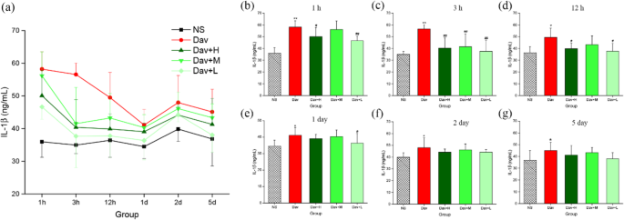
黄柏皮质碳点(PCCC-CDs)对肾组织中IL-1β水平的影响。 一 IL-1β 水平在 1 h、3 h、12 h、1 天、2 天和 5 天发生变化。 (b 中的 IL-1β 水平 ) 1 h, (c ) 3 小时, (d ) 12 小时, (e ) 1 天,(f ) 2 天,以及 (g ) 5天。用生理盐水 (NS)、D.acutus 处理小鼠 毒液(Dav,1 mg/kg)和高(H)、中(M)和低(L)剂量的 PCCC-CDs + Dav(1 mg/kg)。数据表示为每组六只动物的平均值 ± SD。 *p <0.05 和 ** p <0.01 与用生理盐水 (NS) 处理的对照组相比。 # p < 0.05 与 Dav 组相比
抗炎细胞因子IL-10的水平在D中显着增加。尖锐的 不同时间点毒液处理组 (23.27 ± 0.72 ng/mL, P <0.01; 22.03 ± 0.96 ng/mL,P <0.05; 21.76 ± 1.99 ng/mL,P <0.05; 26.31 ± 6.55 ng/mL,P <0.01;与 NS 治疗组相比,分别为第 3 小时、12 小时、第 1 天和第 2 天)(图 9)。与此形成鲜明对比的是,使用高剂量和中剂量 PCCC-CD(分别为 17.17 ± 4.04 和 17.25 ± 5.64 ng/mL,均 P <0.05) 在 3 小时抑制毒液诱导的 IL-10 分泌,而低剂量 PCCC-CDs 在 3 小时、12 小时和第 1 天抑制水平 (17.17 ± 5.24、17.83 ± 4.81.11, 17.83 ± 4.81.13) mL,分别为所有 P <0.05)。 PCCC-CDs组与NS组无显着差异。
<图片>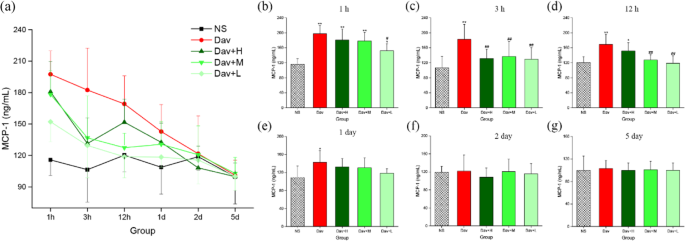
黄柏皮质碳点(PCCC-CDs)对肾组织中IL-10水平的影响。 一 IL-10 水平在 1 h、3 h、12 h、1 天、2 天和 5 天发生变化。 (b 中 IL-10 的水平 ) 1 h, (c ) 3 h, (d ) 12 h, (e ) 1 天, (f ) 2 天和 (g ) 5天。用生理盐水 (NS)、D.acutus 处理小鼠 毒液(Dav,1 mg/kg)和高(H)、中(M)和低(L)剂量的 PCCC-CDs + Dav(1 mg/kg)。数据表示为每组六只动物的平均值 ± SD。 *p <0.05 和 ** p <0.01 与用生理盐水 (NS) 处理的对照组相比。 # p < 0.05 与 Dav 组相比
另外,图10显示了D的注入。尖锐的 毒液显着增加模型组MCP-1的释放(1 h:197.45 ± 22.34 ng/mL,P <0.01; 3 小时:182.42 ± 12.94 ng/mL,P <0.01; 12 小时:169.20 ± 26.74 ng/mL,P <0.01; 24 小时:142.81 ± 25.85 ng/mL,P <0.05)与NS处理的对照(1个小时相比:115.82±14.80毫微克/毫升; 3小时:106.46±13.76毫微克/毫升; 12小时:120.35±15.75毫微克/毫升;和24h:108.81±25.60毫微克/毫升)。更值得注意的是,用三种 PCCC-CD 剂量治疗中毒小鼠抑制了 MCP-1 水平的增加。培养基 - (3 h:136.84 ± 39.94 ng/mL, P <0.01; 12 小时:127.48 ± 13.75 ng/mL,P <0.01) 和低- (1 h:152.13 ± 18.89 ng/mL, P <0.05; 3 小时:129.54 ± 30.85 ng/mL,P <0.01; 12 小时:118.75 ± 19.96 ng/mL,P <0.01) 剂量的 PCCC-CDs 在 1、3 和 12 h 抑制 MCP-1 水平的增加,除了中等剂量,在 1 h (178.20 ± 22.79 ng/mL)。仅在 3 h (131.42 ± 24.62 ng/mL, P <0.01).
<图片>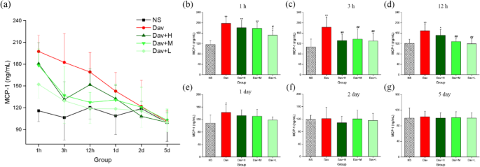
黄柏皮质碳点 (PCCC-CDs) 对肾组织中 MCP-1 水平的影响。 一 MCP-1 水平在 1 h、3 h、12 h、1 天、2 天和 5 天发生变化。 (b 中的 MCP-1 水平 ) 1 h, (c ) 3 h, (d ) 12 h, (e ) 1 天, (f ) 2 天,以及 (g ) 5天。用生理盐水 (NS)、D.acutus 处理小鼠 venom (Dav, 1 mg/kg) and high (H), medium (M), and low (L) doses of PCCC-CDs + Dav (1 mg/kg). Data are represented as mean ± SD of six animals of each group. *p < 0.05 and ** p < 0.01 compared with control group treated with normal saline (NS). # p < 0.05 compared with Dav group
In contrast, IL-1β, IL-10, and MCP-1 levels of the PCCC-CDs-treated groups at certain time points were not significantly different from those of the model group, and showed a decreasing tendency.
Effect of PCCC-CDs on the Inhibition of Thrombocytopenia
PLTs have a specific role in the pathogenesis of AKI; therefore, the PLT count was investigated and the results are shown in Fig. 11 [26]. Compared with the PLT values of the control group (1 h:[1228 ± 51] × 10 9 ; 3 h:[1120 ± 36] × 10 9 ; 12 h:[1245 ± 111] × 10 9 ; day 1:[1177 ± 69] × 10 9 ; day 2:[1195 ± 51] × 10 9 ; and day 5:[1181 ± 46] × 10 9 ), a drastic reduction occurred as early as 1 h after D. acutus venom administration, with a nadir occurring at 3 h. Subsequently, the PLT steadily increased up to day 5 (1 h:[354 ± 70] × 10 9 , P <0.01; 3 h:[315 ± 77] × 10 9 , P <0.01; 12 h:[435 ± 91] × 10 9 , P <0.01; day 1:[663 ± 226] × 10 9 , P <0.01; day 2:[941 ± 248] × 10 9 , P <0.05; day 5:[1083 ± 89] × 10 9 )。 Of note, even at this time interval, the PLT values were significantly lower than those of control mice. In addition, administration of PCCC-CDs at a dose of 8 mg/kg markedly inhibited the venom-induced thrombocytopenia induced at 1 h ([435 ± 91] × 10 9 , P < 0.05), 3 h ([599 ± 290] × 10 9 , P < 0.05), 12 h ([929 ± 92] × 10 9 , P < 0.01), day 1 ([1028 ± 248] × 10 9 , P < 0.01), and day 2 ([1183 ± 89] × 10 9 , P <0.01)。 This inhibitory effect was also observed at a dose of 2 mg/kg PCCC-CDs at 3 h and day 2 and 4 mg/kg PCCC-CDs at day 2, in addition to increased PLT. Although there was no significant difference between the model and medium-dose groups, an increasing tendency was observed at other different time points.

Effects of Phellodendri Chinensis Cortex Carbonisata-carbon dots (PCCC-CDs) on platelet (PLT) counts in the blood. 一 PLT changes in 1 h, 3 h, 12 h, 1 day, 2 days, and 5 days. PLT in (b ) 1 h, (c ) 3 h, (d ) 12 h, (e ) 1 day, (f ) 2 days, and (g ) 5 days. Mice were treated with normal saline (NS), D.acutus venom (Dav, 1 mg/kg) and high (H), medium (M), and low (L) doses of PCCC-CDs + Dav (1 mg/kg). Data are represented as mean ± SD of six animals of each group. *p < 0.05 and ** p < 0.01 compared with control group treated with normal saline (NS). # p < 0.05 compared with Dav group
Histopathological Observations
The renal injury in the venom group was histologically evaluated. As shown in Fig. 12a, in contrast to the NS-treated group, which showed normal glomeruli and tubular cellularity, marked changes were observed in the renal parenchyma of the D. acutus venom-treated group. These changes include marked haemorrhage, renal tubular dilation, and degeneration. Cotreatment with PCCC-CDs prevented D. acutus venom-induced renal damage, and the histopathological examination of the architecture of the renal tissues was almost normal with mild haemostasis, renal tubular dilation, and degeneration.

Effects of Phellodendri Chinensis Cortex Carbonisata-carbon dots (PCCC-CDs) on histopathological changes in kidney tissues in D.acutus venom (Dav)-induced AKI mice. After D.acutus venom challenged, kidney tissues from each experimental group were prepared for histological evaluation. Histological changes of kidney obtained from mice of different groups normal saline (NS), D.acutus venom (Dav, 1 mg/kg) and high (H), medium (M), and low (L) doses of PCCC-CDs + Dav (1 mg/kg) in (a ) 1 h, (b ) 3 h, (c ) 12 h, (d ) 1 day, (e ) 2 days, and (f ) 5 days
Discussion
As an emerging nanomaterial, CDs are beginning to occupy an important niche as innovative materials for next-generation nanomedicines. Compared to traditional heavy-metal-based quantum dots, CDs are good candidates for biomedical application because of their unique characteristics that have considerable potential advantages in the development of novel medicines with relatively low toxicity [27, 28].
The derived PCCC-CDs particles were quasi-spherical and well-dispersed in water, with abundant functional groups present on the surface. This observation is consistent with previously reported findings [23]. In addition, the as-prepared CDs showed low toxicity against L02 and 237 T cells, which indicated its suitability for biomedical applications.
The current study is the first, to the best of our knowledge, to demonstrate the remarkable bioactivities of PCCC-CDs on AKI induced by D. acutus snakebite. D. acutus is widely called “five pacer” or “hundred pacer” in Chinese folk medicine on account of the folkloric description that the people or animals bitten by D. acutus could not walk more than 100 steps. More than 90% of the population of D. acutus is found in China, and the frequency of critical conditions and even death related to the bite of this snake is higher than that by many other venomous snakes [29]. AKI is the most serious systemic effect and common complication, which leads to secondary renal ischemia and failure. An enhanced knowledge of relevant information on AKI induced by D. acutus envenomation would contribute to the development of novel therapeutic approaches. However, in contrast to the considerable knowledge available on the nephrotoxicity of snake venom in general [30, 31], information on the AKI induced by D. acutus venom is rare, which led us to investigate this potential medicine that is still in the early stages of development.
In this study, we established an AKI model by intraperitoneally injecting D. acutus venom into mice to assess the complex and multifactorial pathogenesis of venom-induced AKI. Furthermore, the model provided a tool for investigating the protective effects of PCCC-CDs against AKI induced by D. acutus venom.
Current experiments have shown the development of substantial AKI with distinct changes in inflammatory cytokines and serum and urinary biochemical index, as well histopathological evidence of renal injury after intraperitoneal injection of snake venom [31]. These findings indicated the possible factors that may mainly contribute to the venom toxicity are [32] (1) direct venom cytotoxicity against the kidney and (2) renal inflammatory reactions.
Specifically, renal insufficiency was confirmed approximately 24 h post venom injection based on oliguria associated with proteinuria and elevated serum biomarkers (SCR and BUN). We further affirmed the renal involvement in the D. acutus venom-treated group using biochemical analysis, which showed significantly elevated UTP and MALB levels compared with those of the control group. These findings indicated the presence of glomerular malfunction and tubular reabsorption in the venom-treated group [33], which were supported by evidence of histopathological change. In contrast, a significant reduction in the levels of UTP and MALB was observed in the envenomated medium-dose PCCC-CD-treated group. In addition, serum biochemical indicators (SCR and BUN) are other vital parameters used to determine the elevation of renal dysfunction in AKI and they remain clinical indicators in its diagnosis [34]. Injecting snake venom from day 1 to 5 also dramatically increased the levels of SCR and BUN, whereas treatment with PCCC-CDs reversed these effects, resulting in a faster recovery than that of the control group. More importantly, the distinct changes in the kidney tissue, marked haemorrhage, renal tubular dilation, and degeneration further indicated the direct impairment of the kidney by the venom. The attenuating effects of PCCC-CDs on the histopathological changes were demonstrated in this study. These results suggested that PCCC-CDs inhibited the AKI-induced abnormal manifestation of urinary and serum biochemical markers associated with kidney dysfunction as well as renal histological damage. Furthermore, these effects may be attributable to the amelioration of the direct nephrotoxicity of the D. acutus venom by the PCCC-CDs [35]. The protective effects of PCCC-CDs were evidenced by the inhibition of D. acutus venom-induced direct kidney damage.
An intense inflammatory response is a common feature induced by envenomation by venomous animals such as snakes and caterpillars [35,36,37]. The signs of systemic inflammation with mononuclear cell infiltration, neutrophilic leukocytosis, tubular epithelial cell degeneration, and necrosis have also been shown in kidney impairment induced by injecting snake venom. MCP-1 is a small molecule protein that plays a vital role in recruiting and activating leukocytes during inflammatory responses [38]. In addition, mononuclear phagocytes and lymphocytes may contribute to acute renal cell injury by different mechanisms such as the secretion of proinflammatory mediators, which may then induce resident renal cells to express chemokines [39]. The involvement of MCP-1, inflammatory cytokines (IL-1β) and anti-inflammatory cytokine (IL-10) in the inflammatory response in the pathogenesis of AKI in mice injected with D. acutus venom only was demonstrated in the present study. This observation indicated that the underlying mechanism of the D. acutus venom-induced AKI may be associated with the renal inflammatory response. The evidence that exposure to PCCC-CDs significantly reduced levels of IL-1β, IL-10, and MCP-1 suggests that CDs may exhibit renoprotection by inhibiting renal inflammatory reactions.
Furthermore, PLTs play a crucial role in acute haemostatic and inflammatory processes and are associated with diverse inflammatory pathologies [40, 41]. They are highly sensitive and respond quickly to biological changes when an organism is injured or bleeding, as the first cells to arrive at the site of acute injury to interact with endothelial cells and leukocytes [42]. PLTs are involved in the pathogenesis of AKI [43], and are considered a prognostic marker that is significantly associated with a worse outcome of AKI [44]. This study provided evidence that D. acutus venom conspicuously decreased the PLT count, which was consistent with the results of studies reporting that thrombocytopenia can be induced by snake venom [34, 45]. We observed that exposure to PCCC-CDs significantly elevated the PLT counts, which was consistent with the findings of a previous study [23].
The abnormalities of AKI induced by D. acutus venom were, to our knowledge, demonstrated for the first time in the current study and mainly included renal dysfunction associated with proteinuria, oliguria, elevated BUN and SCR levels, pathological kidney damage, inflammatory responses, and thrombocytopenia.
Remarkably, the PCCC-CDs demonstrated protective activity against D. acutus venom-induced AKI by inhibiting the associated impairments, as evidenced in this study for the first time. This study was a preliminary evaluation of the beneficial effects of PCCC-CDs on AKI induced by D. acutus venom, and further investigations of the underlying mechanism would be the focus of future studies.
Conclusion
The impressive protective effects of PCCC-CDs on D. acutus venom-induced AKI have been demonstrated in this study, for the first time, to the best of our knowledge. The AKI-related effects were mainly manifested as renal dysfunction, pathological kidney damage, inflammatory responses, and thrombocytopenia. These results indicated that PCCC-CDs have potential application prospects for use as a complementary medicine for the treatment of abnormalities induced by D. acutus venom-induced AKI. Furthermore, this provides a novel strategy for the study of active ingredients of traditional Chinese medicine formulations, and further broadens the biomedical applications of CDs.
数据和材料的可用性
本研究期间生成或分析的所有数据均包含在这篇已发表的文章中。
缩写
- AKI:
-
Acute kidney injury
- BUN:
-
Blood urea nitrogen
- CD:
-
碳点
- D. acutus :
-
Deinagkistrodon acutus
- HE:
-
Haematoxylin and eosin
- IL:
-
Interleukin
- MALB:
-
Microalbuminuria
- MCP-1:
-
Monocyte chemotactic protein 1
- PCC:
-
Phellodendri Chinensis Cortex
- PCCC-CDs:
-
Phellodendri Chinensis Cortex Carbonisata-carbon dots
- PLT:
-
Platelet counts
- SCR:
-
Serum creatinine
- UTP:
-
Urinary total protein
纳米材料
- 锻造对碳钢的影响
- 高效独立激发蓝色发光碳点
- 来自废棉手套的具有分级纳米结构的活性炭纤维作为超级电容器的高性能电极
- 合成富含吡啶的 N、S 共掺杂碳量子点作为有效的酶模拟物
- 磁性碳微球作为可重复使用的吸附剂从水中去除磺胺
- 从豆腐废水中合成荧光碳量子点的简单方法
- 利用柠檬汁通过水热反应制备的荧光碳量子点的材料和光学特性
- 源自静电纺丝和原位热解的高活性和稳定的 Fe-N-C 氧还原电催化剂
- 吸入多壁碳纳米管对血压和心脏功能的影响
- 从石榴皮提取物中合成的银纳米粒子的抗菌和细胞毒性作用
- 用于光热疗法的聚多巴胺碳点的简便一锅法合成
- Carbon Dots @ Platinum Porphyrin Composite 作为用于有效光动力癌症治疗的治疗诊断纳米剂