

CKAP4 抗体共轭硅量子点胶束用于肺癌靶向成像
摘要
目前,已经设计并合成了各种荧光纳米材料作为手术导航的光学造影剂。然而,目前还没有关于使用硅量子点(Si QDs)制备用于肺癌手术导航的荧光造影剂的报道。该研究改进和修改了 Pi 等人报道的水分散性 Si QD 胶束。制备 Si QD 胶束-CKAP4。数据表明,Si QD 胶束-CKAP4 是平均水直径约为 78.8 nm 的球形颗粒。 Si QD 胶束-CKAP4 的紫外可见吸收范围为 200 到 500 nm。激发波长为 330 nm,在荧光发射光谱中观察到 640 nm 处的强荧光。激光共聚焦显微镜和荧光显微镜实验表明,Si QD 胶束-CKAP4 在体外对肺癌细胞和肺癌组织表现出良好的靶向能力。体内荧光成像分析表明,Si QD 胶束-CKAP4 经肝脏代谢并经肾脏排泄。此外,与健康肺组织相比,Si QD 胶束-CKAP4 在体内特异性靶向肺癌组织。细胞毒性和苏木精和伊红染色试验表明,Si QD 胶束-CKAP4 在体外和体内表现出很高的生物安全性。 Si QD micelles-CKAP4是肺癌特异性靶向显像剂,有望成为未来肺癌手术导航的荧光造影剂。
介绍
癌症是全球威胁人类健康的主要疾病之一[1]。其中,肺癌的发病率和死亡率居所有恶性肿瘤之首,且呈逐年上升趋势[2]。手术切除是实体瘤的主要治疗方法。然而,区分恶性组织与健康组织的方法主要限于触觉和视觉线索以及外科医生的经验[3]。
近年来,荧光引导手术在实时术中引导肿瘤切除研究领域变得流行[4]。借助荧光造影剂和荧光成像系统,外科医生可以使用实时图像引导切除原发肿瘤并识别手术腔内的残留肿瘤。与传统的生物医学成像方法相比,荧光成像系统具有无电离辐射和高空间分辨率等优点[3, 5, 6]。
目前,研究人员已经报道了各种用于生物成像的荧光纳米材料[7, 8]。其中,硅量子点(Si QDs)已在生物成像领域流行起来[9,10,11]。然而,目前还没有关于利用Si QDs制备肺癌手术导航荧光造影剂的报道。
2014 年,Pi 等人。报道了一种新型水分散性 Si QD 胶束的合成方法 [12]。研究数据表明,Si QD 胶束具有尺寸可控、荧光量子效率高、光稳定性好等优点[12]。本研究旨在改进和修改 Pi 等人报道的水溶性 Si QD 胶束。制备新型荧光造影剂,有望在未来作为肺癌手术导航的荧光造影剂使用。
2018 年,柳田等人。等报道CKAP4在肺癌患者的血清和癌组织中高表达,可作为肺癌早期诊断的新生物标志物[13]。因此,CKAP4抗体偶联的Si QD胶束可能是一种新型的荧光造影剂,可作为肺癌手术导航的荧光造影剂。
在本研究中,DSPE-PEG-COOH 用于制备表面带有羧基的 Si QD 胶束。然后我们使用 EDC 将 CKAP4 抗体与 Si QD 胶束结合。结果表明,Si QD 胶束-CKAP4 在水溶液中分散良好。 Si QD 胶束-CKAP4 在 365 nm 紫外光照射下表现出强烈的红色荧光。由于抗体的结合,Si QD micelles-CKAP4在体外和体内均显示出对肺癌细胞和组织的良好靶向能力。此外,Si QD 胶束-CKAP4 在体外和体内均表现出高生物安全性。本研究制备了一种潜在的用于肺癌手术导航的荧光造影剂。
方法
试剂
牛血清白蛋白 (BSA) 购自 Sigma(分析 ≥ 98%;美国密苏里州);来自 Adamas-beta 的四氢呋喃 (THF)(纯度:99.9%;瑞士巴塞尔);来自阿拉丁(中国上海)的 DSPE-PEG-COOH;来自 Abcam 的 CKAP4 抗体(0.55 mg/mL;英国剑桥);来自 Hyclone(美国犹他州)的 Dulbecco 改良鹰培养基 (DMEM) 和磷酸盐缓冲液 (PBS);胎牛血清(FBS)和来自 Gibco(纽约,美国)的酪氨酸;多聚甲醛 (4%),使用 Triton X-100 的免疫染色通透缓冲液和来自 Beyotime(中国上海)的用于免疫染色的封闭缓冲液; Solarbio 的甘油(纯度 ≥ 99%;中国北京);和来自 Kingmorn 的胰蛋白酶(0.25%;中国上海)。
仪器
使用 JEM-2100F 电子显微镜(JEOL,东京,日本)拍摄透射电子显微镜 (TEM) 图像。 Hydrodiameter 使用 JEM Zetasizer Nano-ZS90 (Malvern Instruments, Malvern, UK) 测定。使用 Cary 50 分光光度计(Varian,Palo Alto,CA,USA)获得 UV-Vis 吸收光谱。使用 F-182 4500 分光光度计 (Hitachi Ltd., Tokyo, Japan) 获得荧光发射光谱。 X 射线光电子能谱 (XPS) 使用 RBD 升级的 PHI-5000C ESCA 系统(Perkin Elmer,Waltham,MA,USA)进行。使用荧光光谱仪(FLS1000,Edinburgh Instruments,Livingston,England)估计光致发光(PL)量子产率(QY)。傅里叶变换红外光谱 (FTIR) 使用 Thermo IS50 (Thermo Fisher Scientific, Massachusetts, USA) 进行。 X射线衍射(XRD)由SmartLab(Rigaku Corporation,Tokyo,Japan)测试。
细胞
A549 和 293 T 细胞系购自上海细胞生物学研究所(中国上海)。
Si QD 胶束的制备
首先,根据先前报道的方法 [12],通过氢化硅烷化反应制备十二烷基硅量子点并将其溶解在四氢呋喃中以制备十二烷基硅量子点溶液 (15 mg/mL)。然后,将 20 mg DSPE-PEG-COOH 溶解并混合在 10 mL 去离子水中以制备 DSPE-PEG-COOH 溶液(2 mg/mL,10 mL)。随后,将十二烷基-Si QDs 溶液(15 毫克/毫升,66 微升)缓慢滴加到 10 毫升 DSPE-PEG-COOH 溶液中。超声搅拌 3 分钟后,得到澄清透明的溶液。然后将溶液置于通风橱中并在黑暗中搅拌 12 小时。随后,将溶液透析 24 小时,然后冷冻干燥 3 天以获得 Si QD 胶束。
Si QD 胶束-CKAP4 的制备
在这项研究中,CKAP4 抗体通过 EDC 激活羧基与 Si QD 胶束结合 [14]。方法如下:将 4 mg Si QD 胶束分散到 900 µL PBS 中以制备 Si QD 胶束溶液。将 EDC (10 mg) 添加到 100 µL PBS 中以制备 EDC 溶液。然后,将 100 µL EDC 溶液缓慢加入 900 µL Si QD 胶束中,超声搅拌 5 分钟。此外,缓慢加入 20 µL CKAP4 抗体,并将溶液混合物在室温下避光搅拌 2 小时。然后将产物以 4000 rpm 超滤 15 分钟。 PBS洗涤3次后所得产物为Si QD胶束-CKAP4。
细胞毒性测试
在这项研究中,台盼蓝用于测试纳米材料的生物安全性 [15]。 A549 和 293 T 细胞在 24 孔板(1 × 10 6 /well) 在 37°C 下放置 12 小时。弃去培养基后,将一系列浓度的 Si QD 胶束或 Si QD 胶束-CKAP4(Si 浓度:0–152 µg/mL)添加到 24 孔板中,并在 37°C 下孵育 2 小时。弃去培养基后,收集细胞并用PBS洗涤两次。台盼蓝与细胞悬液以 1:1 的比例混合。使用 Countstar 检测和记录活细胞和死细胞的数量。细胞活力(%) =活细胞数/(活细胞数 + 死细胞数) × 100。使用三个生物学重复。 Prism 软件(7.01 版;GraphPad Software,San Diego,CA,USA)用于统计分析。使用带有 Dunnett 事后检验的双向方差分析对数据进行分析。统计显着性设为P <0.05。 *P <0.05; **P <0.01;和***P <0.001.
体外靶向肺癌细胞的纳米材料
Si QD胶束和Si QD胶束-CKAP4体外对肺癌细胞的靶向能力检测如下:首先,A549细胞在激光共聚焦培养皿中培养(3 × 10 5 细胞/培养皿)在 37°C 下放置 12 小时。弃去培养基后,用PBS洗涤细胞3次。随后,将 1 mL 多聚甲醛 (4%) 添加到细胞中并在 25°C 下孵育 10 分钟。弃去多聚甲醛后,用PBS洗涤细胞3次。接下来,将含有 Triton X-100 的 1 mL 免疫染色通透缓冲液添加到细胞中,并在 25°C 下孵育 10 分钟。用 Triton X-100 丢弃免疫染色通透缓冲液后,用 PBS 洗涤细胞 3 次。随后,将用于免疫染色的 1 mL 封闭缓冲液添加到细胞中,并在 25°C 下孵育 10 分钟。弃去免疫染色用的封闭缓冲液后,用PBS洗涤细胞3次。然后,将 200 μL Si QD 胶束或 Si QD 胶束-CKAP4(Si 浓度 150 μg/mL)添加到细胞中,并在 37°C 下孵育 2 小时。弃去纳米材料后,用PBS洗涤细胞3次。使用激光共聚焦显微镜(Leica,Wetzlar,Germany)拍摄照片。激发光405 nm,发射波长630 nm。
Si QD胶束和Si QD胶束-CKAP4体外对正常细胞的靶向能力检测如下:首先,在1.5 mL Eppendorf管中收集293个T细胞,密度为1 × 10 6 细胞/管。以 1000 rpm 离心 5 分钟后,弃去上清液。接下来,将 1 mL 多聚甲醛 (4%) 添加到细胞中并在室温下孵育 10 分钟。以 1000 rpm 离心 5 分钟后,弃去上清液。然后用PBS洗涤细胞三次。然后,将含有 Triton X-100 的 1 mL 免疫染色通透缓冲液添加到细胞中,并在 25°C 下孵育 10 分钟。然后将细胞以 1000 rpm 离心 5 分钟。弃去上清液后,用PBS洗涤细胞3次。然后,将用于免疫染色的 1 mL 封闭缓冲液添加到细胞中,并在 25°C 下孵育 10 分钟。然后将细胞以 1000 rpm 离心 5 分钟。弃去上清液后,用PBS洗涤细胞3次。随后,将 200 μL Si QD 胶束或 Si QD 胶束-CKAP4(Si 浓度 150 μg/mL)添加到细胞中并在 37°C 下孵育 2 小时。以 1000 rpm 离心 5 分钟后,弃去上清液,用 PBS 洗涤沉淀物 3 次。收获细胞并制备显微镜载玻片。使用激光共聚焦显微镜进行显微镜检查。激发光405 nm,发射波长630 nm。
进行了三个独立的实验。使用 ImageJ 软件 (Bethesda, MD, USA) 进行平均荧光强度的定量分析。 Prism 软件(7.01 版)用于统计分析。使用单向方差分析和 Dunnett 事后检验分析数据。统计显着性设为P <0.05。 ***P <0.001.
荷瘤小鼠模型
十只雄性裸鼠(5-6 周龄)购自上海 SLAC 实验动物有限公司(中国上海)。所有小鼠均饲养在同济大学(中国上海)实验动物中心的无特定病原体(SPF)条件下。为建立荷瘤小鼠模型,小鼠皮下注射1 × 10 6 A549 细胞。当肿瘤直径达到6毫米时,在实验中使用荷瘤小鼠。所有小鼠实验均按照机构指南进行,并经同济大学机构动物使用和护理委员会批准。
Si QD 胶束-CKAP4 体外靶向肺癌组织
在麻醉下处死荷瘤小鼠。收集肿瘤和正常肺组织制备石蜡切片。在脱水、抗原修复和血清封闭后,将 Si QD 胶束或 Si QD 胶束-CKAP4(Si 浓度 150 µg/mL)添加到组织中,并在 37°C 下孵育 2 小时。弃去纳米材料后,用 PBS 洗涤组织 3 次。组织用 4', 6-二脒基-2-苯基吲哚 (DAPI) 染色 10 分钟。用 PBS 洗涤 3 次后,向组织中加入抗褪色培养基并在 25°C 下孵育 5 分钟。弃去抗褪色培养基后,用 PBS 洗涤组织 3 次。使用荧光显微镜(Nikon,Tokyo,Japan)拍摄照片,激发波长为 405 nm,发射波长为 630 nm。进行了三个独立的实验。使用 ImageJ 软件对平均红色荧光强度进行定量分析。 Prism 软件(7.01 版)用于统计分析。使用单向方差分析和 Dunnett 事后检验分析数据。统计显着性设为P <0.05。 ***P <0.001.
体内荧光成像
实验中将9只荷瘤小鼠分为三组(Si QD胶束-CKAP4注射组、Si QD胶束注射组、生理盐水注射组)。 Si QD 胶束-CKAP4 注射组的小鼠静脉注射 200 µL Si QD 胶束-CKAP4(Si:4 mg/kg)。 Si QD 胶束注射组的小鼠静脉注射 200 µL Si QD 胶束(Si:4 mg/kg)。生理盐水注射组的小鼠静脉注射 200 µL 生理盐水。在不同时间点(0、0.25、0.5、1、1.5、2、2.5、3、3.5 和 4 小时)获取荧光图像。 Si QD 胶束-CKAP4 注射组的小鼠在 4 h 时被麻醉和安乐死,以检测其心脏、肝脏、脾脏、肺、肾脏和肿瘤中的荧光信号。在这项研究中,使用带有 PE 滤光片组 (λ) 的 VISQUE Invivo Smart-LF(VIEWORKS,京畿道,韩国)进行荧光成像 激发 450–500 nm,λ 发射 600-650 纳米)。 Prism 软件(7.01 版)用于统计分析。使用单向方差分析和 Dunnett 事后检验分析数据。统计显着性设为P <0.05。 *P <0.05; **P <0.01.
苏木精和曙红 (H&E) 染色
在这项研究中,12 只荷瘤小鼠注射了生理盐水(200 µL)、Si QD 胶束(Si:4 mg/kg,200 µL)或 Si QD 胶束-CKAP4(Si:4 mg/kg,200 µL)通过尾静脉。 24 小时后,将小鼠安乐死。对他们的心脏、肝脏、脾脏、肺和肾脏进行 H&E 染色。光学显微镜观察病理改变[14, 16]。
结果
Si QD 胶束的制备和表征
首先,根据先前发表的报告(方案 1,步骤 1)[12],通过氢化硅烷化制备了十二烷基-Si QD。与报道的数据相似,我们小组合成的十二烷基-Si QDs 在甲苯中分散良好 [12]。 TEM 表明十二烷基-Si QD 是球形的,尺寸相对均匀。使用 ImageJ 软件测量 TEM 图像中十二烷基-Si QD 的尺寸。数据显示十二烷基-Si QD 的直径范围为 4.1 到 5.3 nm,平均直径约为 4.7 nm(图 1a)。在十二烷基-Si QD 的放大图像中,可以观察到明显的晶格条纹(图 1a)。在这项研究中,晶格的平面距离是使用 ImageJ 软件测量的。数据显示,晶格条纹之间的平均距离约为 0.35 nm,与之前报道的十二烷基-Si QD(0.34 nm)几乎相同。因此,本研究中合成的十二烷基-Si QD 显示出良好的结晶度 [12]。动态光散射 (DLS) 显示十二烷基-Si QD 的流体动力学直径范围为 4.8 到 13.5 nm,平均为 6.5 nm(图 1b)。
<图片>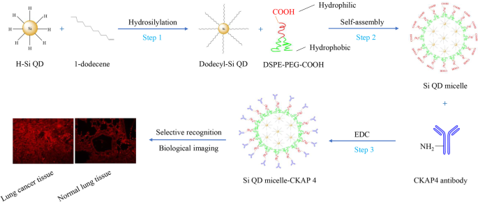
Si QD胶束-CKAP4制备及肺癌组织靶向生物成像示意图
<图片>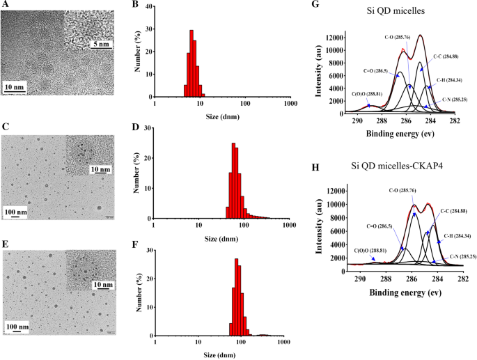
一 十二烷基-Si QD 的 TEM 图像; b 十二烷基-Si QDs的水直径分布; c Si QD胶束的TEM图像; d Si QD胶束的水直径分布; e Si QD胶束-CKAP4的TEM图像; f Si QD胶束-CKAP4的水径分布; g Si QD胶束的C1s X射线光电子能谱(XPS)光谱; h Si QD胶束-CKAP4的C1s XPS光谱
为了制备水分散性 Si QD 胶束,十二烷基-Si QD 通过 DSPE-PEG-COOH 的自组装进行改性(方案 1,步骤 2)[12]。在这里,使用 DSPE-PEG-COOH 代替 F127 来制备表面具有羧基的 Si QD 胶束。结果表明Si QD胶束在水中分散良好。 TEM 图像显示 Si QD 胶束是插入了 Si QD 的球形颗粒。 Si QD 胶束的直径范围为 22.7 至 54.6 nm,平均直径约为 34.3 nm(图 1c)。 DLS 显示 Si QD 胶束的流体动力学直径范围为 43.8 至 615.9 nm,平均为 58.7 nm(图 1d)。 Si QD 胶束的平均 zeta 电位约为 - 20.7 mV。在本研究中,FTIR 用于研究 Si QD 胶束的组成(附加文件 1:图 S1A)。数据表明,N-H、C-H、O-H和C=O的伸缩振动吸收峰分别位于3430、2920、2850和1640 cm -1 , 分别; C-H和O-H的弯曲振动吸收峰分别在1460和1400 cm -1 , 分别; Si–C、C–O 和 P–O–C 的伸缩振动吸收峰分别位于 1250、1100 和 951 cm -1 , 分别; Si-H和O=C-N的弯曲振动吸收峰分别在850和526 cm -1 , 分别。这些数据表明,DSPE-PEG-COOH 的特征基团(如 N-H、O-H、C=O、C-O、P-O-C 和 O=C-N)和在 Si QD 胶束的 FTIR 光谱中可以发现十二烷基-Si QDs(如 Si-C 和 Si-H),表明十二烷基-Si QDs 成功地封装在由 DSPE-PEG-COOH 自组装的胶束中。此外,Si QD 胶束通过 XRD 进行表征(附加文件 1:图 S1B)。 Si QD胶束的衍射峰尖锐,表明Si QD胶束的结晶度良好。在 XRD 图中,峰值在 2θ 27.073、28.451、56.598 和 73.145 对应于硅的 (100)、(002)、(112) 和 (203) 晶面 (PDF #80-0005),表明 Si QD 成功封装在Si QD 胶束。 2θ 处的峰值 31.692、45.431、66.200、75.260 和 83.955 对应于 NaCl 的 (200)、(220)、(400)、(420) 和 (422) 晶面 (PDF # 77-2064)。 Si QD胶束中存在NaCl的合理解释可能如下:Si QD胶束制备后,我们将Si QD胶束溶解在PBS中,导致Si QD胶束中存在NaCl。
紫外可见吸收光谱显示 Si QD 胶束的吸收范围为 200 到 500 nm(图 2a)。激发波长为 330 nm,在 Si QD 胶束的荧光发射光谱中观察到 650 nm 处的强荧光(图 2b)。 Si QD 胶束水溶液(Si 浓度:80 µg/mL)在自然光下透明透明;然而,它在 365 nm 紫外光下发出强烈的红色荧光(图 2c)。在本研究中,使用荧光光谱仪 (FLS1000) 测试了绝对 PL QY。激发波长为 365 nm。数据表明,Si QD胶束的绝对PL QY约为14.49%。
<图片>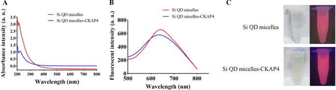
一 Si QD胶束和Si QD胶束-CKAP4的紫外-可见吸收光谱; b Si QD胶束和Si QD胶束-CKAP4的荧光发射光谱; c Si QD 胶束和 Si QD 胶束-CKAP4 暴露在自然光(左)和 365 nm 紫外光(右)下的图像
Si QD 胶束-CKAP4 的制备和表征
我们使用 EDC(方案 1,步骤 3)将 CKAP4 抗体与 Si QD 胶束结合 [14]。 TEM 图像显示 Si QD 胶束-CKAP4 是插入了 Si QD 的球形颗粒。 Si QD 胶束-CKAP4 的直径范围为 24.4 至 67.7 nm,平均直径约为 35.5 nm(图 1e)。 DLS 显示 Si QD 胶束-CKAP4 的流体动力学直径范围从 58.7 到 824.9 nm,平均为 78.8 nm,略大于 Si QD 胶束(图 1f)。这种增加可能是由于 CKAP4 抗体的结合。 C1s XPS 光谱显示 C1s 区域有六种类型的碳原子:C(O)O (288.81 eV)、C=O (286.5 eV)、C–O (285.76 eV)、C–N (285.25 eV)、C –C (284.88 eV) 和 C–H (284.34 eV)(图 1g)。然而,Si QD胶束-CKAP4的XPS光谱中C(O)O的峰几乎消失,表明羧基与伯胺之间的共轭成功;因此,CKAP4 抗体成功地与 Si QD 胶束结合(图 1h)。 Si QD 胶束-CKAP4 的平均zeta 电位约为- 10.7 mV,高于Si QD 胶束。对上述现象的解释可能是:Si QD胶束表面有很多羧基;因此,Si QD 胶束表现出很强的负电性。然而,在Si QD胶束-CKAP4中,CKAP4抗体的偶联导致Si QD胶束-CKAP4表面的羧基减少;因此,Si QD胶束-CKAP4的平均zeta电位增加。
紫外可见吸收光谱显示 Si QD 胶束-CKAP4 的吸收范围为 200 到 500 nm,与 Si QD 胶束的结果相似(图 2a)。激发波长为 330 nm,在荧光发射光谱中从 Si QD 胶束-CKAP4 观察到 640 nm 处的强荧光(图 2b)。这些结果表明抗体偶联对 Si QD 胶束-CKAP4 的荧光特性没有显着影响。 Si QD 胶束-CKAP4 水溶液(Si 浓度:80 µg/mL)在自然光下是透明的。在 365 nm 紫外光下,它可以发出强烈的红色荧光,与 Si QD 胶束的结果一致(图 2c)。 PL QY测试表明Si QD胶束-CKAP4的绝对PL QY约为16.96%。
Si QD 胶束-CKAP4 体外细胞毒性试验
以人肺癌细胞系 A549 和人肾上皮细胞系 293 T 为靶细胞,研究 Si QD 胶束和 Si QD 胶束-CKAP4 的生物安全性。在本研究中,不同浓度的 Si QD 胶束和 Si QD 胶束-CKAP4(Si 浓度为 0、2.375、4.75、9.5、19、38、76 和 152 µg/mL)首先在 37 °C 2 小时。使用台盼蓝检测细胞活力(图 3)[15]。 Si QD 胶束和 Si QD 胶束-CKAP4 的 ID50 是使用 GraphPad 软件获得的。数据显示,在 A549 和 293 T 细胞中,Si QD 胶束的 ID50 大于 152 µg/mL。 Si QD 胶束-CKAP4 的 ID50 在 A549 细胞中为 25.94 µg/mL,在 293 T 细胞中为 72.69 µg/mL。这些结果表明Si QD胶束-CKAP4的细胞毒性显着高于Si QD胶束,并且Si QD胶束-CKAP4在A549细胞中的细胞毒性显着高于293 T细胞。这种现象可能是由于补体依赖性细胞毒性[17]。此外,这些结果还表明Si QD胶束-CKAP4在正常细胞中表现出比在肺癌细胞中更高的生物安全性,为其在体内的应用奠定了基础。
<图片>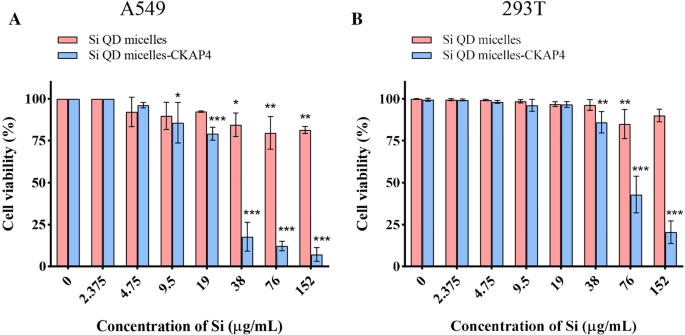
A549 a 的细胞活力 和 293 T b 用不同浓度的 Si QD 胶束和 Si QD 胶束-CKAP4(Si 浓度:0、2.375、4.75、9.5、19、38、76 和 152 µg/mL)孵育 2 小时 (n =3;平均值 ± 标准差;带有 Dunnett 后检验的双向方差分析; *P <0.05; **P <0.01;和***P <0.001)
Si QD 胶束-CKAP4 体外靶向肺癌细胞
为了进一步研究 Si QD 胶束-CKAP4 在体外肺癌细胞中的靶向功能,使用 A549 和 293 T 细胞作为靶细胞。 Si QD 胶束和 Si QD 胶束-CKAP4 首先与 A549 和 293 T 细胞在 37°C 下孵育 2 小时。使用激光共聚焦显微镜检测靶向效应(图 4)。结果表明,Si QD胶束-CKAP4孵育A549的红色荧光显着高于Si QD胶束-CKAP4和293 T共孵育组、Si QD胶束和A549共孵育组以及Si QD胶束和 293 T 联合孵化组。这些结果表明Si QD胶束-CKAP4可以在体外特异性靶向A549细胞。
<图片>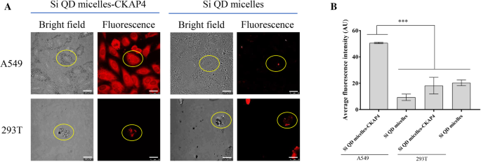
检测 Si QD 胶束-CKAP4 体外靶向 A549 细胞的能力。 一 A549 和 293 T 与 Si QD 胶束-CKAP4 和 Si QD 胶束(Si 浓度 150 µg/ml)在 37 ℃下孵育 2 小时。弃去上清液并洗涤两次后,通过共聚焦激光扫描显微镜(Bar,25 µm)对细胞进行拍照。 b 平均荧光强度的定量分析由 ImageJ (n =3;平均值 ± 标准差;使用 Dunnett 后检验的单向方差分析; ***P <0.001)
Si QD 胶束-CKAP4 体外靶向肺癌组织
为了探索Si QD胶束-CKAP4靶向肺癌组织的可能性,我们设计了一个体外实验来验证Si QD胶束-CKAP4对A549组织的靶向特性。在这项研究中,A549 细胞被皮下接种到小鼠体内。肿瘤形成后,收集 A549 肿瘤组织和正常肺组织,并与 Si QD 胶束和 Si QD 胶束-CKAP4 在 37°C 下孵育 2 小时。使用激光共聚焦显微镜检测靶向效应(图 5)。结果表明,Si QD 胶束-CKAP4 孵育的 A549 组织可以在 405 nm 的激发波长下发出强烈的红色荧光。然而,在 Si QD 胶束孵育的 A549 组织、Si QD 胶束-CKAP4 孵育的正常肺组织和 Si QD 胶束孵育的正常肺组织中仅观察到微弱的红色荧光。 Si QD胶束-CKAP4培养的A549组织中的红色荧光显着高于Si QD胶束培养的A549组织、Si QD胶束-CKAP4培养的正常肺组织和Si QD胶束培养的正常肺组织的红色荧光。这些数据表明Si QD胶束-CKAP4在体外可以有效靶向肺癌组织,有望成为一种潜在的肺癌荧光造影剂。
<图片>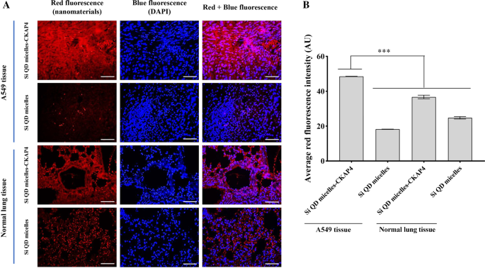
检测 Si QD 胶束-CKAP4 体外靶向 A549 组织的能力。 一 A549 组织和正常肺组织与 Si QD 胶束-CKAP4 和 Si QD 胶束(Si 浓度:150 μg/ml)在 37 ℃下培养 2 h。洗涤两次后,组织用 DAPI 染色以观察细胞核。通过共聚焦激光扫描显微镜(Bar,200 µm)对组织进行成像。 b 通过 ImageJ (n =3;平均值 ± 标准差;使用 Dunnett 后检验的单向方差分析; ***P <0.001)
体内荧光成像
9只荷瘤小鼠被分为三组(Si QD胶束-CKAP4注射组、Si QD胶束注射组和生理盐水注射组)。 Si QD 胶束-CKAP4 注射组的小鼠静脉注射 200 µL Si QD 胶束-CKAP4(Si:4 mg/kg)。 Mice in the Si QD micelle injection group were intravenously injected with 200 µL of Si QD micelles (Si:4 mg/kg). Mice in the saline injection group were intravenously injected with 200 µL saline. Fluorescence images were acquired at different time points (0, 0.25, 0.5, 1, 1.5, 2, 2.5, 3, 3.5, and 4 h). The significant fluorescence signals were not observed in lung cancer tissues among all the mice in the three groups (data not shown). This might be because of the weak fluorescence penetration of the Si QD micelles-CKAP4 and Si QD micelles.
The mice in the Si QD micelles-CKAP4 injection group were anesthetized and euthanized at 4 h to detect the fluorescence signal in their heart, liver, spleen, lung, kidney, and tumor. Data showed that a significant fluorescence signal could be observed in the liver, kidney, and tumor tissues. However, there was no significant fluorescence signal in the heart, spleen, or healthy lung tissues (Fig. 6). The significant fluorescence signal in the liver and kidney indicated that the Si QD micelles-CKAP4 was metabolized by the liver and excreted by the kidney. In addition, the fluorescence signal in A549 tumor tissues was significantly higher than that in healthy lung tissues. This phenomenon contributed to the targeting ability of the Si QD micelles-CKAP4 to lung cancer tissues. These results indicate that Si QD micelles-CKAP4 specifically targets lung cancer tissue, which is expected to be a fluorescent contrast agent for lung cancer surgical navigation in the future.

一 Bright photograph of the heart, liver, spleen, lung, kidney, and tumor in Si QD micelles-CKAP4 injected mice. b Fluorescence photograph of the heart, liver, spleen, lung, kidney, and tumor in Si QD micelles-CKAP4 injected mice. c Relative fluorescence intensities of heart, liver, spleen, lung, kidney and tumor tissue (n =3; mean ± SD; one-way ANOVA with Dunnett’s post test; *P < 0.05; **P < 0.01)
Biosafety Evaluation of Si QD Micelles-CKAP4 In Vivo
Finally, we investigated the acute toxicity of the Si QD micelles-CKAP4 in vivo. In this study, tumor-bearing mice (n = 12) were intravenously injected with saline (200 µL), Si QD micelles (Si:4 mg/kg, 200 µL), or Si QD micelles-CKAP4 (Si:4 mg/kg, 200 µL) and then euthanized 24 h later. H&E staining images of the heart, liver, spleen, lungs, and kidneys are shown in Fig. 7. Compared with saline-injected mice, there were no evident pathological changes in the major organs of Si QD micelles-injected mice and Si QD micelles-CKAP4-injected mice. The above data indicated that Si QD micelles (Si:4 mg/kg) and Si QD micelles-CKAP4 (Si:4 mg/kg) showed no significant toxicity in vivo.

H&E staining images of the heart, liver, spleen, lungs, and kidneys in tumor-bearing mice injected with saline, Si QD micelles (Si:4 mg/kg), and Si QD micelles-CKAP4 (Si:4 mg/kg) for 24 h (magnification:200×)
Discussion
At present, various fluorescent nanomaterials have been designed and synthesized as optical contrast agents for surgical navigation [3, 4]. In 2020, On et al. prepared fluorophore [indocyanine green (ICG) or cyanine 5.5 (Cy5.5)]-conjugated glycol chitosan nanoparticles (CNPs) as a fluorescent contrast agent in lung cancer surgery navigation [18]. However, there are no reports on the preparation of fluorescent contrast agents for lung cancer surgery navigation using Si QDs.
This study successfully synthesized Si QD micelles-CKAP4, which could target the imaging of lung cancer tissues. Si QD micelles-CKAP4 is expected to be used as an optical contrast agent for navigation in lung cancer surgery in the future. The advantages of Si QD micelles-CKAP4 are as follows:(1) We synthesized a fluorescent contrast agent for lung cancer surgery navigation using Si QDs for the first time, which expands the toolbox of the lung cancer surgery navigation tool library. (2) This study optimized water-dispersible Si QD micelles reported by Pi et al. to add targeting ability to Si QD micelles [12]. Thus, it provides a new possibility for applying Si QDs in the biomedical field. (3) The principle of Si QD micelles-CKAP4 targeting lung cancer is based on the high expression of CKAP4 protein in lung cancer tissue. Owing to the conjugation of CKAP4 antibody, Si QD micelles-CKAP4 could actively target lung cancer tissue. Compared with the nanomaterials that passively target tumors via the EPR effect, Si QD micelles-CKAP4 showed stronger specificity to lung cancer tissue. (4) Si QD micelles-CKAP4 showed selective toxicity to tumor cells in a specific range of concentrations. Therefore, Si QD micelles-CKAP4 showed high biological safety in the range of rational drug concentrations, which exhibited its potential clinical application value.
In this study, PL QY tests showed that the absolute PL QY of Si QD micelles was approximately 14.49% and the absolute PL QY of the Si QD micelles-CKAP4 was approximately 16.96%. A study by Pi et al. showed that the PL QY of dodecyl-Si QDs was approximately 26%, which was very high for Si QDs that emit red light with a PL peak at less than 700 nm [12, 19, 20]. However, the PL QY of the Si QD micelles in this study was approximately 14.49%, which was lower than that of dodecyl-Si QDs. This might be because of the surface defects introduced during the emulsion process, which was nearly consistent with the decrease in PL QY in Si-QD micelles (dodecyl-passivated Si QDs were encapsulated in the micelles self-assembled from F127 in the emulsion) in the study reported by Pi et al. [12, 21]. In this study, the PL QY of Si QD micelles-CKAP4 was approximately 16.96%, which was slightly higher than that of Si QD micelles (14.49%). This could be because in the Si QD micelles-CKAP4, the coupling of the CKAP4 antibody repaired some surface defects of the Si QD micelles, leading to a slight enhancement of the PL QY.
As we all know, there is a challenge in Si QDs biological imaging applications:Although Si exhibits little cytotoxicity, Si particles may exhibit cytotoxicity as the size decrease to a certain extent. In this study, cytotoxicity tests showed that Si QD micelles-CKAP4 exhibited selective cytotoxicity to lung cancer cells at 9.5–19 µg/mL. Therefore, as a result of CKAP4 antibody conjugation, the cytotoxicity of Si QD micelles-CKAP4 to normal cells was significantly less than that of lung cancer cells, which provides a powerful condition for the biological application of Si QD micelles-CKAP4.
In this study, human lung cancer cell line A549 and human renal epithelial cell line 293 T were used as target cells to investigate the targeting ability of Si QD micelles and Si QD micelles-CKAP4. A549 and 293 T cells were adherent and semi-adherent, respectively. During the experiment, the number of 293 T cells decreased because of repeated centrifugation and washing. Therefore, as shown in Fig. 4, the number of 293 T cells was lower than that of A549 cells.
In addition, it should be noted that A549 cells belong to the p53 wild-type lung cancer cell line. Therefore, only A549 cells cannot fully reflect the targeting ability of the Si QD micelles-CKAP4 to lung cancer. Further studies will be conducted to test the targeting ability and imaging effect of Si QD micelles-CKAP4 on p53-negative cell lines (such as H1299) to provide a solid experimental basis for the clinical application of Si QD micelles-CKAP4 [22].
结论
This study improved and modified the Si QD micelles reported by Pi et al. to prepare water-dispersible Si QD micelles-CKAP4. Si QD micelles-CKAP4 were spherical particles with a mean hydrodiameter of approximately 78.8 nm. UV–visible absorption of the Si QD micelles-CKAP4 ranged from 200 to 500 nm. With an excitation wavelength of 330 nm, strong fluorescence at 640 nm was observed in the fluorescence emission spectra. Si QD micelles-CKAP4 exhibited good targeting ability to lung cancer cells and lung cancer tissues both in vitro and in vivo. In addition, the in vivo fluorescence-imaging assay further showed that the Si QD micelles-CKAP4 was metabolized by the liver and excreted by the kidney. Cytotoxicity and H&E staining assays showed that the Si QD micelles-CKAP4 exhibited high biosafety both in vitro and in vivo. Si QD micelles-CKAP4 is a specifically targeted imaging agent for lung cancer and is expected to be a fluorescent contrast agent for lung cancer surgical navigation in the future.
数据和材料的可用性
The data and the analysis in the current work are available from the corresponding authors on reasonable request.
缩写
- Si QD:
-
Si quantum dot
- Si QD micelles-CKAP4:
-
CKAP4 antibody-conjugated Si quantum dot micelles
- Dodecyl-Si QDs:
-
Dodecyl-passivated Si QDs
- BSA:
-
牛血清白蛋白
- THF:
-
四氢呋喃
- DMEM:
-
Dulbecco’s modified eagle’s medium
- PBS:
-
Phosphate buffer saline
- FBS:
-
胎牛血清
- TEM:
-
透射电子显微镜
- DLS:
-
动态光散射
- XPS:
-
X射线光电子能谱
- FTIR:
-
傅里叶变换红外光谱
- XRD:
-
X射线衍射
- SPF:
-
Specific pathogen free
- DAPI:
-
4′, 6-Diamidino-2-phenylin-dole
- H&E:
-
苏木精和伊红
纳米材料
- 用于癌症治疗的纳米粒子:当前的进展和挑战
- 用于癌症应用的基于细胞的药物递送
- S、N 共掺杂石墨烯量子点/TiO2 复合材料用于高效光催化制氢
- 变质 InAs/InGaAs/GaAs 量子点异质结构光电压的双极效应:光敏器件的表征和设计解决方案
- 消除 InAs/GaAs 量子点中的双峰尺寸,用于制备 1.3-μm 量子点激光器
- 介观四(对磺基苯基)卟啉对半胱氨酸包覆的 CdSe/ZnS 量子点发光的刺激
- 用于近红外光声成像的可调谐纵横比金纳米棒的种子介导合成
- 适体修饰的磁性纳米增敏剂,用于表达 HER2 的癌症的体内 MR 成像
- 用于超声介导多柔比星靶向递送的生物相容性壳聚糖纳米泡
- 用于高效结肠癌基因治疗的基于阳离子胶束的 siRNA 递送
- 具有用于白光源的量子点转换器的高均匀性平面微型芯片级封装 LED
- 环境兼容的生物共轭金纳米粒子作为炎症诱导的癌症成像的有效造影剂