

CpG 的改性 Fe3O4 磁性纳米颗粒递送抑制肿瘤生长和自发性肺转移以增强免疫治疗
摘要
作为一种新型的 toll 样受体 9 (TLR9) 激动剂,合成的未甲基化胞嘧啶-磷酸-鸟嘌呤 (CpG) 寡脱氧核苷酸可以刺激 Th1 免疫反应,并有可能用作治疗癌症的治疗剂或疫苗佐剂。然而,CpG 的一些缺点限制了它们的应用,例如通过核酸酶介导的降解快速消除和细胞吸收不良。因此,治疗需要重复大剂量给药。在这项工作中,首次设计和研究了基于 3-氨基丙基三乙氧基硅烷 (APTES) 修饰的 Fe3O4 纳米粒子 (FeNPs) 的 CpG 递送系统,以实现更好的 CpG 生物活性。在我们的结果中,我们通过将 CpG 加载到 FeNP 中,设计了 FeNP 递送的 CpG 颗粒 (FeNP/CpG),其平均尺寸较小,约为 50 nm。 FeNP/CpG 颗粒递送系统在体外和通过瘤内注射增强骨髓衍生树突细胞 (BMDC) 中 CpG 的细胞摄取,通过刺激 C26 结肠癌和 4T1 乳腺癌更好的体液和细胞免疫反应显示出显着的抗肿瘤能力体内癌症异种移植模型优于游离 CpG。此外,经 FeNP/CpG 颗粒处理的小鼠肿瘤生长延迟,抑制率高达 94.4%。此外,C26 模型中大约 50% 的肿瘤似乎完全消退。同样,与未治疗的对照组相比,4T1 乳腺癌肿瘤模型的肺转移率较低,肿瘤抑制率为 69%。 FeNP/CpG颗粒除具有有效性外,制备简单、安全性和稳定性高,也使其成为一种极具吸引力的抗肿瘤免疫疗法。
背景
恶性肿瘤是威胁人类健康和生命的重大疾病之一,肿瘤的发病率不断上升[1, 2]。不幸的是,传统疗法如放疗、化疗和手术治疗恶性肿瘤的效果并不显着。与针对快速增殖的细胞进行死亡的化疗和放疗不同,免疫疗法旨在增强宿主自身的免疫防御系统,以靶向和消灭肿瘤细胞。
值得注意的是,免疫治疗性 CpG 因其在肿瘤预防和消退方面的功效而被广泛研究 [3]。尽管临床数据令人鼓舞,但使用免费 CpG 仍然有几个缺点。首先,CpG 在生物条件下对核酸酶介导的降解敏感。其次,它们在全身给药后缺乏对靶细胞的特异性并且细胞摄取差。因此,可能需要改进。因为 TLR9s 位于内体上,我们假设肿瘤相关炎症细胞对 CpG 的吸收不良导致临床反应较弱。因此,增强CpG内化的方法可能会增强其免疫刺激反应。
除了游离 CpG 的缺点外,CpG 的给药途径影响抗肿瘤能力和毒性。通过静脉注射给药的游离 CpG 和其他稳定的硫代磷酸酯寡核苷酸被迅速清除并具有广泛的组织分布 [4, 5]。此外,全身给药游离 CpG 可诱导非特异性免疫激活,导致严重的副作用,包括免疫细胞耗竭、淋巴滤泡破坏、肝损伤和自身免疫性疾病恶化 [6,7,8]。这些特性可能是人类患者全身给药游离 CpG 失败的原因 [9]。以往的研究表明,CpG 的瘤内注射通过将免疫刺激“集中”在肿瘤部位,具有很好的抗肿瘤作用[10]。如何控制注射到肿瘤中的CpG的保留时间是一个问题。
通过使用 toll 样受体 9,树突状细胞、巨噬细胞和 NK 细胞可以识别细菌 DNA 中常见的 CpG [11]。 CpG 诱导免疫细胞产生趋化因子和细胞因子,以及上调 T 细胞的共刺激细胞表面分子 [12,13,14,15]。因此,CpG 已成为有效的 1 型极化佐剂 [16, 17]。此外,将CpG直接注射到肿瘤中是一种免疫形式,它使用原位肿瘤作为抗原来源并引入CpG作为佐剂以激活肿瘤内的免疫反应。因此,我们假设瘤内注射CpG会通过招募和激活局部树突状细胞来消除肿瘤的免疫特权,这取决于1型抗肿瘤T细胞反应途径。
为了克服上述问题,我们开发了一种基于 Fe3O4 颗粒的改良磁性颗粒平台,通过肿瘤部位的肿瘤内注射来指导和控制 CpG 的释放。由于 Fe3O4 磁性颗粒的一些独特特性,特别是其良好的组织相容性 [18]、超顺磁性 [19, 20]、低毒性 [21] 以及易于制备和外磁场靶向给药 [22, 23],它们的可以通过外部磁场控制体内的运动和浓度,使 Fe3O4 颗粒可作为基因药物的载体,提高基因转染效率 [24]。此外,通过共价键在 Fe3O4 颗粒上涂覆 APTES 可防止聚集体的形成,并提供更多的修饰位点(氨基)[25, 26] 以进一步帮助结合 CpG。在这项研究中,Fe3O4 磁性颗粒是通过共沉淀法制备的 [27] 并用 APTES 改性。 CpG 通过静电相互作用牢固地结合到改性 Fe3O4 颗粒的表面,形成直径约为 50 nm 的 FeNP/CpG 颗粒。进一步的研究表明,与树突细胞中游离 CpG 相比,FeNP 颗粒递送系统对 CpG 的吸收效率更高,并且 FeNP/CpG 颗粒的瘤内注射刺激了有效的抗肿瘤 Th1 型免疫反应,不仅可以抑制肿瘤原位生长还能抑制体内 C26 结肠癌和 4T1 乳腺癌模型的肿瘤转移。因此,纳米制剂治疗可能是一种潜在的肿瘤免疫治疗策略。
材料和方法
材料和动物
3-氨基丙基三乙氧基硅烷(APTES)购自阿拉丁公司。六水氯化铁(FeCl3∙6H2O)和四水氯化亚铁(FeCl2∙4H2O)购自天津光复精细化工研究院。以下 CpG 由 Invitrogen Co. 合成:5'-TCGTCGTTTTGTCGTTTTGTCGTT-3'。异硫氰酸荧光素 (FITC) 购自 Sigma-Aldrich (St Louis, MO, USA)。
从美国典型培养物保藏中心(ATCC)获得人胚肾细胞系(293T)、鼠乳腺肿瘤细胞系(4T1)和结肠癌细胞系(C26),并获得骨髓来源的树突状细胞(BMDCs)来自 BALB/c 小鼠。雌性6-8周龄BALB/c小鼠购自北京华富康实验动物有限公司。小鼠在无病原体条件下饲养和饲养。所有动物实验方案均按照美国国立卫生研究院实验动物护理和使用指南进行。
FeNP的制备
共沉淀法用于合成 Fe3O4 颗粒 [29]。在实验过程中,将 11.68 g FeCl3∙6H2O 和 4.30 g FeCl2∙4H2O 在 80 °C 下加入含有 200 mL 去离子水的三颈烧瓶中,然后加入 15 mL 25% NH3·H2O。在1小时反应过程中,混合物用N 2 吹扫并搅拌。通过超声振动 30 分钟,将 30 毫克制备的 Fe3O4 分散到 60 毫升去离子水和无水乙醇的混合物中。通过超声振动 30 分钟,将 0.3 g 制备的 Fe3O4 分散到 4 mL 去离子水和 600 mL 无水乙醇的混合物中。然后,在持续机械搅拌 7 小时的情况下,将 1.2 mL 的 APTES 添加到混合物中。得到的功能化 APTES 修饰的 Fe3O4 (FeNP) 纳米颗粒通过磁铁与上清液磁性分离,然后用乙醇洗涤并在 40°C 下真空干燥 24 小时。最后制备了FeNP沉淀物以备进一步使用。
FeNP/CpG 粒子的表征
Fe3O4、FeNP 和 FeNP/CpG 颗粒的粒度分布光谱使用 Zetasizer Nano ZS90 激光粒度分析仪(Malvern Instruments,Malvern,UK)在 25°C 下测定。每个试验进行 3 次,取平均值。使用透射电子显微镜(H-6009IV;Hitachi Ltd.,Tokyo,Japan)和扫描电子显微镜(SEM)观察制备的颗粒形貌。
进行琼脂糖阻滞试验以评估 FeNP 颗粒的 CpG 结合能力。简而言之,将功能化的 APTES 修饰的 Fe3O4 颗粒 (FeNP) 与 1 μg CpG 以不同的比例 (FeNP:CpG, w /w ) 在蒸馏水中。在室温下共孵育 30 分钟后,混合物在 1% (w /v ) 120 V 琼脂糖凝胶 30 分钟。然后将凝胶用 Golden View™ (0.5 mg/mL) 染色,并通过紫外线照明器 (Bio-Rad ChemiDox XRS, USA) 拍照。
MTT 检测
使用 MTT 测定法评估 FeNP 颗粒对 C26、4T1 和 293T 细胞系的细胞毒性。 C26、4T1和293T细胞以5 × 10 3 的密度接种 在 96 孔板中每孔含有 10% FBS 的 100 μL RPMI 1640 培养基中培养 24 小时或 72 小时。将制备的细胞暴露于一系列不同浓度的 FeNP 颗粒:1.25、0.625、0.3、0.15、0.07、0.03、0.01 和 0 mg/ml,一式六份。培养指定时间后,将 100 μl 完全培养基和 10 μl MTT 移入每个孔中,并在 37°C 下孵育 4 小时。将 DMSO 添加到溶质中 30 分钟,形成甲臜。然后,使用 Spectramax M5 微量滴定板发光计(Molecular Devices,美国)在 570 nm 处测量吸光度。未处理细胞的细胞活力为100%。
体外转染
BMDCs 由 BALB/c 小鼠制备。简而言之,使用注射器用含有游离 FBS 的 1640 培养基冲洗出来自股骨和胫骨的骨髓细胞。细胞以 1500 rpm 离心 3 分钟,用 ACK 裂解缓冲液(Lonza Inc.)处理以去除红细胞,并重悬于添加了 10% FBS、青霉素(100 U/mL)和链霉素的 RPMI-1640 培养基中( 100 U/mL),37°C,5% CO2 和 20 ng/mL 粒细胞-巨噬细胞集落刺激因子 (GM-CSF)。然后将细胞以10 6 的密度接种到六孔板中 每孔细胞数,每 2 天更换一次生长培养基。第7天收获树突细胞。
在第 7 天,将质量比为 10:1 的 0.2 μg 游离 FITC 偶联 CpG (CpG-FITC) 或 FeNP 的微粒当量添加到预先设计的孔中。转染后 1 小时或 3 小时,除去生长培养基,用 DAPI 染色细胞 10 分钟,用生理盐水洗涤细胞 3 次。用激光扫描共聚焦显微镜(LSCM)对每孔拍照,并通过流式细胞术(NovoCyte Flow Cytometer,ACEA Biosciences,USA)测量转染效率。
体内肿瘤抑制试验
将 10 万个 C26 或 4T1 细胞皮下注射到每只 BALB/c 小鼠(6-8 周龄)的背部。随后使用数显卡尺测量肿瘤体积并通过以下公式计算:肿瘤体积=0.5 × 长度(mm) × [宽度(mm)] 2 .一旦肿瘤达到大约 50 毫米 3 在大小上,应用了处理。老鼠 (n =10) 每 3 天接受相当于 20 μg CpG 的 CpG/FeNP(比例为 10:1)颗粒的瘤内注射或单独 CpG 进行四次治疗。接受等量生理盐水 (NS) 或 FeNP 颗粒的小鼠被视为对照组。每 3 天监测肿瘤大小和动物体重。在第31天,所有小鼠通过颈椎脱位处死。肿瘤组织及其他重要器官用4%中性福尔马林固定,石蜡切片HE染色评价形态差异,免疫荧光检测肿瘤微环境。在4T1模型中,我们测量肿瘤重量并计算肺转移结节数。
肿瘤再攻击实验在C26模型中进行。简而言之,用 5 × 10 5 再次攻击用 FeNP/CpG 治疗治愈的小鼠 C26 或 4T1 细胞 s.c.侧翼另一侧的两个独立位置,无需进一步处理。当 4T1 肿瘤的体积长到 200-300 毫米时处死小鼠 3 或 20 天。
细胞毒性 T 淋巴细胞检测
CTL 测定是根据已发表的方法进行的。详细地,作为效应细胞的脾淋巴细胞被收获并用氯化铵去除红细胞,并在处理实验结束时通过尼龙羊毛。 4T1或C26作为靶细胞用300 mci的Na2 51 标记 CrO4 保持 2 小时,然后用 PBS 洗涤并分配到 96 孔板中。制备的效应细胞与靶细胞以不同的 E:T 比例共孵育。用闪烁计数器测量含有来自靶细胞的放射性的上清液。特异性裂解如下测定:%特异性裂解=100[(在CTL自发释放的存在下释放)/(最大释放自发释放)]。在所有实验中,自发释放均小于最大释放量的30%。
ELISpot 检测
在实验结束时从对照或 CpG 处理的小鼠中收获脾细胞,并根据制造商的说明使用小鼠 IFN-γ/IL-4 双色 ELISpot 试剂盒进行 ELISpot 检测。将收获的脾细胞与其各自处理共孵育96小时,滴加悬液,用洗涤缓冲液洗涤细胞3次,然后加入IFN-γ和IL-4的混合抗体。显色剂处理后,显微镜下计数斑点形成细胞(SFCs)数。
ELISA 检测
根据制造商的说明,进行 ELISA 以确定 Ct 特异性血清抗体滴度的水平。简而言之,用含有 0.05% Tween 20 (PBST) 的 PBS 溶液冲洗用纯化的全 Ct 蛋白(BestBio Biotechnology Co.,China)包被的微孔板,然后用 100 μl 封闭缓冲液(PBST 中的 5% 脱脂牛奶)封闭在 37°C 下保持 1 小时。用 PBST 冲洗五次后,将板与免疫血清(在封闭缓冲液中以 1:1000 稀释,100 μl/孔)或阴道冲洗液(在封闭缓冲液中以 1:10 稀释,100 μl/孔)在 37 ℃孵育°C 2 小时。用 PBST 冲洗五次后,将板与 HRP 偶联的山羊抗小鼠 IgG 抗体(在封闭缓冲液中以 1:1000 稀释,100 μl/孔)或 HRP 偶联的山羊抗小鼠 IgA 抗体(稀释于1:2000 在封闭缓冲液中,100 μl/孔)在 37°C 下保持 1 小时。
组织学分析
从体内抑制研究中收获的肿瘤组织和主要器官被固定并包埋在石蜡中。脱蜡和再水化后,蜡包埋的组织切片用 Mayer 的 HE 染色。为了分析每组肿瘤组织内的浸润淋巴细胞 (TIL),切片进行高压抗原修复,用抗小鼠 FITC-CD4、PE-CD8 和 PE-CD49b(作为 NK 标记)(BD,美国)在 4°C 下染色 1 小时,然后在 PBS 洗涤成像前用 DAPI 染色 10 分钟。通过正荧光显微镜(Olympus,Japan)拍摄每组的图像。
统计分析
数据表示为平均值 ± 标准偏差。使用 Prism 5.0c 软件(GraphPad Software,La Jolla,CA,USA)SPSS 17 软件通过单向方差分析(ANOVA)进行统计分析。使用 Tukey 检验进行单因素方差分析 (ANOVA) 进行组间比较。 P <0.05 的值被认为具有统计学意义。
结果
FeNP/CpG 颗粒的制备和表征
为了开发一种具有低细胞毒性的用于 CpG 递送的磁性颗粒,使用共沉淀法合成了 Fe3O4 颗粒,如图 1a 所示。在 Fe3O4 表面接枝 APTES 的氨基丙基硅烷基团 (-O)3Si-CH2-CH2-CH2-NH2,形成 FeNP 颗粒(图 1b)。带负电荷的 CpG (CpG) 与 APTES 提供的有效官能团结合,通过静电相互作用形成 FeNP/CpG 颗粒,如图 1c 所示。基于动态光散射测量,制备的 Fe3O4 颗粒的尺寸为 10.7 ± 4.1 nm(图 2a)。通过 TEM(图 2b)和 SEM(图 2c)观察到,在这项工作中开发的 Fe3O4 显示出均匀且接近球形的形状。 APTES修饰的Fe3O4粒子的动态直径为34.5 ± 5.0 nm(图2d),与TEM结果(图2e)吻合良好。
<图片>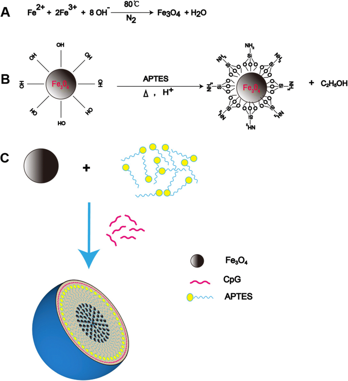
FeNP/CpG 颗粒的制备。 一 使用共沉淀法合成 Fe3O4 磁性颗粒的方程式。 b 水解和缩合反应产生硅烷聚合物和 APTES 在磁铁矿表面的硅烷化反应形成 FeNP 颗粒。 c FeNP/CpG 颗粒示意图。 CpG通过静电相互作用与FeNPs表面结合形成FeNP/CpG颗粒
<图片>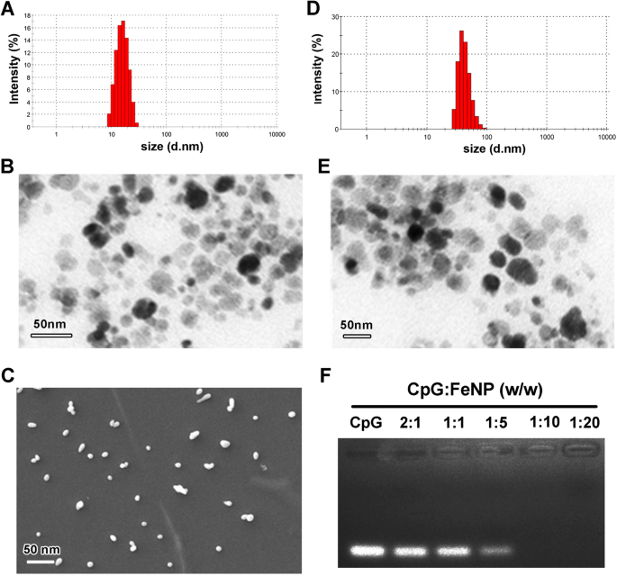
FeNP/CpG 颗粒的表征。 一 Fe3O4 颗粒的尺寸分布谱。 b Fe3O4 的透射电子显微镜图像 (TEM)。 c Fe3O4 颗粒的扫描电子显微镜图像。 d APTES 包覆的 Fe3O4 (FeNP) 颗粒的尺寸分布。 e FeNP颗粒的TEM图像。 f FeNPs 的 CpG 结合能力由凝胶阻滞试验确定。所有数据代表三个独立实验
为了说明 FeNP 颗粒与 CpG 的结合能力,进行了凝胶阻滞试验(图 2f)。当 FeNP 与 CpG 的摩尔比为 10:1 时,没有观察到明亮的 CpG 带,表明带负电荷的 CpG 可以通过电泳后的静电相互作用被 FeNP 颗粒完全吸附。因此,我们选择了该处方比例以在我们的研究中进一步应用。总之,这些结果表明CpG可以通过静电相互作用在FeNPs表面成功结合,表现出稳定性和小尺寸。
FeNP/CpG 粒子的体外细胞活力和转染
为了进一步描述体外生物物理表征,我们在 24 小时或 72 小时时检测了 FeNPs 在 293T、4T1 和 C26 细胞中的细胞毒性(图 3a)。在 293T、C26 和 4T1 细胞中,细胞活力与 FeNP 颗粒之间没有明显的剂量依赖性关系。三种细胞的细胞存活率在 24 小时时超过 80%,在 72 小时时超过 60%,即使 FeNP 浓度为 1.25 毫克/毫升。这些结果证明了FeNP颗粒对正常细胞(293T)或肿瘤细胞(C26、4T1)的生物相容性。
<图片>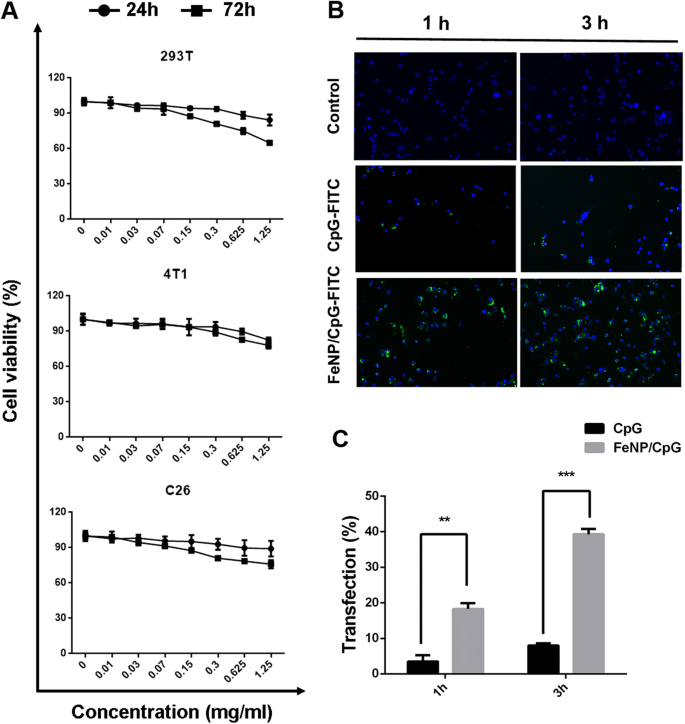
FeNP/CpG 颗粒的体外细胞毒性和吸收活性。 一 MTT 测定。在用不同浓度的 FeNP 颗粒处理 24 小时或 72 小时后,我们检测了 293T、4T1 和 C26 细胞的活力。在任何剂量的 FeNP 下,细胞活力没有显着差异。 b , c BMDCs 中 FeNP/CpG 摄取的检测。 GM 后制备的 BMDCs -脑脊液 处理 7 天后,用 CpG-FITC 或 FeNP 递送的 CpG-FITC 转染 1 或 3 小时,然后密集洗涤、固定并用 DAPI 标记。通过激光扫描共聚焦显微镜 (LSCM) 捕获图像。与游离 FITC 标记的 CpG 相比,FeNP/CpG 具有增强的荧光强度(b ) 和转染效率有统计学意义 (c )。这些数据代表三个独立实验,平均值 ± 标准误差; *P < 0.05,*P < 0.01 和 ***P < 0.001 vs 未治疗组
树突状细胞作为 CpG 的主要受体细胞,在 TLR9 介导的抗肿瘤免疫反应中发挥重要作用。 CpG 作为一种有效的 1 型极化佐剂,通过分泌趋化因子和细胞因子诱导 DCs 激活肿瘤内的免疫反应。因此,将 CPG 有效递送到 DC 细胞中对于实现抗肿瘤反应至关重要。在我们的研究中,FITC 偶联的 CpG 用于研究 FeNP 颗粒在体外向 DCs 的递送能力。转染后 1 小时或 3 小时后,通过激光扫描共聚焦显微镜 (LSCM),FITC 共轭 CpG 包覆 FeNP 颗粒 (FeNP/CpG-FITC) 处理组的细胞荧光强度高于等量的游离 CpG 基团(图 3b)。 FeNP/CpG-FITC 颗粒能够在 1 小时后转染多达 18.3% 的 DC。然后,转染后 3 小时,阳性细胞的比例增加到 39.2%(图 3c)。同时,对于游离 FITC 缀合的 CpG 处理的细胞,即使在转染后 3 小时也能观察到最小的荧光,转染率低于 10%。因此,表明FeNP颗粒递送CpG增加了细胞对CpG的摄取。
FeNPs 将 CpG 递送至异种移植物并抑制体内肿瘤生长和自发性肺转移
CpG/FeNP 颗粒的抗癌活性首先在体内 C26 结肠癌和 4T1 乳腺癌皮下移植肿瘤模型中进行评估。当肿瘤体积约为 50 mm 3 , 将小鼠随机分为四组开始治疗 (n =10)。在整个实验过程中,小鼠用 CpG/FeNP 颗粒瘤内注射 3 次(图 4a)。在我们的研究中,与对照组相比,肿瘤内注射 FeNP/CpG 颗粒导致异种移植肿瘤生长的显着抑制,抑制率高达 69% (P <0.05 与 NS)(图 4b)。与生理盐水 (NS) 治疗组 (0.90 ± 0.08 g) 和 FeNP 组 (0.81 ± 0.03 g) 相比,FeNP/CpG 颗粒治疗组的肿瘤重量显着减少 (0.38 ± 0.03 g,P <0.001)。相比之下,游离 CpG 组显示出最小的抗癌能力(0.68 ± 0.03 g)(图 4c)。此外,PBS 对照组的小鼠发生了广泛的肺转移,相比之下,用 FeNP/CpG 颗粒治疗的小鼠肺中肿瘤结节的数量减少了 64.1%(图 4d、e),证明了强大的力量FeNP/CpG 颗粒在治疗转移性肿瘤中的作用。如图 4f 所示,与其他组相比,FeNP/CpG 治疗后肺的 H&E 染色显示肺负荷减少,肺转移明显。这些结果表明 FeNP 将 CpG 递送到 4T1 细胞中不仅显着抑制了 4T1 肿瘤的生长 (P <0.001 vs NS),但也抑制了乳腺癌向肺部的转移。
<图片>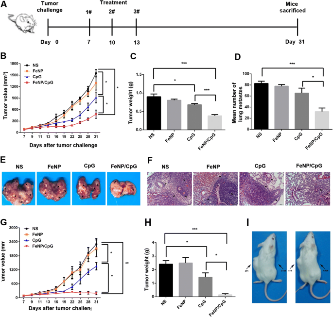
FeNP/CpG 颗粒在体内的抗肿瘤能力。 一 FeNP/CpG 颗粒在 C26 结肠癌和 4T1 乳腺癌异种移植模型中通过体内肿瘤内注射的抗肿瘤治疗方案。 b –d FeNP/CpG 粒子处理抑制 4T1 肿瘤的生长。 b 4T1 肿瘤模型的代表性肿瘤生长曲线。 c 平均肿瘤重量。 d FeNP/CpG瘤内注射后,每组平均肺肿瘤结节清楚地显示肺转移。 e , f 肺的明场成像和 H&E 染色:e 可见肺转移的数量,箭头表示肺转移。 f 肺转移瘤的 H&E 染色。比例尺,100 μm 用于 H&E 染色。 g , h FeNP/CpG 颗粒显着抑制 C26 异种移植肿瘤的生长:e 实验和f各组肿瘤生长曲线 平均肿瘤重量。每组有10只小鼠。 g 肿瘤再攻击实验。 我 在C26模型中,用FeNP/CpG治疗治愈的小鼠再次用5 × 10 5 攻击 C26(右侧)或 4T1 细胞(左侧)s.c.侧翼另一侧的两个独立位置,无需进一步处理。显示的图像是肿瘤再攻击后 20 天的代表性小鼠。所有数据均代表三个独立实验,平均值 ± SEM; *P < 0.05,*P < 0.01 和 ***P < 0.001
类似地,在 C26 孵育小鼠的皮下模型中观察到 FeNP/CpG 治疗组的抗肿瘤作用显着增强。如图 4g 所示,对照小鼠、载体组和 CpG 组的肿瘤生长迅速,平均肿瘤体积约为 2300 ± 239.4 mm 3 , 2116.7 ± 360.9 mm 3 , 和 1353.3 ± 158.9 mm 3 分别在第 31 天。相比之下,在 FeNP/CpG 组中,肿瘤生长受到极大抑制,平均肿瘤体积约为 153.7 ± 62.7 mm 3 (P <0.01 vs NS) 和 94.4% 的抑制率。初次治疗后肿瘤似乎生长缓慢,3次治疗结束后部分肿瘤开始逐渐消失。更重要的是,在 FeNP/CpG 粒子治疗组中,近一半小鼠的肿瘤在实验结束时完全消失。如图 4h 所示,FeNP/CpG 治疗组的平均肿瘤重量远低于其他组(P <0.001):FeNP/CpG治疗组(0.14 ± 0.08g)、NS组(2.41 ± 0.26g)、FeNP组(2.50 ± 0.4g)和CpG组(1.44 0.26g)。为了确定 FeNP/CpG 颗粒是否足以介导特定的抗肿瘤反应,在 C26 模型中进行了肿瘤再攻击实验(图 4i)。对C26结肠癌模型中经FeNP/CpG粒子处理后肿瘤完全消退的小鼠,分别在BALB/c小鼠不同部位皮下接种4T1和C26肿瘤,不再给予进一步处理。所有 4T1 肿瘤都迅速生长,到攻击后第 20 天它们的体积超过 1000 mm3。相比之下,C26 模型中存在肉眼看不见的实体瘤,表明 FeNP/CpG 颗粒组刺激了特异性抗肿瘤反应。这些结果表明,FeNP/CpG 颗粒的瘤内注射有效抑制了肿瘤的生长。皮下异种移植。
FeNP/CpG 粒子刺激增强的系统性抗肿瘤免疫反应
进一步研究了上述两种模型中FeNP/CpG颗粒的体内抗肿瘤机制。为了研究免疫反应的类型,我们应用 ELISpot 分析来分析小鼠脾脏的 IFN-γ/IL-4 水平。如果脾淋巴细胞中的IFN-γ水平(ELISpot板显示红斑)显着高于IL-4水平(ELISpot板显示蓝点),则刺激的免疫反应类型为Th1,即激活CD8 + T 淋巴细胞,主要是身体的 CTL 反应。否则,免疫反应类型为Th2,即主要激活CD4 + T 淋巴细胞,从而刺激 B 淋巴细胞并产生抗原特异性抗体。进行双色 ELISpot 测定以测量不同处理中 IFN-γ 和 IL-4 的表达(图 5a)。与其他三组相比,FeNP/CpG颗粒处理组小鼠脾淋巴细胞分泌的IL-4和IFN-γ斑点形成细胞(SFCs)数量呈上升趋势,SFCs数量呈上升趋势。分泌 IFN-γ 是分泌 IL-4 的 1.5 倍(P <0.05),表明FeNP/CpG颗粒瘤内注射后抗肿瘤细胞免疫反应增强。此外,使用 ELISA 测定法分析来自 NS、FeNP、CpG 和 FeNP/CpG 处理小鼠的血清,以确定在 3 次免疫后是否存在总肿瘤特异性 IgG。在 C26 肿瘤模型中,虽然 FeNP/CpG 免疫小鼠的肿瘤特异性 IgG 滴度显着高于其他组(图 5b),但 FeNP/CpG 免疫小鼠和 CpG 免疫小鼠之间没有统计学差异.
<图片>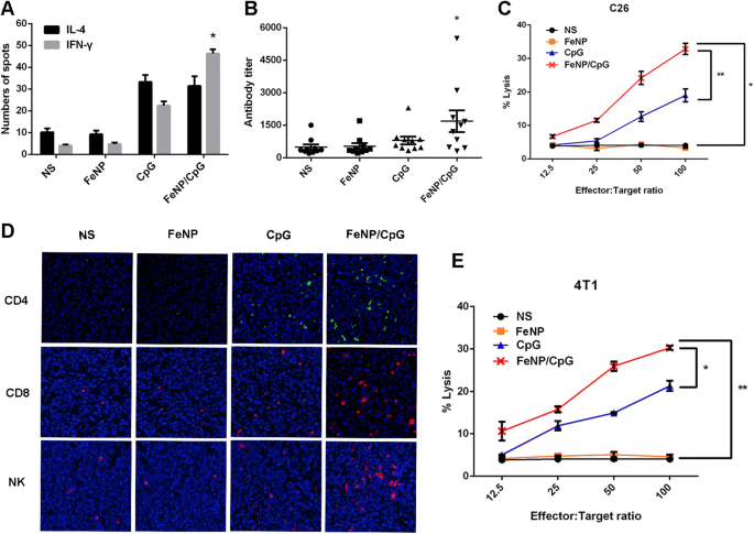
抗肿瘤免疫反应的机制。 一 ELISpot 检测。 Splenocytes were harvested from NS or treated mice after the final treatment, and ELISpot assays were performed using the mice IFN-γ/IL-4 Dual-Color ELISpot kit in the C26 xenograft model. b In the C26 model, serum antibody titers in tumor-bearing mice treated by FeNP/CpG particles were tested using an ELISA assay. Each symbol represents an individual mouse, and horizontal lines indicate the median as determined by Mann–Whitney U 测试。 c CTL assays. The cytotoxicity of splenocytes against C26 cells were examined in a 4-h 51Cr-release assay. d Representative immunofluorescence images showing infiltrating immune cells in tumor tissues in each group. The FeNP/CpG particles treatment groups with enhanced CD4 + T, CD8 + T and NK (CD57 + ) cell infiltration in tumors compared to that of other groups. e The cytotoxicity of splenocytes against 4T1 cells were examined in a 4-h 51Cr-release assay
To assess the cell-mediated immune response stimulated by the FeNP/CpG, CTL activity was assessed using the 51Cr release assay on C26 and 4T1 cells ex vivo (Fig. 5c, e). When the killing activity of CTL was at a 100:1 ratio of effector cells to target cells, in the C26 cells, the effectors in the FeNP/CpG groups showed 8.2-fold more tumor-killing activity than that in the NS group and 9.7-fold and 1.7-fold more than that in the FeNP and CpG groups (P < 0.05 versus NS) (Fig. 5c). Similarly, this observation was coincident with the result in the 4T1 cells; the FeNP/CpG group had 7.4-fold more tumor-killing activity than that of the NS group and 6.6-fold and 1.4-fold more than that of the FeNP and CpG groups (P < 0.01 versus NS). These results illustrated that compared to that of free CpG, the use of APTES-coated Fe3O4 to deliver CpG can stimulate a more intense humoral immune response and result in a better antitumor efficiency in vivo.
The FeNP/CpG Increases Infiltrating Lymphocytes in Tumors and Alters Tumor Microenvironments
Tumor-infiltrating lymphocytes (TILs), a primary immune component infiltrating solid tumors, are considered the manifestation of the host antitumor reaction. Immunofluorescence analyses were performed to study TILs in tumors. The intensity of infiltration by CD4 + T cells, CD8 + T cells and CD49B + NK cells was studied (Fig. 5d). There was a significant increase in the intensity of infiltration of NK cells, CD4 + T cells, and CD8 + T cells in tumors from the FeNP/CpG groups compared with that in the FeNP, CpG, and NS groups (P < 0.005), which suggests that FeNP/CpG not only stimulates a higher immune response but also alters the tumor microenvironment by activating more immune cells into the tumor tissues. During the entire experiment, the mouse body weight and state were observed, and there was no piloerection, poor appetite, weight loss, or abnormal behavior in the C26 xenograft model (Fig. 6a). As shown in Fig. 6b, no significant pathological changes in heart, liver, spleen, lung, or kidney were observed through HE analysis. No histopathological changes and changes of body weight were observed in the 4T1 subcutaneous xenograft model (data not shown). Overall, our data suggested that the developed FeNP particles for CpG delivery were capable of treating cancer and inhibiting tumor metastasis with high safety.

Safety and toxicity evaluation of FeNP/CpG formulation. (a ) Body weight changes in NS, FeNP, CpG, and FeNP/CpG groups. b The section of the heart, liver, spleen, lung, and kidney in each group was collected and stained with H&E staining. No significantly pathological changes were observed in any group (magnification, × 200)
讨论
Synthetic CpG, identified as an effective drug in immunotherapy by simulating the immune activation of natural bacterial DNA [28,29,30], have been characterized in a variety of tumor models. CpG intratumoral injection is one of treatments in clinical trials on CpG. Molenkamp and his colleagues showed that the intratumoral injection of CpG alone or combined with radiotherapy could obviously increase the tumor-specific CD8 + T cell immune response in lymphoma or melanoma patients, causing systemic tumor regression [31]. So far, CpG combined with other treatments showed both a great antitumor effect and good security, which suggests that CpG are quite effective in tumor immunotherapy. However, the greater challenge of CpG truly coming into clinical application is if they can efficiently be delivered into the cells to combine with receptors such as TLR9 to stimulate an immune response. Nanotechnology can address such concerns by enhancing the delivery of CpG to antigen-presenting cells, and a number of nanocarriers have been explored for this purpose [32,33,34]. The nanoparticle formulations explored include gelatin particles, liposomes [35, 36], and DNA origami structures, but these were only explored in the context of combination treatments.
In this study, we first developed a non-viral delivery system, FeNPs, which were easily formed via APTES-modified Fe3O4, to deliver CpG into BMDCs. The CpG surrounded the surface of the modified magnetic Fe3O4 particles via electrostatic interactions to form FeNP/CpG particles with a small size distribution. The FeNP/CpG particles not only showed enhanced transfection efficiency compared to that of free CpG but also had a great antitumor effect in subcutaneous tumor models of C26 colon cancer, with 94.5% tumor inhibition, and 4T1 breast cancer, with a 64.3% inhibitory rate, though intratumoral injection in vivo. Moreover, FeNP/CpG treatments significantly stimulate an effective antitumor immune response and alter the tumor microenvironment by recruiting a number of immune cells to tumor tissues.
We further discuss possible reasons to account for the enhanced transfection and therapeutic efficiency of FeNP/CpG particles over that of free CpG. First, the developed FeNPs take advantage of characteristics such as their good histocompatibility, superparamagnetism, and a strong positive charge (for the Fe3O4 particles) to assist in delivering the CpG into the intracellular space, resulting in drug-carrier particles focusing on the tumor site to increase the drug concentrations in the area, reduce the drug loss, and improve drug utilization. Simultaneously, APTES was used to modify Fe3O4, providing more CpG binding sites to firmly bind the CpG onto the carrier. FeNPs exerted higher cell viability on 293T, C26, and 4T1 cells in vitro, and there was no remarkable pathological change after FeNP/CpG treatment by intratumoral injection that could avoid the side effects due to CpG entering into circulation by systemic administration. Second, the FeNP load CpG into DCs with a higher transfection efficiency than that of free CpG, increasing opportunities for integration with intracellular TLR9, which is widely expressed in macrophages, NK cells, DCs, and so on. The combination of CpG and TLR9 induces the secretion of local cytokines and chemokines, recruiting and activating immune cells of the innate immune response to stimulate the antitumor immune response; additionally, NK cells and CTLs could kill tumor cells directly or use IFN-γ or granzyme B to kill tumor cells [37, 38]. We suspected that FeNP/CpG may be taken up by the recruited immune cells to trigger the secondary cascade amplification of the immune response. Third, some studies have indicated that CpG can react with tumor cells expressing TLR9, aiming to induce tumor cell autophagy [39]. Autophagy may enhance the sensitivity of tumor cells to the immune response, and the ATP dependent on autophagy could be released extracellularly and used to recruit DCs into the tumor tissue, activating the tumor-specific T cell immune response. This therapy is a form of immunization that uses the in situ tumor as a source of antigen and introduces CpG as an adjuvant to activate an immune response within the tumor. In our study, although the FeNP/CpG was injected without any specific tumor antigen, a cell-specific and systematic immune response could be elicited, and the contralateral tumors appear regressive, as shown in the tumor re-challenge experiment and the two-tumor site model. In addition, being coated with the lysate of tumor cells in a 96-well plate, the ELISA assay showed after FeNP/CpG intratumoral injection in mice that the antitumor antibody titer in the serum appeared to significantly increase. The ELISpot assay suggested that FeNP/CpG treatment, to a great degree, increased the spleen lymphocyte secretion of IFN-γ and IL-4, especially that of IFN-γ , which indicated that the humoral immunity responses and cellular immunity responses were both enhanced by treatment with FeNP/CpG in mice. In addition, immunohistochemistry analyses were performed to study TILs in the tumor microenvironment. Compared to that in the other groups, in the FeNP/CpG group, the number of CD4 + T, CD8 + T, and NK cells in the tumors showed a significant increase. Taken together, our study suggested that FeNP/CpG particle intratumoral injection may be a potential tumor immunotherapy strategy.
In recent years, treatment strategies based on nucleic acids have become one of the important areas of tumor treatment. The negative charge of the lipid bilayer of cell membranes makes it difficult for nucleic acid drugs with the same charge to pass through the cell membrane into the cell. Thus, APTES-modified Fe3O4 as a safe and efficient delivery system is expected to be used for other nucleic acid drugs to contribute to the carrier solution.
Conclusion
In summary, magnetic APTES-modified Fe3O4 particles (FeNP) were used to deliver CpG adjuvants via intratumoral injection to treat C26 colon carcinoma and 4T1 breast cancer, with enhanced antitumor ability over that of free CpG. The prepared FeNPs showed high stability, low toxicity, and high transfection ability for CpG in vitro and in vivo. Our results demonstrated the potential capacity of FeNP particles in non-viral nucleic acid drug delivery and offered an alternative strategy for CpG immunotherapy.
缩写
- APTES:
-
3-Aminopropyltriethoxysilane
- BMDC:
-
Bone marrow-derived dendritic cell
- CpG:
-
Cytosine-phosphate-guanine
- CpG-FITC:
-
FITC-conjugated CpG
- FeNP:
-
3-Aminopropyltriethoxysilane-modified Fe3O4 nanoparticle
- FeNP/CpG:
-
FeNP-delivered CpG particles
- FeNP/CpG-FITC:
-
FeNP-delivered CpG-FITC particles
- GM -CSF :
-
Granulocyte-macrophage colony-stimulating factor
纳米材料
- 用于增强药物递送的纳米纤维和细丝
- 钴掺杂 FeMn2O4 尖晶石纳米粒子的制备和磁性
- 青蒿琥酯的纳米颗粒递送通过激活线粒体介导的细胞凋亡来增强抗肿瘤效率
- 用 6-巯基嘌呤和神经元穿透肽修饰的金纳米颗粒促进 SH-SY5Y 细胞生长
- 探测 Ag n V (n =1-12) 簇的结构、电子和磁特性
- CoFe2O4/Fe3O4 和 Fe3O4/CoFe2O4 核/壳纳米颗粒中的深刻界面效应
- 基于叶酸和 gH625 肽的 Fe3O4 磁性纳米颗粒功能化增强细胞内化的比较
- 纳米粒子毒性对其物理和化学性质的依赖性
- 磁性金纳米粒子标记乙酰肝素酶单克隆抗体及其在肿瘤磁共振成像中的后续应用
- Fe3O4@C 混合纳米粒子的水热合成和磁性吸附性能以去除水溶液中的重金属离子
- 单分散 CoFe2O4@Ag 核壳纳米粒子的一锅法合成及其表征
- 具有疏水修饰的普鲁兰多糖纳米颗粒的米托蒽醌新型递送抑制膀胱癌细胞以及纳米药物大小对抑制效率的影响