

酸性胶溶剂对 TiO2 纳米颗粒锐钛矿-金红石比和光催化性能的影响
摘要
通过使用硫酸、硝酸和乙酸的简单胶溶方法,从异丙醇钛合成 TiO2 纳米颗粒。研究了胶溶酸对TiO2粉体理化和光催化性能的影响。采用XRD、TEM、N2-物理吸附、拉曼、DR UV-vis等手段对合成的TiO2粉末的结构性质进行了分析。 、FTIR 和 X 射线光电子能谱技术。表征结果表明,醋酸胶溶促进了 500°C 热处理后纯锐钛矿相的形成;相比之下,硝酸胶溶导致主要的金红石相形成 (67%)。有趣的是,使用硫酸胶溶的样品产生了 95% 的锐钛矿相和 5% 的金红石相。评价合成的 TiO2 纳米粒子的光催化活性,用于降解选定的有机染料(结晶紫、亚甲蓝和 p -硝基苯酚)水溶液。结果证实,使用硝酸胶溶的 TiO2 样品(金红石和锐钛矿相的比例为 3:1)提供了最高的有机染料降解活性,尽管使用硫酸和乙酸胶溶的 TiO2 样品具有较小的粒径,较高的带隙能量和高表面积。有趣的是,用硝酸胶溶的 TiO2 样品具有相对较高的理论光电流密度 (0.545 mAcm −2 ) 和孔径 (150 Å),它们是光化学降解过程中有机反应物的高电子-空穴分离效率以及扩散和传质的原因。用硝酸胶溶的 TiO2 样品的优异活性是由于光生电子在金红石和锐钛矿相之间的有效转移。
背景
二氧化钛 (TiO2) 是一种广为人知的半导体材料,可用于许多应用,包括太阳能转换、污染控制和光催化 [1,2,3]。 TiO2一般具有三种多晶型物,即锐钛矿、金红石和板钛矿。据报道,锐钛矿和板钛矿在高温(<610°C)下热处理后可以转化为金红石[4, 5]。众所周知,TiO2 锐钛矿是一种用于降解有机污染物的活性光催化剂 [1, 5,6,7,8]。据观察,TiO2 样品的尺寸、晶相和孔隙率对其应用有很大影响[9]。多孔纳米二氧化钛的低温合成需要更长的合成时间[10,11,12]。李等人。 [13] 通过对无定形 TiO2 进行热处理,合成了纯锐钛矿以及金红石和锐钛矿相的混合物。纯锐钛矿的形成涉及高温(500°C)热处理[14],这通常会导致 TiO2 纳米结构的烧结。在较低温度下合成纯结晶锐钛矿是一个有趣的研究课题 [15]。溶胶-凝胶和水热合成方法[16]被用于在低温和短反应时间下制备结晶良好的二氧化钛[17]。王等人。 [12] 通过水热 HNO3 胶溶 TiO2 溶胶合成了高度结晶的锐钛矿和金红石纳米颗粒。然而,水热法需要特殊的合成条件和昂贵的设备,可以承受高pH值和温度[18]。
溶胶-凝胶合成方法是在温和的温度(<100°C)下利用钛醇盐作为 Ti 前体,并产生高度分散的纳米级 TiO2 样品 [16]。化学胶溶方法适用于合成稳定的金属氧化物纳米结构,包括二氧化钛 [19],其中凝结的悬浮液溶解并重结晶为纳米粒子与胶溶剂的稳定溶液 [20]。据报道,胶溶酸的性质对物理化学特性有影响,例如微晶尺寸、组成和颗粒的形态 [21]。扎班等人。 [22] 在水热条件下用 HNO3 和 CH3COOH 合成了 TiO2 胶体,并观察到两种情况下锐钛矿和板钛矿混合物的形成。刘等人。 [23]在不同的胶溶剂作用下从偏钛酸中得到TiO2水溶胶,并研究了胶溶条件对TiO2水溶胶的结构和光催化性能的影响。 Kanna 和 Wongnawa [24] 采用溶胶-凝胶合成法,通过使用不同的酸,如 HCl、HNO3、H2SO4、H3PO4 和 CH3COOH,获得无定形锐钛矿-金红石。作者观察到硫酸盐和磷酸盐基团的存在是抑制金红石相生长的原因。后来,阿尔方斯等人。 [25] 通过在强酸性介质中水解异丙醇钛合成了由锐钛矿和板钛矿相组成的 TiO2 聚集体。帕拉等人。 [26] 研究了用乙酸合成锐钛矿纳米颗粒的反应途径。他们使用 FTIR 和 NMR 技术得出结论,乙酸根离子作为两个 Ti 中心之间的双齿配位体。
周等人。 [27] 研究了 HCl、HNO3 和 CH3COOH 在溶剂热法中对合成不同形貌的 3D TiO2 结构的影响。作者得出结论,用 0.68 M HCl 合成的样品同时具有锐钛矿/金红石相,并且由于其独特的形态和光学特性而具有最高的光催化活性。托巴尔迪等人。 [28] 采用异丙醇钛与 HNO3、HBr 和 HCl 的受控水解/胶溶来合成 TiO2 纳米颗粒。观察到卤化物离子增强了锐钛矿到金红石的相变,样品在 450°C 煅烧后含有高达 77 wt% 的金红石和 5 wt% 的板钛矿。
在较早的出版物 [29] 中,通过在大气湿度条件下对干凝胶进行酸性胶溶来合成纳米尺寸的 TiO2 粉末。观察到伴随超声振动的酸性胶溶作用对二氧化钛结构特性有影响。然而,只有少数研究致力于研究胶溶条件对金红石相形成的影响及其对二氧化钛纳米粒子光催化活性的影响。在本文中,我们研究了胶溶酸(H2SO4、HNO3 和 CH3COOH)的性质对金红石相形成的影响及其对 TiO2 纳米颗粒在降解三种不同有机污染物(结晶紫(CV)中的光催化效率的影响) )、亚甲蓝 (MB) 和 p -硝基苯酚 (p -NP)).
方法
使用不同胶溶酸制备 TiO2 纳米颗粒
四异丙醇钛[Ti(OPri)4]用作Ti前体,Ti(OPri)4的水解在标准大气条件下进行[29]。典型的合成程序可描述如下:将 50 mL Ti(OPri)4 置于深色玻璃瓶中,并将瓶子置于通风橱中 15 天。通风柜的温度和湿度分别测量为 25 ± 5°C 和 50 ± 10%。 Ti前体的水解在15天内完成,所得溶液转化为凝胶,然后将其干燥以获得干凝胶。将胶溶酸(100 mL 的 1 N CH3COOH 或 HNO3 或 H2SO4)移入玻璃烧杯中,并在持续搅拌下将已知量的无定形干凝胶粉末(2.0g)缓慢加入胶溶酸中。然后,将烧杯放入保持在 40°C 的超声波浴中,并将混合物进行 10 分钟的超声波处理。离心后收集胶溶的 TiO2 纳米颗粒。然后,将材料用蒸馏水洗涤并在 500°C 下煅烧 3 小时。合成的样品在胶溶酸后分别标记为“ace”、“nit”和“sul”,分别对应CH3COOH、HNO3和H2SO4。
材料表征
使用带有 Cu Kα 辐射的 Philips PW1700 衍射仪和带有自动发散狭缝的石墨单色仪收集粉末 X 射线衍射图谱。 XRD 谱用标准 JCPDS 数据索引。 Spurr 和 Myers [30] 公式 [Eq. (1)]用于测定锐钛矿相和金红石相的重量分数。
$$ {X}_{\mathrm{R}}=1/\left[1+k\ \left({I}_{\mathrm{A}}/{I}_{\mathrm{R}}\右)\右] $$ (1)其中我 A 和 I R 分别是锐钛矿的 (101) 反射和金红石的 (110) 反射的积分强度。经验常数 k 在这项工作中取为 0.80。合成样品的微晶尺寸使用 Scherrer 公式 [Eq. (2)] 和锐钛矿 (101) 和金红石 (110) 反射。
$$ D=B\lambda /{\beta}_{1/2}\cos \theta $$ (2)其中 D 是相的平均晶粒尺寸,B 是谢乐常数 (0.89),λ 是 X 射线辐射的波长 (1.54056 Å),β 1/2 是反射的半高宽,θ 是衍射角。
使用配备有 200 kV 场发射枪的 Philips CM200FEG 显微镜对样品进行 TEM 分析。应用球差系数Cs =1.35 mm。像素大小为 0.044 nm 的 HRTEM 图像是用 CCD 相机拍摄的。样品的激光拉曼光谱分析使用配备 FRA106/S FT-拉曼模块和液氮冷却的 Ge 检测器的 Bruker Equinox 55 FT-IR 光谱仪进行,使用 Nd:YAG 激光器的 1064-nm 线和输出激光功率为 200 mW。
使用美国 Micromeritics Instrument Corporation 的 ASAP 2010 仪器进行 N2 物理吸附测量。比表面积 (S 样品的 BET) 使用 N2 吸附值和 BET 方程进行测量。采用BJH法测定样品的孔宽和孔容。
漫反射 UV-vis 使用 Thermo Scientific Evolution 分光光度计在 220-700 nm 波长范围内记录合成 TiO2 样品的光谱。样品的带隙能量使用 Kubelka-Munk 变换 (K ) 如方程式所示。 (3).
$$ K=\frac{{\left(1-R\right)}^2}{2R} $$ (3)其中 R 是反射率。波长 (nm) 被转换为能量 (eV),以及 \( {\left(\mathrm{Kh}\upnu \right)}^{0.5} \)vs 的图。 hν 被绘制。带隙能量(eV)估计为绘制曲线的两个斜率的交点。
使用 Thermo Scientific Escalab 250 Xi XPS 仪器收集样品的 X 射线光电子能谱,Al Kα X 射线的光斑尺寸为 650 毫米。使用C1s的结合能校正电荷补偿引起的峰移 顶峰。数据是使用 100 eV 的通过能量、200 ms 的驻留时间、0.1 eV 的步长和 10-30 次扫描获得的。
结晶紫、亚甲蓝和p的光催化降解 -硝基苯酚
CV、MB和p的光催化降解 -NP 实验在玻璃反应器中进行,使用合成的 TiO2 样品作为光催化剂,在紫外线照射下进行不同的反应时间。总共使用了六个具有 18 W 功率和 60 × 2.5 厘米尺寸的黑色紫外线灯 (F20 T8 BLB)。使用Newport 918DUVOD3检测器测量有机染料水溶液表面紫外线照射的总功率,功率计测量为13 Wm -2 .将 100 毫克催化剂加入 100 毫升有机污染物水溶液 (10 ppm) 中。在评估催化剂的光催化效率之前,通过搅拌 45 分钟使有机染料溶液与催化剂平衡,以稳定有机染料在催化剂表面的吸附。 CV、MB和p的光催化降解 -NP 通过使用 Thermo Fisher Scientific Evolution 160 UV-vis 定期测量有机染料的吸光度来监测 分光光度计。使用表达式计算降解百分比
$$ \eta =\left(1-C/{C}_0\right)\times 100 $$ (4)C 在哪里 0 为光照前有机染料的浓度,C 为一定反应时间后的浓度。
通过可重复使用性实验分析了光催化剂的稳定性。使用简单的程序进行催化剂的再生。在活性测量的第一个循环之后,通过离心从光反应器和等分试样中过滤出催化剂。所得催化剂用蒸馏水和丙酮彻底洗涤。催化剂在 50°C 下干燥 2 小时,然后重新用于下一个光催化测量循环。同理,重复多次循环实验以研究催化剂的稳定性。
结果与讨论
X 射线粉末衍射
煅烧的 TiO2-ace、TiO2-nit 和 TiO2-sul 样品的 X 射线衍射图如图 1 所示。样品中 TiO2 相的 XRD 峰位置和强度与 JCPDS 数据库相辅相成。已知锐钛矿相在 2θ 处显示主要衍射峰 24.8°、37.3°、47.6°、53.5°、55.1° 和 62.2° 的值与 (101)、(004)、(200)、(105)、(211) 和 (204) 晶面匹配 [JCPDS第 21-1272 号]。另一方面,金红石相在 2θ 处显示主要衍射峰 27.0°、35.6°、40.8°、54.0°、53.9°、56.1°和61.0°的值对应于(110)、(101)、(200)、(111)、(210)、( 211)、(220)、(002) 和 (310) [JCPDS 第 21-1276 号]。样品中锐钛矿相和金红石相的微晶尺寸和重量分数分别使用 Scherrer 公式和 Spurr 和 Myers 方法确定。 TiO2-ace 样品的粉末 XRD 谱表明它由纯锐钛矿相(100%)组成,粒径为 48 nm(表 1)。
<图片>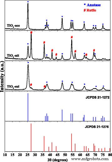
煅烧后的 TiO2 样品的粉末 XRD 图(经 [29] 许可转载。版权所有 @ 2017 Elsevier)
TiO2-sul 样品主要具有锐钛矿相 (95%),粒径约为 23 nm;然而,在该样品中可以看到对应于金红石相 (110) 面的小衍射峰。相比之下,TiO2-nit 样品显示锐钛矿和金红石相的 XRD 反射,微晶尺寸分别为 41 nm 和 50 nm。据观察,金红石是该样品中的主要相 (67%)。这些结果表明胶溶酸的性质在TiO2相的形成中起作用。
高分辨率透射电子显微镜
进行 TEM 以检查合成的 TiO2 纳米粉末的颗粒尺寸、结晶度和形态。合成的 TiO2 纳米粉末的 TEM 和 HRTEM 图片显示在图 2 中。可以看出,TiO2-sul 样品由紧密堆积的附聚锐钛矿颗粒组成,估计平均粒度约为 7 nm。 TiO2-nit 样品具有大小在 10 到 20 纳米之间的球形纳米颗粒和宽度为 20 纳米的大片。相比之下,TiO2-ace 样品由 TiO2 纳米颗粒(15-20 纳米)组成,主要由球形形态组成。 Vinogradov 和 Vinogradov [31] 也观察到类似的结果,当使用强胶溶酸(如 HNO3 和 H2SO4)进行胶溶时,检测到小尺寸聚集体。与使用 TEM 分析测量的晶粒尺寸相比,由 Scherer 公式测量的晶粒尺寸导致更大的晶粒尺寸。如前所述,微晶尺寸与晶粒尺寸不同;然而,在某些情况下,微晶尺寸可能与晶粒尺寸相匹配 [32]。可以观察到,TiO2-sul 和 TiO2-ace 样品的 HRTEM 图像显示颗粒包含对应于具有 d 的锐钛矿晶格平面的条纹 -(101) 面 [33] 的间距为 0.356 nm,而 TiO2-nit 样品的 HRTEM 图像显示具有 d 的金红石晶格面 (110) 的晶格条纹的颗粒 -与锐钛矿晶格 (101) 面的间距为 0.325 nm。
<图片>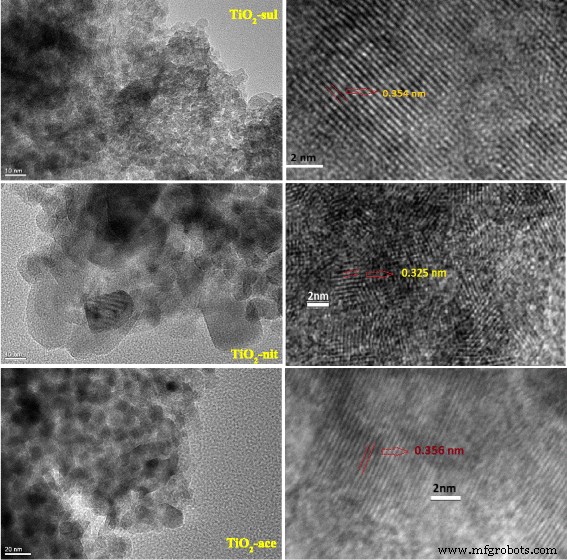
煅烧后的TiO2样品的TEM和HRTEM图像
拉曼光谱
拉曼光谱也用于探测合成的 TiO2 样品中的相形成。图 3 显示了在 500°C 下煅烧的三个 TiO2 样品的拉曼光谱。据报道,锐钛矿相和金红石相分别具有 6 个和 5 个活跃的拉曼谱带(锐钛矿 143、195、395、512 和 638 cm -1 ;金红石 145、445、611 和 826) [34]。从图 3 可以清楚地看出,所有三个样品都显示出高度强烈的锐拉曼带 (E g) 在 141–146 cm −1 范围内 ,这是由于锐钛矿相的存在的特征带。在插图中可以清楚地观察到由于锐钛矿和金红石相引起的低强度拉曼谱带。由于锐钛矿相和金红石相,TiO2-nit 和 TiO2-sul 样品显示出拉曼谱带;然而,在 TiO2-nit 样品的情况下,由于金红石相的存在,拉曼谱带的强度很高。相比之下,TiO2-ace 样品仅由于锐钛矿相而表现出拉曼谱带。
<图片>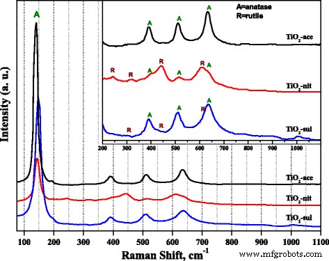
煅烧后TiO2样品的拉曼光谱
据报道,拉曼光谱结果可用于研究 TiO2 纳米粒子的粒径,因为拉曼谱带的异常带移可能与样品粒径的减小有关 [35]。在图 3 中,TiO2-ace 样品表现出 E g 波段在 141.5 cm −1 ;然而,波段转移到 146 和 150 cm -1 分别是 TiO2-nit 和 TiO2-sul 样品。拉曼光谱观察表明,TiO2-sul样品的粒径小于其他两种样品,这与XRD和TEM观察结果一致。
漫反射 UV-vis
DR UV-vis 合成的 TiO2 样品在 500°C 下热处理的光谱如图 4 所示。DR UV-vis 导数中峰值的位置 三个样品的光谱显示在图的插图中。它清楚地表明样品在紫外区具有很强的电子反射。使用三种不同酸合成的样品的反射峰最大值不同。 TiO2-sul 样品在 372 nm 处显示出峰值,而在 TiO2-ace 和 TiO2-nit 样品中分别移至 383 nm 和 402 nm。据报道,锐钛矿和金红石的带隙能量分别为 3.2 eV (380 nm) 和 3.0 eV (415 nm) [1]。反射率最大值的差异可归因于样品微晶尺寸和相结构的变化[36]。对于具有更多金红石相百分比的样品,吸收最大值向更高波长移动。通过确定 hν 和 (αhν) [2] [附加文件 1:图 S1] 之间的关系,计算煅烧样品的带隙能量 (eV)。数据显示,与 TiO2-ace (2.99 eV) 和 TiO2-nit (2.97 eV) 相比,TiO2-sul (3.12 eV) 的带隙能量更高。当样品中金红石相占主导地位时,TiO2 的带隙减小。据报道,锐钛矿相和金红石相的价带 (VB) 主要是由于 O2p 状态;另一方面,导带 (CB) 由 Ti 3d 组成 状态 [37]。 TiO2 的带隙能量是由 CB 和 VB 位置建立的,这主要受相组成的影响。因此,同时含有锐钛矿相和金红石相的样品的带隙能量应介于纯锐钛矿和金红石相之间。
<图片>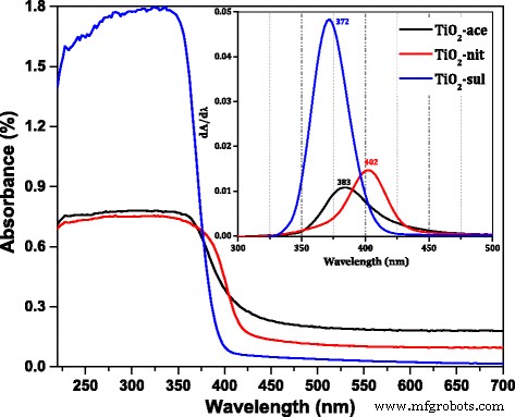
DR UV-可见光 煅烧 TiO2 样品的光谱(插图;DR UV-vis 的导数 光谱)
N2-物理吸附测量
三个合成样品的氮吸附-解吸等温线如图 5a 所示。对于通过用乙酸 (TiO2-ace) 和硫酸 (TiO2-sul) 胶溶合成的样品,观察到具有 H2 型滞后回线的 IV 型等温线。这表明这两个样品具有由 TiO2 纳米颗粒聚集体产生的介孔。然而,对于 TiO2-nit 样品,观察到具有窄 H3 型滞后回线的典型 IV 型等温线(开放和/或狭缝形孔的特征)。还可以观察到滞后回线在较高的相对压力 (P/P 0 = 1) 并且这一观察结果表明存在大尺寸的孔[38]。
<图片>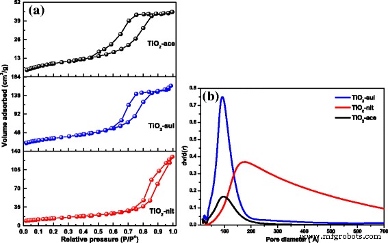
一 N2 吸附-解吸等温线。 b 煅烧后的 TiO2 样品的孔径分布(经 [29] 许可转载。版权所有 @ 2017 Elsevier)
合成材料的 BJH 模型孔径分布是从等温线的吸附分支值获得的。样品的 BJH 孔径分布如图 5b 所示。对于 TiO2-ace 和 TiO2-sul 样品,观察到窄的单峰孔径分布。然而,对于用硝酸制备的样品,观察到更宽的孔径分布,这可能是由于较大颗粒之间存在空隙空间。样品的质地特性如表 1 所示。结果表明,高表面积 (115 m 2 g −1 ) 观察到在 500°C 下煅烧的 TiO2-ace 样品。 S 的顺序 BET 变化为 TiO2-ace> TiO2-sul> TiO2-nit。观察结果清楚地表明,合适的胶溶条件在制备具有多孔结构的纳米粒子方面非常有效。
傅立叶变换红外光谱
TiO2 的光催化活性取决于结晶度、微晶尺寸、组成、电子-空穴复合率、表面积以及表面羟基的密度[39]。 FTIR 和 XPS 光谱技术用于研究煅烧 TiO2 样品中存在的 –OH 基团的性质。图 6 显示了三个 TiO2 样品在 1600-4000 cm -1 范围内的 FTIR 光谱 .据报道,TiO2载体可以具有不同类型的表面羟基;它们可以归类为孤立的Ti-OH、羟基通过氢键和化学键合的H2O分子相互键合[40]。
<图片>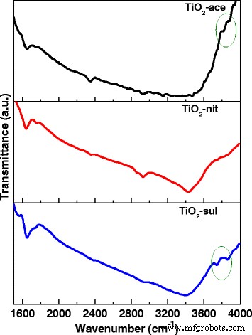
煅烧后的TiO2样品的FTIR光谱
三个样品显示出以 3408 cm -1 为中心的宽带 ,这归因于 O-H 基团(水分子和自由表面 -OH 基团)的伸缩振动。在 2340 和 1640 cm −1 处也出现了额外的波段 ,这可以分别归因于 O-H 伸缩振动和分子吸附的 H2O [41]。在锐钛矿情况下存在两个 –OH 伸缩振动(在 3715 和 3675 cm -1 ) 和 3680 cm −1 处的一个弱波段 以前曾报道过金红石 [42]。在合成的 TiO2 样品中可以看到非常相似的结果。
X 射线光电子能谱
图 7 显示了去卷积的 Ti2p 和 O1s 合成的 TiO2 样品的 XP 光谱。三个样品在 457.2 和 463.1 eV 处显示了两个主要峰,对应于 2p 3/2 和 2p 1/2 Ti 4+ 在二氧化钛中 [43]。在 Ti 2p 中观察到非常相似的结合能值 所有三个 TiO2 样品的区域表明这些样品中的 Ti 原子以相同的氧化态存在。对于所有样品,还观察到 455.8 和 458.7 eV 处的两个小肩峰。 455.8 eV 的肩峰可以分配给 Ti 3+ 状态,由于 TiO2 [44] 缺氧,而 458.7 eV 的另一个肩峰来自 Ti 4+ Ti-OH 物质的状态 [45]。从 Ti2p 可以看出 光谱表明,TiO2-nit 中缺氧 TiO2 物质的贡献高于 TiO2-sul 和 TiO2-ace 样品。所有样本均显示 O1s XP 在 528.4、529.3 和 531.3 eV 处达到峰值。 528.4 eV 处的 XPS 峰可归因于 O-Ti 4+ TiO2 晶格中的氧物种,而 529.3 和 531.3 eV 的其他两个峰可归为表面吸附羟基中存在的氧物种 [46]。
<图片>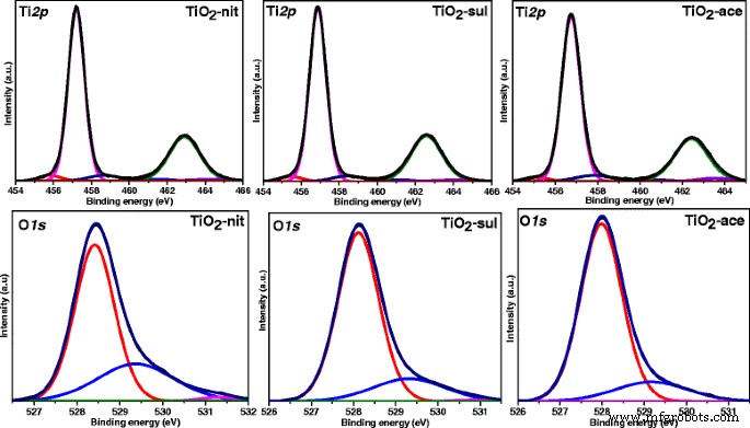
Ti 2p 和 O1s 煅烧后的TiO2样品的X射线光电子能谱
McCafferty [47] 还观察到 O1s 峰在高结合能值处具有尾峰,这可能是由于 Ti-OH 基团的存在。由于物理吸附表面,Ti-OH 基团可以在用于操作 XPS 仪器的超高真空下轻松去除 [48]。样品中出现的这些-OH 基团一定是由于 Ti-OH 化学键合到 TiO2 的表面缺陷,其中-OH 基团在 TiO2-nit 样品的总氧物种中的百分比略高于 TiO2- sul 和 TiO2-ace(表 2)。
结晶紫、亚甲蓝和Para的光催化降解 -硝基苯酚染料
煅烧后的 TiO2 纳米颗粒对 CV、MB 和 p 降解的光催化活性 -NP 被估计。据报道,光催化降解反应一般遵循Langmuir-Hinshelwood 动力学[1]。因此,有机染料的光催化降解可表示为
$$ -\mathrm{dc}/\mathrm{dt}=\mathrm{kC} $$ (5)并在积分后,方程。 (4) 可导出
$$ C={C}_0{\exp}^{\left(-\mathrm{kt}\right)} $$ (6)其中 C 0 是有机染料的初始浓度(ppm),k 是速率常数,它取决于反应时间、温度和溶液 pH 值。通常情况下,催化剂的光催化效率随着运行时间的延长而增加。
进行空白实验以确认光催化剂和紫外线照射的重要性。当单独施加催化剂和紫外线照射时,没有进行任何反应。在我们之前的发现中观察到类似的结果 [49]。如实验部分所述,TiO2 光催化剂与有机染料溶液平衡 45 分钟,以确定有机染料对合成 TiO2 样品的吸附。紫外光可见光 CV、MB和p的吸收光谱 -NP 在光催化剂平衡后记录。附加文件 1:图 S2、S3 和 S4 显示了 UV-vis 的变化 CV、MB 和 p 的吸收光谱 -NP 溶液 (10 ppm) 分别在 TiO2-ace、TiO2-sul 和 TiO2-nit 样品上具有不同的反应时间。对应于 CV、MB 和 p 的吸收峰强度 -NP随着反应时间的增加而降低。紫外光可见光 反应产物的光谱表明有机染料在光反应过程中降解。与 TiO2-sul 和 TiO2-ace 样品相比,发现 TiO2-nit 样品是最有效的光催化剂。 p 50% 的降解 TiO2-nit 样品在 60 分钟内观察到 -NP,而降解 50% p 需要 75 和 100 分钟 TiO2-ace 和 TiO2-sul 样品在相似条件下的 -NP。对MB和CV染料的降解观察到相似的光催化活性模式。
所研究的催化剂的百分比降解效率使用方程计算。 (4).图 8 显示了 CV、MB 和 p 的百分比变化 -NP 水溶液在室温下存在煅烧 TiO2 样品。反应仅 10 分钟后,TiO2-nit 样品的 CV 降解效率为 29%,而 TiO2-ace 和 TiO2-sul 样品的 CV 降解效率分别仅为 17% 和 9%。三个样品的光催化活性随着反应时间的增加而急剧增加。然而,120 分钟后,TiO2-nit 和 TiO2-ace 样品显示出 99% 的效率;然而,TiO2-sul样品的效率仅为65%。
<图片>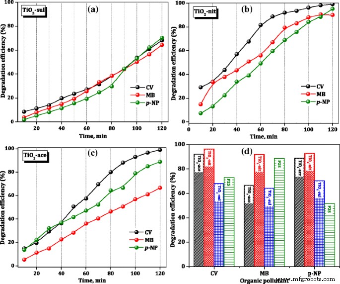
TiO2催化剂的光催化降解效率
为了比较合成 TiO2 样品的光催化性能,商业 P25 样品在 120 分钟后对有机染料的降解效率包含在图 8d 中。 It is clear that TiO2-nit sample showed better performance than the P25 sample in the degradation of three organic dyes; however, TiO2-ace and TiO2-sul samples showed lower activity than the P25 catalyst in case of p -NP degradation. These results are suggesting that the performance of catalysts is influenced by the physicochemical characteristics of the TiO2 samples and nature of the organic dye.
The rate constants for photocatalytic degradation of CV, MB, and p -NP over synthesized TiO2 samples and commercial P25 sample were determined from the slope of the straight line which is plotted between ln(C 0/C t) and t , and the results are presented in Table 3. The observed results are indicating that the photocatalytic activity of the degradation of organic dyes was greatly influenced by the composition of TiO2 sample and amount of the surface hydroxyl groups. The activity indeed is not influenced by the particle size, crystallinity, and surface area of TiO2 synthesized in this work. This observation is not consistent with the results observed by Fujishima et al. [8] that the catalyst which possesses lower particle size offered high photocatalytic efficiency.
Previously, it was reported that anatase is a better photocatalyst than rutile due to its high band gap energy and a large number of surface OH groups [50]. It was thought that TiO2-nit sample would offer low photocatalytic activity due to the presence of more rutile phase (67%). However, Masahashi et al. [51] claimed that rutile exhibited higher performance than anatase MB degradation due to its superior crystalline nature.
Determination of photocurrent values was carried out to obtain a better insight responsible factor for the superior photocatalytic performance of the samples containing more rutile. It was reported that photocatalytic activity is directly related to the electron-hole separation efficiency of a catalyst which is influenced by the photocurrent density [52]. Theoretical photocurrent density of the TiO2 samples was calculated from the absorption edge of the TiO2 samples (obtained from DR UV-vis spectroscopy measurements) and theoretical equations (supporting information) presented in the literature [53]. The results of photocurrent of TiO2 samples are presented in Table 4 along with the percentage of rutile and photocatalytic efficiency values. The photocurrent density of TiO2-nit (0.545 mA/cm 2 ) is higher than other two synthesized TiO2 samples and also commercial P25 sample (0.401 mA/cm 2 ), manifesting the beneficial role of rutile phase in improving the photoactivity of TiO2 samples.
Previously, Melcher et al. [54] reported that photocatalytic capability of the commercial P25 material originates due to the presence of a mixture of rutile and anatase phases in the sample (75% anatase and 25% rutile). Hirakawa et al. [55] indicated that pure rutile itself is not a powerful photocatalyst, and it is also reported that the light with a wavelength of 380 nm is not powerful enough to generate charge carriers in the pure anatase [56]. Based on XPS spectroscopy results and theoretical calculations, Scanlon et al. [57] concluded that electrons were moved from rutile to anatase and the holes were transported from anatase to rutile, which inhibited the electron-hole recombination.余等人。 [58] reported a similar observation that TiO2 sample with mixed phases was beneficial to decrease the rate of h + -e − recombination and thus enhance the photocatalytic efficiency of the catalyst.
In literature reports, two possible transfer mechanisms have been proposed for anatase-rutile composite samples [59]. The first mechanism is the interfacial electron transfer from CB of anatase to that of the rutile [60], and the second one is an electron transfer from CB of rutile to lower energy anatase active sites [61]. It is known that the anatase CB possesses higher negative potential than the rutile CB due to the fact that anatase has a higher band gap (3.12 eV) than rutile. Therefore, it is not possible for an electron to move from the rutile CB to the anatase CB because it would have to overcome the energetic barrier between the two bands. The band gap of anatase VB is also slightly higher, than the rutile VB, so the generated holes could be moved to the anatase VB, to achieve an effective charge separation. Most probably, the electron-hole pair is formed in the composite of rutile and anatase in case of TiO2-nit and TiO2-sul samples (Fig. 9), and this rate is much higher in TiO2-nit sample due to predominant rutile formation.

Plausible model of a generation of electron-hole pairs and b effective charge carrier separation via transfer of the generated holes into the anatase valence band
It was reported that increase of crystal growth of initial phase is possible by increasing the mobility of ions presented in precursor solution [62]. Several researchers added small volumes of mineral acids (such as hydrochloric acid and sulfuric acid) to improve the mobility of dissociated ions [63]. Their role is not only to increase the rate of diffusion of ions in a solution but also to alter the surface charge. Under humidity conditions, titanium isopropoxide can subsequently undergo hydroxylation and polymerization to TiO2.
$$ \mathrm{Ti}{\left(\mathrm{OPri}\right)}_4+4{\mathrm{H}}_2\mathrm{O}\to \mathrm{Ti}{\left(\mathrm{OH}\right)}_4+4\ \mathrm{PriOH}\kern2.5em \left(\mathrm{hydroxylation}\right) $$ (7) $$ \mathrm{Ti}{\left(\mathrm{OH}\right)}_{4\kern0.5em }\to {\mathrm{TiO}}_{2.}x{\mathrm{H}}_2\mathrm{O}+\left(2\hbox{-} x\right)\ {\mathrm{H}}_2\mathrm{O}\kern5em \left(\mathrm{condensation}\right) $$ (8)Depending on the nature of peptizing acid, the transformation of TiO2 leads to anatase or rutile phase [64]. Formation of amorphous TiO2 or metastable anatase phase was observed when the condensation initiated before hydrolysis of Ti precursor. Under highly acidic conditions, the rutile phase formation is favorable as the rate of condensation is slow. Accordingly, the rutile phase was obtained when sulfuric and nitric acids were used for the peptization. The use of weak acid (acetic acid) as a peptizing agent allows the control of both the degree of condensation and oligomerization and persuades the preferential crystallization of TiO2 in the anatase phase.曾等人。 [20] used polycarboxylic acid as a peptizing agent and observed the formation of nanoparticles of anatase which they attributed to chelation effect of organic acid.
It is known that TiO6 octahedra are a fundamental structural unit for both anatase and rutile phases (D 4h system), and the only difference between these two structures is the assembly of the octahedral chains [65]. Face-shared linking of TiO6 units results in anatase structure, while edge-shared linking results in rutile structure [66]. It is clear that NO 3− anions possessed weaker affinity to Ti atoms in an aqueous solution than CH3COO − and SO4 2− 阴离子。 The strong affinity of CH3COO − and SO4 2− anions with Ti atoms is responsible for the inhibition of the phase transformation.
In the previously reported studies, many of the photocatalysts have not been tested for reuse mainly due to undergo photocorrosion; hence, their photostability is reduced for further usage. The reusability of the calcined TiO2 samples was examined to study the effectiveness of these photocatalysts. It was observed that the used photocatalyst offered 90% efficiency for three consecutive cycles. The efficiency of the catalyst was reduced to 80 and 75% during fourth and fifth cycle, respectively. The decrease is due to the loss of some amount of catalyst during the filtration and regeneration procedures.
Conclusions
A simple peptization method was adapted to synthesize TiO2 nanoparticles by using sulfuric, nitric, and acetic acid as peptizing agents and titanium isopropoxide as Ti precursor. The influence of acid species on the crystal phase, morphology, textural, and surface composition of TiO2 were studied in detail. The TiO2 sample peptized with acetic acid possessed pure anatase phase, while the formation of minor (5%) and major (67%) of rutile phase was observed in case of samples peptized with sulfuric acid and nitric acid, respectively. It is observed that TiO2 peptized with nitric acid showed sheet-like structures along with nanoparticles, while TiO2 samples peptized with sulfuric and acetic acids possessed near spherical nanoparticles. The photocatalytic properties of synthesized TiO2 nanostructures were evaluated for photodegradation of aqueous CV, MB, and p -NP solutions. The TiO2 peptized using nitric acid showed the best photocatalytic activity than commercial P25 and other two peptized samples, and its photodegradation efficiency was reached to 95% in 120 min for p -NP degradation. Although TiO2 samples peptized using sulfuric acid and acetic acid possessed smaller particle size, higher band gap energy, and high surface area, TiO2 sample peptized with nitric acid possessed a higher percentage of rutile and photocurrent density. The observed photocurrent density is dominated by the photoactivity of TiO2. The results indicate a direct correlation between the photocatalytic activity and the photocurrent density of the TiO2 samples. The superior activity of TiO2 sample peptized with nitric acid is due to the effective transfer of photogenerated electrons between rutile and anatase phases, and large pore diameter could have enhanced the diffusion and mass transportation of reacting molecules and OH radicals during the photochemical reaction. The synthesized TiO2 photocatalysts can be recycled with a minor change in the activity.
缩写
- CV:
-
Crystal violet
- DR UV-vis :
-
Diffuse-reflectance ultraviolet-visible spectroscopy
- FTIR:
-
傅里叶变换红外光谱
- HRTEM:
-
高分辨透射电子显微镜
- MB:
-
Methylene blue
- NMR:
-
Nuclear magnetic resonance
- p –NP:
-
para –nitro phenol
- TEM:
-
透射电子显微镜
- TiO2 :
-
Titanium oxide
- XPS:
-
X射线光电子能谱
- XRD:
-
X-ray powder diffraction
纳米材料
- SrTiO3 改性金红石型 TiO2 纳米纤维的一步静电纺丝路线及其光催化性能
- 两种石墨烯改性二氧化钛复合光催化剂的高光催化性能
- 纳米颗粒作为外排泵和生物膜抑制剂,恢复常规抗生素的杀菌效果
- 迈向 TiO2 纳米流体——第 2 部分:应用和挑战
- TiO2 中金纳米粒子分布对染料敏化太阳能电池光学和电学特性的影响
- Li/Nb 比对 Li-Nb-O 化合物制备和光催化性能的影响
- Au-Plasmonic 纳米粒子在涂有 MoO3 的 TiO2 纳米管光电极上增强光催化活性
- 中空结构LiNb3O8光催化剂的制备和光催化性能
- 用于有色冷色颜料的 Cr 掺杂 TiO2 的结构和可见近红外光学特性
- TiO2 纳米管阵列:由软硬模板制造和场发射性能的晶粒尺寸依赖性
- 使用表面光谱分析测定过渡金属掺杂的 TiO2 纳米颗粒的催化活性
- 圆形金纳米粒子:粒径和浓度对拟南芥根系生长的影响